
t PowerPoint PPT Presentation
1 / 42
Title: t
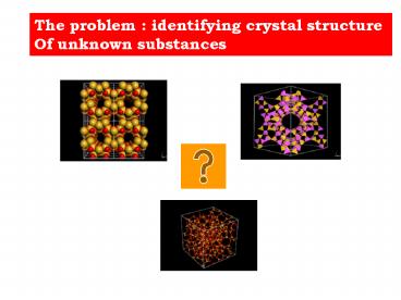
1
The problem identifying crystal structure Of
unknown substances
t
2
Powder diffractometry is the solution for phase
identification but NOT for ab-initio structure
analysis
3
TEM microscopy may resolve crystal structures
using high resolution images of individual
crystals
120 kV
200 kV
300 kV
300 FEG kV
4
However, high resolution imaging IS NOT
directly interpretable in terms of crystal
structure
Through-focus series of YBa2Cu3O8 along 100
Coene et al. 1992, PEO Bull. 132
200 kV FEG S-TWIN
5
Image interpretation IS NOT straightforward Sever
al corrections are needed....
6
Grain boundary atomic resolution Focus-series
reconstruction
atomistic interpretation of the grain boundary
structure
Image courtesy of C.L. Jia A. Thust,
Forschungszentrum Juelich
200 kV FEG S-TWIN
7
High resolution imaging HR-STEM
Si lt110gt
SrTiO3
1.4 Å
Ti
Sr
Sample courtesy Dr. B. Wiedenhorst, University
of Cologne, Germany
8
How to solve a structure ? The classic phase
problem
Phases
Amplitudes
FT
IFT
- We measure IF(k)I, the modulus
- r ( r ) exp (2pik.r) I F(k) I exp (
i f (k)) dk - Phase information , f (k) is lost
9
ELECTRON CRYSTALLOGRAPHY X-RAY
CRYSTALLOGRAPHY
To reconstruct a TEM image,Intensities and
phases of all reflections are needed FT
Ad exp (-ifd ) FT AC exp (-ifC ) FT
Ad exp (-ifC ) Phases of cat amplidude of
duck Cat In general, phases of reflections
are more important than amplitudes
IFT
10
ELECTRON CRYSTALLOGRAPHY X-RAY CRYSTALLOGRAPHY
infinite number of possible arrangements of
atoms
DIRECT METHODS
Intensities are known, phases are unknown ...
Finite
R, c2, structure and chemical criteria
Structure resolved
11
ELECTRON CRYSTALLOGRAPHY X RAY
CRYSTALLOGRAPHY
12
ELECTRON DIFFRACTION X RAY DIFFRACTION
Electron diffraction scattering is much
stronger than X-Ray for fx 10 exp -11 cm,
fe 10exp -8 cm
Ix / Ie / In fn
10 exp -12 cm 1 / 10 exp 6
/ 10 exp 2
Ix fx exp2 Ie fe exp2
In fn exp2
- (xyz)
- electron density
DETECTABILITY OF LIGHT ATOMS Electron
diffraction amplitudes are only weakly dependent
on Z fe (0) Z exp 1/3 fx (0)
z fe (senq/l) z fx ( senq/l
) z exp 3/2 Contribution of light atoms to the
total intensity compared to heavy atoms is
greater in electron diffraction
X-RAYS
ELECTRONS
- (xyz)
- atomic potential
13
ELECTRON DIFFRACTION ADVANTAGES OVER X-RAY
DIFRACTION DETECTABILITY OF LIGHT ATOMS
(Vainstein , 1964 ) If we define w
f(0) Zlight / f (0) zheavy as
detectability then w ( z light / z heavy )
exp 0.75 for electron diffraction
w ( z light / z heavy ) exp 1.25 for
X-Ray diffraction Electron diffraction is most
valuable for detecting light atoms in presence
of heavy ones This can be expressed by
w/ w ( Z heavy / Z light ) exp ½ For
example , in organic structures (Z carbon / Z
hydgogen ) exp 0.5 2.5 2.5 times easier to
detect H in organic crystals with electron
diffraction than by X-Ray diffraction
14
Many structures have been solved by electron
diffraction recently ( Dorset,Gilmore,Hovmoller,Z
ou,Marks,Sinkler,Terasaki,Weirich)
Electron diffraction is much more sensitive
than X-Ray diffraction
15
Electron diffraction is much more sensitive
for light atom detection than X-Ray
H atoms detected accurately
16
Electron diffractometry in EDC
Disadvantages beam size (1 micron 1 mm)
,time of analysis several hours
17
Precise electron diffractometry Measurements
performed only at ED cameras, NEVER at TEMs
up to now ...
18
Electron diffractometry in TEM
Any TEM (100 300 KV) can be used...
19
Electron diffractometry in TEM
Measure accurately Intensities (lt1)
Any crystal from 1 nm 1 micron can be studied
Convert intensities to Structure Factors
Correct for dynamical diffraction effects
Find structure by using direct methods to
assign phases for all reflections
20
ED dynamic range of intensities
I 10 exp 6
I 1
to find crystal structure ,all intensities need
to be measured with precision lt 1-3 , specially
the weak ones
21
Electron diffractometry in TEM challenges
Dynamical scattering Ihkl k I Fhkl I
Wrong model light atoms do not appear Atomic
positions displaced
Kinematical scattering Ihkl k I Fhkl I exp
2
Correct model
22
Electron diffractometry in TEM
23
TEM
Gun
Condenser stigmator coils (CSC)
C1
Deflection and beam tilt coils (DBTC)
C2
C2 aperture isolated to record beam current
Obj Up
Specimen
Obj Lo
Objective stigmator and alignment coil (OSDAC)
Diff
Diffraction stigmator coil (DSC)
Int
Diffraction and Intermediate Alignment coil
(DIAC)
Proj
35 mm camera port
Diffractometer with retractable Faraday cage
(FC) In case of installed at 35 mm camera port
Web cam or CCD camera takes diffraction image
through The window of the projection chamber
Fluorescent screen
Mechanical interface for diffractometer (MID)
MID
Faraday cage (FC) accumulation preamplifier
FC
24
Precession of beam
ED pattern is scanned above a fixed detector
f
PM, Faraday cage, CDEM
Vincent- Midgley method
Electron beam is tilted and precessed
equivalent of tilting Laue zone Much decrease of
dynamical diffraction contribution
25
Precession
Special scan-descan interface available even if
STEM not present in a TEM
e
Upper coil drives
Specimen plane
Lower coil drives
To obtain a stationnary ED pattern scan and
descan must be in exact antiphase
No precession
26
CONTROL UNIT DIFFRACTOMETER
Unit to control hollow cone for precession
27
NdAl3(BO3)4 without precession
28
NdAl3(BO3)4 with precession
29
NdAl3(BO3)4 without precession
30
NdAl3(BO3)4 with precession
31
POWDER BaF2 without precession
32
POWDER BaF2 with precession
33
Preview CCD CAMERA
34
Selection of scanning parameters
35
ELECTRON DIFFRACTOMETER SPECIFICATIONS
Beam blanking during area selection by the user
Integral Intensity of reflections and background
substraction is measured automatically,
Prescan Any area of reciprocal space ( r, f
coordinates ) can be selected for scanning
linescan or individual hkl reflections Scanning
variable step and pixel sixe scan for better
resolution Acumulation mode all intensities in
ED pattern are measured with SAME accuracy lt 1
36
New software for correction of dynamical
diffraction calculation of many-beam intensity
for the hollow-cone (precession) mode of
electron diffraction. New program for this
calculation will use algorithms which have been
elaborated by Avilov in two works Avilov A.S.,
and Parmon V.S. Nonsystematic many beam
interaction in high energy electron difraction.
Sov.Phys.Crystallography. 1983. Vol.28. No.2.
P.294-295 AvilovA.S., et all. Calculation of
reflected intensities in multiple-beam
diffractionof fast electrons by polycrystalline
specimens. Sov.Phys.Crystallography. 1984.
Vol.29. No.1. P. 5-7. The algorithm allows to
calculate the excitation errors for all
diffracted intensities for any arbitrary
orientation of crystals relatively the primary
beam. In the case of the hollow-cone mode of the
electron diffraction we will have consistent
positions of the primary beam, relatively the
surface of crystal, when the beam moves along
the surface of cone having constant angle between
the primary beam and the axe of the cone. It is
necessary to calculate many beam intensities for
every position of the PrB and to sum all
intensity for the full cicle of the PrB. The
calculation of the many beam intensity can be
made by the well known way using the matrix
formulation of the theory M ?
? x?,
. P0 n0 nog Where dynamic matrix
M nog ph nhg , nondiagonal
elements are the structure
nog v gh pg
amplitudes and diagonal are connected with the
excitation errors. Then the problem with
eigen-functions and eigen-values is solved by
known methods.
37
Electron diffractometry in TEM Application
example Lix Mn2 O4 cubic
spinel compound
a 0.82 nm Position and stoichiometry of
Li atoms directly linked to electrochemical
properties. Howerer , is impossible to localize
Li atoms with X-Ray diffraction
(powder). Electron diffractometry can give
solution to this problem.
Using electron diffraction the relative
detectability of Li atoms in presence of
heavier Mn is ( Z heavy / Z light ) exp ½
2.886 times easier to detect Li with electron
diffraction than by X-Ray diffraction
38
SAED of LixMn2O4 ( sample 100) along 111
a
b
2 2 0
2 2 0
2 0 -2
2 0 -2
No precession
With precession
Precise measurement for all intensities
(accumulation mode) with DI/Ilt 3 , collection
time 90 min , dynamic range 10 exp 5
39
Projection of the Density map of LixMn2O4 (100)
along 111, Corresponding model is depicted.
2.9 Å
Mn, Li, O
Mn, O
ED precession mode
40
Projection of the potential map of LixMn2O4
along 110
41
CONCLUSION Li atomic positions can be
localized with electron diffractometer
o
o
Li
Li
Mn
Mn
Mn
Li
Li
Mn
Mn
Precession mode no dynamical diffraction
Mn
Mn
Li
Li
Mn
Mn
Mn
Li
Li
42
ELECTRON DIFFRACTOMETRY
can be used in any TEM e.g 100-120-200-300-400
kV LaB6 / FEG GET ATOMIC STRUCTURE FROM
ELECTRON DIFFRACTION LIKE X-RAY
DIFFRACTOMETRY Correction of dynamical
diffraction using beam precession and
software of many-beam
interaction
Accurate intensities can also be used in HREM
for phase extension
Recommended
