
Syndesmosis Injuries PowerPoint PPT Presentation
Title: Syndesmosis Injuries
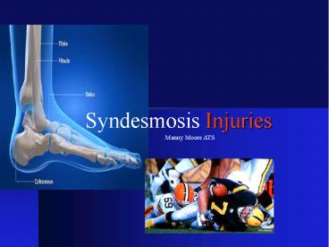
1
Syndesmosis Injuries
Manny Moore ATS
2
Syndesmosis Injuries
11-18 of all ankle sprains Longer recovery v.s.
Lateral sprains Men v.s. Women?
3
Bone Anatomy
- Tibia
- Articular Surface
- Fibula
- Articular Surface
- Talus
- Dome
Provides Stability Proper Ankle Function
Articular Surface
4
Snydesmosis Ligaments
- AIFL- Chaputs Tubercle
- Most Vulnerable
- PIFL- Wagstaffes Tubercle
- Strongest
- ITFL- Thickening of PIFL
- IM- Fibrous tissue
- Transmit force
- IL- Thickening of IM
5
Biomechanics
- Mechanism of Injury
- Eversion
- Dorsiflexion
- Pronation
- Closed Pack Position
- Forces the talus against the fibula
- Widening of mortise
- 1mm lateral shift increases joint
surface pressure by 42
Associated injuries?
6
Clinical Examination
- History
- ER with DF
- Contact
- None Contact
- Acute v.s. Chronic
- Observation
- Edema
- Eccymosis
- Antalgic gait
- Possible Deformity?
7
Clinical Examination
- Palpation
- Tenderness Length
Nussbaum et al.
- Special Test
Squeeze Test Dorsiflexion Test Kleigers
Test Cross-leg Test
8
Imaging Techniques
X-RAY
- Radiographs
- AP, Lateral, Mortise Views
- AP View
- Fractures
- Tibiofibular clear space
widening of 6 mm - Tibiofibular overlap gt 42
Fibula Width - Medial clear space widening gt 4mm
- Lateral View
- Non weight bearing ER
- Fractures
9
Imaging Techniques
X-RAY
Tibiofibula overlap
Tibiofibula clearance space
Medial clear space
10
Imaging Techniques
X-RAY
Tibiofibula overlap
Tibiofibula clearance space
Medial clear space
11
Imaging Techniques
X-RAY
- Lateral View
12
Imaging Techniques
X-RAY
- AP View
Heterotopic Ossification
13
Imaging Techniques
MRI CT
- MRI (Magnetic Resonance Imaging)
- Frontal, Axial, Saggital Views
- High sensitivity and specificity
- More reliable detecting disruptions
- CT (Computed Tomography)
- More effective detecting minor disruptions
- Less Cost v.s. MRI
14
Imaging Techniques
MRI
- Axial Views
15
West Point Instability Scale
Grade I
Grade II
Grade III
Edema Ecchymosis Localized Mild Localized Moderate Diffuse Severe
Weight Bearing Ability Full or Partial Without Significant Pain Difficult Without Crutches Impossible Significant Pain
Ligament Damage Ligament Stretch Partial Tear Complete Tear
Ligament Involvement AIFL AIFL IL Possible AD AIFL/PIFL IL AD
16
Treatment Criteria
Based on Patients Goals Length of
Symptoms Severity of Injury
- Conservative
- Non Conservative
Grade I Non-Fractures Stable Grade II
Grade III Unstable Grade II Fractures Chronic
Injury
17
Conservative Protocols
- Results vary patient to patient
- Grade I Injuries 2-4 Weeks RTP
- Grade II Injuries 6-8 Weeks RTP
- Without Instability or Fractures
18
Conservative Protocols
- Phase I (0-5 Days) or (5-14Days)
- Immobilize
- Reduce Pain
- Reduce Inflammation
- Cryotherapy
- E-Stim
- Increase ROM
- Manual 30 PF Stretch
- Ankle Pumps
- Toe Curls
- Towel Stretch
19
Conservative Protocols
- Phase II (6-10 Days) or (2-4 weeks)
- Immobilize Grade II
- Reduce Pain
- Reduce Inflammation
- Proprioception
- Increase Flexibility
- Increase ROM
- Increase Strength
- CV Endurance
20
Conservative Protocols
- Phase III (18-25 Days) or (4-8 Weeks)
- Protect Injury
- Reduce Pain
- Increase Pain free Activity
- Sports Specific
- Proprioception
- Increase Strength
- Increase Flexibility
- CV Endurance
21
Conservative Protocols
- Phase III (18-25 Days) or (4-8 Weeks)
- Sports Specific
Drill2
Drill1
22
Conservative Protocols
Return To Play Criteria
- Full Strength
- Full ROM
- Functional Test
- Physician Clearance
- Protect Injury
23
Operative Treatment
Arthroscopy
- Goal is to restore structures, and mobility
- Open Reduction Internal Fixations
- Autographs
- Modified Brostrum Technique
- 4.5 mm Cortical Screws
- Complications
- Screw Breakage
- Screw Type
- Infection
- Calcification Joint Stiffness
24
Operative Treatment
Arthroscopy
Before
After
25
Post-Operative Protocols
Arthroscopy
- Results vary patient to patient
- Grade III Injuries 4-8 Months RTP
- Non Weight Bearing 6-8 Weeks
- Screw Removal _at_ 3 Months
- Follow-up Imaging every 2 weeks
26
Post-Operative Protocols
- Phase I (1-3 Weeks)
- Phase I- Conservative Rehabilitation
- Immobilize Non Weight Bearing
- Protect Wound
- Reduce Pain
- Reduce Inflammation
- Proprioception
- Increase ROM
- Maintain Flexibility
- CV Endurance
27
Post-Operative Protocols
- Phase II (3-8 Weeks)
- Phase I- Conservative Rehabilitation
- Immobilize Partial Weight Bearing
- Protect Wound
- Reduce Pain
- Reduce Inflammation
- Increase ROM
- Increase Strength
- Proprioception
- Increase Flexibility
- CV Endurance
28
Post-Operative Protocols
- Phase III (8-12 Weeks)
- Phase II- Conservative Rehabilitation
- Full Weight Bearing Cam-walker
- Remove Screws
- Reduce Pain
- Increase ROM
- Increase Strength
- Proprioception
- Increase Flexibility
- Sports Specific
- CV Endurance
29
Post-Operative Protocols
- Phase IV (4-8 Months)
- Phase III Conservative Rehabilitation
- Protect Injury
- Increase Pain Free Activity
- Increase ROM
- Increase Strength
- Proprioception
- Increase Flexibility
- Sports Specific
- CV Endurance
30
Post-Operative Protocols
Return To Play Criteria
- Full Strength
- Full ROM
- Functional Test
- Physician Clearance
- Protect Injury
31
Conclusion
- Early Recognition
- Determine Extent of Injury
- Rule out Associated Injuries
- Conservative Treatment (2-8 Weeks)
- Surgical Intervention (4-8 Months)
- Complications
32
Questions
33
References
- Eric Nussbaum, Timothy M. Hosea, Shawn Sieler,
Brian Incremona, Donald Kessler. Prospective
Evaluation of Syndesmotic Ankle Sprains Without
Diastasis. American Journal of Sports Medicine.
2001 2931-35. - David A. Porter. Evaluation and Treatment of
Ankle Syndesmosis Injuries. Editorial. 2009
58575-581. - Cyrus M. Press, Asheesh Gupta, Mark R. Hutchinson
Management of Ankle Syndesmosis Injuries in the
Athlete. American Academy of Sports
Medicine.2009 8228-233. - Marc L Wagener, Annechien Beumer, Bart A
Swierstra. Chronic instability of the anterior
tibiofibular syndesmosis of the ankle.
Arthroscopic Findings and Results of Anatomical
Reconstruction. Bio Med Central Musculoskeletal
disorders 2011 121-7. - Albert Alonso, Lynette Khoury, Roger Adams.
Clinical Tests for Ankle Syndesmosis Injury
Journal of Sports and Physical Therapy. 1998
27276-284.
Recommended
