
Inflammation and Repair - PowerPoint PPT Presentation
1 / 15
Title:
Inflammation and Repair
Description:
... old medical student presents to the Dermatology Clinic with an itchy rash on the ... Leads to edema from increased capillary filtration and ... – PowerPoint PPT presentation
Number of Views:149
Avg rating:3.0/5.0
Title: Inflammation and Repair
1
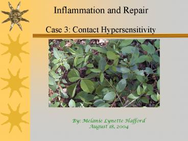
Inflammation and Repair Case 3 Contact
Hypersensitivity
By Melanie Lynette Hafford August 18, 2004
2
Scenario
A 22 year old medical student presents to the
Dermatology Clinic with an itchy rash on the
trunk and extremities that started to appear two
days after a camping trip to the Texas hill
country.
3
Clinical Presentation
- Calor (heat)
- Rubor (redness)
- Dolor (pain)
- Tumor (swelling)
Results of increased blood flow to the irritated
area. Leads to edema from increased capillary
filtration and leakage of plasma proteins out of
capillary. Increased flow pushes lymphocytes out
of vessel into tissue
4
Allergic Contact Dermatitis (Acute Eczematous
Dermatitis)
Red papulovesicular oozing and lesions Edema
localized to perivasuclar spaces in epidermis
5
Types of Hypersensitivity Reactions
Type 1 Anaphylaxis, quick (secs-mins), IgE,
accumulate neutrophils and eosinophils. Ex
asthma hay fever Type 2 Antibody dependent,
hrs-dys, IgG IgM, complement and phagocytosis.
Ex Hemolytic anemia Myasthenia gravis Type
3 Immune complex initiated, hrs-dys, IgG
IgM, Accumulate neutrophils macrophages. Ex
Lupus Type 4 Cell mediated, delayed (days),
reactant T cells, lymphocytes, macrophages,
monocytes. Ex leprosy, contact dermatitis, organ
graft rejection
6
Type IV Hypersensitivity Reaction Delayed Type
Hypersensitivity (DTH)
- Antigen introduced to the tissue modifies
extracellular - and cell surface proteins
- Macrophages process antigen, present to Th1 cells
- Th1 effector cell recognizes the antigen and
releases - cytokines which act on local vascular
endothelium and - further activate macrophages (increase in size,
microbicidal - activity, lysosome content)
- T cell recruitment (CD4 CD8), fluid and protein
- absorption by interstitum (edema).
7
The Process
Langerham cells (APCs) uptake allergen, process
MHCII, travel to node, present to T cells (CD4
CD8)
T cells proliferate and migrate
Th1 cells release chemokines (macrophage
recruitment) and cytokines (macrophage
activation monocyte production)
Macrophages release cytokines to further drive T
cell differentiation
T cells release cytokines to increase
microvasuclar permeablilty (activate Endothelial
cells) leading to edema
The Lesion becomes apparent
8
This Process Takes A Lot of Time!!!
The delay in the appearance of a type IV
hypersensitivity reaction (2-3 days) is due to
the time it takes to recruit antigen-specific T
cells and other cells to the site of antigen
localization and to develop the inflammatory
response.
9
Chemical Factors Involved
- IL-12 (macrophages). Drives differentiation of T
cells, induces IFN- - gamma secretion
- IFN-gamma (T cells). Further activates macrophages
- IL-2 (T cells). Increases T cell proliferation
within tissue
- IL-3 (T cells). Stimulates monocyte production
- TNF (T cells). Increase secretion of Nitric Oxide
Prostacyclins by - endothelial cells, local tissue destruction, and
increase - expression of adhesion molecules on vessels
- E- selectins vascular adhesion molecule,
increases mononuclear cell - attachment
10
So what type of inflammation are we seeing here?
Both Chronic Acute Inflammation
- Chronic
- Large numbers of mononuclearcytes (macrophages,
plasma cells,etc) - Cell mediated immunity- see eosionophils
- Accounts for delayed hypersensitivity
- Acute
- Vascular changes, leading to increased
permeability - Edema
11
Destruction and Repair
Damage to the Extracellular Matrix and
destruction of skin cells that present modified
antigen peptides occurs via CD8 T cells,
monocytes, and macrophages.
Typically in chronic inflammation, healing
proceeds simultaneously, and often a scar is
formed from fibrogenesis.
However, in contact dermatitis, it is relatively
unlikely that a scar will form
12
Why is this?
Eventually lymphatic drainage and
macrophage ingestion of damaged cells and debris
leads to clearance of inflammatory cells and
edema as seen in acute inflammation.
Additionally epidermal cells are labile, and will
readily regenerate.
13
Treatment Options
- Generally Rxs arent indicated, most OTC
medications - will relieve itching.
- Hydrocortisone
- Calamine Lotion
- Oral Antihistamines
- If rash is covers a significant portion of the
body, or is - infected, the following Rxs are often given
- Cortisone
- Oral corticosteroids
- Antibiotics
14
Conclusion
Rash can last up to 3 weeks even with treatment
Recognizing poison ivy and minimizing exposure if
you come in contact with the plant are the only
forms of prevention.
15
References
- Kumar, Cotran, Robbins. Basic Pathology 7th
Edition. W B Saunders, - 2003.
- Parham, P. The Immune System 2nd Edition. Garland
Publishing, 2004. - http//edcenter.med.cornell.edu/CUMC_PathNotes/Imm
unopathology/ - Immuno_02.html
- http//www.mayoclinic.com/invoke.cfm?idAN00539
Recommended
CrystalGraphics Presentations


