
Sequential Events of Contraction - PowerPoint PPT Presentation
1 / 21
Title:
Sequential Events of Contraction
Description:
Aerobic respiration. Muscle Fatigue ... Intense exercise produces rapid muscle fatigue (with rapid recovery) ... Low-intensity exercise produces slow-developing ... – PowerPoint PPT presentation
Number of Views:75
Avg rating:3.0/5.0
Title: Sequential Events of Contraction
1
Sequential Events of Contraction
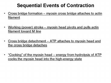
- Cross bridge formation myosin cross bridge
attaches to actin filament - Working (power) stroke myosin head pivots and
pulls actin filament toward M line - Cross bridge detachment ATP attaches to myosin
head and the cross bridge detaches - Cocking of the myosin head energy from
hydrolysis of ATP cocks the myosin head into the
high-energy state
2
Sequential Events of Contraction
3
Motor Unit The Nerve-Muscle Functional Unit
- A motor unit is a motor neuron and all the muscle
fibers it supplies - The number of muscle fibers per motor unit can
vary from four to several hundred - Muscles that control fine movements (fingers,
eyes) have small motor units (i.e. few muscle
fibers per motor neuron)
4
Muscle Twitch
- A muscle twitch is the response of a muscle to a
single, brief threshold stimulus - The three phases of a muscle twitch are
- 1. Latent period - first few milliseconds after
stimulation when excitation-contraction coupling
is taking place - 2. Period of contraction cross bridges actively
form and the muscle shortens - 3. Period of relaxation Ca2 is reabsorbed into
the SR, and muscle tension goes to zero
5
Muscle Response to Varying Stimuli
- A single stimulus results in a single contractile
response a muscle twitch - Frequently delivered stimuli (muscle does not
have time to completely relax) increases
contractile force wave summation - More rapidly delivered stimuli result in
incomplete tetanus - If stimuli are given quickly enough, complete
tetanus results
6
Muscle Metabolism Energy for Contraction
- ATP is the only source used directly for
contractile activity - As soon as available stores of ATP are hydrolyzed
(4-6 seconds), they are regenerated by - The interaction of ADP with creatine phosphate
(CP) - Anaerobic glycolysis
- Aerobic respiration
7
Muscle Fatigue
- Muscle fatigue the muscle is in a state of
physiological inability to contract - Muscle fatigue occurs when
- ATP production fails to keep pace with ATP use
- There is a relative deficit of ATP, causing
contractures - Lactic acid accumulates in the muscle
- Ionic imbalances are present
- Intense exercise produces rapid muscle fatigue
(with rapid recovery) - Na-K pumps cannot restore ionic balances
quickly enough - Low-intensity exercise produces slow-developing
fatigue - SR is damaged and Ca2 regulation is disrupted
8
Force of Muscle Contraction
- The force of contraction is affected by
- The number of muscle fibers contracting the
more motor fibers in a muscle, the stronger the
contraction - The size of the muscle the bulkier the muscle,
- greater its strength - Degree of muscle stretch
9
Muscle Fiber Type Functional Characteristics
- Speed of contraction determined by speed in
which ATPases split ATP - The two types of fibers are slow and fast
- ATP-forming pathways
- Oxidative fibers use aerobic pathways
- Glycolytic fibers use anaerobic glycolysis
- These two criteria define three categories slow
oxidative fibers, fast oxidative fibers, and fast
glycolytic fibers
10
Skeletal Muscle Attachments
Most skeletal muscles span joints and are
attached to bones in at least 2 places. When a
muscle contracts, the movable bone (the muscles
insertion), moves toward the immovable or less
movable bone (the muscles origin).
11
Skeletal Muscle / Joint Movements
Angular movements - increase or decrease the
angle between 2 bones Flexion bending movement
usually along the sagittal plane that decreases
the angle of the joint and brings the
articulating bones closer together e.g. bending
the knee from straight to an angled
position Extension the reverse of flexion and
occurs at the same joints. It involves movement
along the sagittal plane that increases the angle
between the articulating bones e.g. straightening
the knee
12
Skeletal Muscle / Joint Movements
13
Skeletal Muscle / Joint Movements
Dorsiflexion and Plantarflexion The up- and
down movements of the foot at the ankle Lifting
the foot to being the superior surface towards
the shin is dorsiflexion Depressing the foot is
plantarflexion.
14
Smooth Muscle
- Composed of spindle-shaped fibers with a diameter
of 2-10?m and lengths of several hundred ?m - Lack the coarse connective tissue sheaths of
skeletal muscle, but have fine endomysium - Organized into two layers (longitudinal and
circular) of closely apposed fibers - Found in walls of hollow organs (except the
heart) - Have similar contractile mechanisms as skeletal
muscle
15
Peristalsis
- When the longitudinal layer contracts, the organ
dilates and contracts - When the circular layer contracts, the organ
elongates - Peristalsis alternating contractions and
relaxations of smooth muscles that mix and
squeeze substances through the lumen of hollow
organs
16
Innervation of Smooth Muscle
- Smooth muscle lacks neuromuscular junctions
- Innervating nerves have bulbous swellings called
varicosities - Varicosities release neurotransmitters into wide
synaptic clefts called diffuse junctions
17
Microscopic Anatomy of Smooth Muscle
- SR is less developed than in skeletal muscle
- T tubules are absent
- Plasma membranes have pouchlike infoldings called
caveoli - Ca2 is sequestered in the extracellular space
near the caveoli, allowing rapid influx when
channels are opened - There are no visible striations and no sarcomeres
- Thin and thick filaments are present
18
Proportion and Organization of Myofilaments in
Smooth Muscle
- Thick filaments have heads along their entire
length - Thick and thin filaments are arranged diagonally,
causing smooth muscle to contract in a corkscrew
manner - Noncontractile intermediate filament bundles
attach to dense bodies (analogous to Z discs) at
regular intervals
19
Contraction of Smooth Muscle
- Whole sheets of smooth muscle exhibit slow,
synchronized contraction - Action potentials are transmitted from cell to
cell - Some smooth muscle cells
- Act as pacemakers and set the contractile pace
for whole sheets of muscle - Are self-excitatory and depolarize without
external stimuli
20
Contraction Mechanism
- Actin and myosin interact according to the
sliding filament mechanism - The final trigger for contractions is a rise in
intracellular Ca2 - Ca2 is released from the SR and from the
extracellular space - Ca2 ultimately activates myosin
Role of Calcium Ion
- Ca2 ultimately activates calmodulin kinase
- Activated kinase transfers phosphate from ATP to
myosin cross bridges - Phosphorylated cross bridges interact with actin
to produce shortening - Smooth muscle relaxes when intracellular Ca2
levels drop
21
Types of Smooth Muscle Single Unit
- The cells of single-unit smooth muscle, commonly
called visceral muscle - Contract rhythmically as a unit
- Are electrically coupled to one another via gap
junctions - Often exhibit spontaneous action potentials
- Are arranged in opposing sheets and exhibit
stress-relaxation response
Types of Smooth Muscle Multiunit
- Multiunit smooth muscles are found
- In large airways to the lungs
- In large arteries
- Their characteristics include
- Rare gap junctions
- Structurally independent muscle fibers
Recommended
CrystalGraphics Presentations


