
Last lecture: Skeletal Muscle Contraction - PowerPoint PPT Presentation
1 / 28
Title:
Last lecture: Skeletal Muscle Contraction
Description:
The ionic concentration of the resting state is restored by the. Na -K pump ... Muscle fatigue the muscle is in a state of physiological inability to contract ... – PowerPoint PPT presentation
Number of Views:103
Avg rating:3.0/5.0
Title: Last lecture: Skeletal Muscle Contraction
1
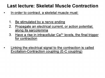
Last lecture Skeletal Muscle Contraction
- In order to contract, a skeletal muscle must
- Be stimulated by a nerve ending
- Propagate an electrical current, or action
potential, along its sarcolemma - Have a rise in intracellular Ca2 levels, the
final trigger for contraction - Linking the electrical signal to the contraction
is called Excitation-Contraction coupling (E-C
coupling)
2
Nerve Stimulus of Skeletal Muscle
- Skeletal muscles are stimulated by motor neurons
- Axons of these neurons travel in nerves to muscle
cells - Axons of motor neurons branch as they enter
muscles - Each axonal branch forms a neuromuscular junction
with a single muscle fiber
3
Neuromuscular Junction
- The neuromuscular junction is formed from
- Axonal endings, which have small membranous sacs
(synaptic vesicles) that contain the
neurotransmitter acetylcholine (ACh) - The motor end plate of a muscle, which is a
specific part of the sarcolemma that contains ACh
receptors and helps form the neuromuscular
junction - Though exceedingly close, axonal ends and muscle
fibers are always separated by a space called the
synaptic cleft
4
Neuromuscular Junction
5
Neuromuscular Junction
- When a nerve impulse reaches the end of an axon
at the neuromuscular junction - Voltage-regulated calcium channels open and allow
Ca2 to enter the axon - Ca2 inside the axon terminal causes axonal
vesicles to fuse with the axonal membrane - This fusion releases ACh into the synaptic cleft
via exocytosis - ACh diffuses across the synaptic cleft to ACh
receptors on the sarcolemma - Binding of ACh to its receptors initiates an
action potential in the muscle
6
Destruction of Acetylcholine
- ACh bound to ACh receptors is quickly destroyed
by the enzyme acetylcholinesterase - This destruction prevents continued muscle fiber
contraction in the absence of additional stimuli
7
Action Potential
- A transient depolarization event that includes
polarity reversal of a sarcolemma (or nerve cell
membrane) and the propagation of an action
potential along the membrane
Role of Acetylcholine (Ach)
- ACh binds its receptors at the motor end plate
- Binding opens chemically (ligand) gated channels
- Na and K diffuse out and the interior of the
sarcolemma becomes less negative - This event is called depolarization
8
Action Potential Electrical Conditions of a
Polarized Sarcolemma
- The outside (extracellular) face is positive,
while the inside face is negative - This difference in charge is the resting membrane
potential - The predominant extracellular ion is Na
- The predominant intracellular ion is K
- The sarcolemma is relatively impermeable to both
ions
9
Action Potential Depolarization and Generation
of the Action Potential
- An axonal terminal of a motor neuron releases ACh
and causes a patch of the sarcolemma to become
permeable to Na (sodium channels open) - Na enters the cell, and the resting potential is
decreased (depolarization occurs) - If the stimulus is strong enough, an action
potential is initiated
10
Action Potential Propagation of the Action
Potential
- Polarity reversal of the initial patch of
sarcolemma changes the permeability of the
adjacent patch - Voltage-regulated Na channels now open in the
adjacent patch causing it to depolarize - Thus, the action potential travels rapidly along
the sarcolemma - Once initiated, the action potential is
unstoppable, and ultimately results in the
contraction of a muscle
11
Action Potential Repolarization
- Immediately after the depolarization wave passes,
the sarcolemma permeability changes - Na channels close and K channels open
- K diffuses from the cell, restoring the
electrical polarity of the sarcolemma - Repolarization occurs in the same direction as
depolarization, and must occur before the muscle
can be stimulated again (refractory period) - The ionic concentration of the resting state is
restored by the Na-K pump
12
Excitation-Contraction Coupling
- Once generated, the action potential
- Is propagated along the sarcolemma
- Travels down the T tubules
- Triggers Ca2 release from the sarcoplasmic
reticulum - Ca2 binds to regulatory proteins and allows
- Actin active binding sites to be exposed
- Myosin cross bridges alternately attach and
detach - Thin filaments move toward the center of the
sarcomere - Hydrolysis of ATP powers this cycling process
- Ca2 is removed into the SR and the muscle fiber
relaxes
13
Excitation-Contraction Coupling
14
Role of Ionic Calcium (Ca2) in the Contraction
Mechanism
- At low intracellular Ca2 concentration
- Myosin cross bridges cannot attach to binding
sites on actin - The muscle fiber is in a relaxed state
- At higher intracellular Ca2 concentrations
- Ca2 binds to regulatory proteins and allows
myosin to bind actin - Myosin head can now bind and cycle
- This permits contraction (sliding of the thin
filaments by the myosin cross bridges) to begin
15
Sequential Events of Contraction
- Cross bridge formation myosin cross bridge
attaches to actin filament - Working (power) stroke myosin head pivots and
pulls actin filament toward M line - Cross bridge detachment ATP attaches to myosin
head and the cross bridge detaches - Cocking of the myosin head energy from
hydrolysis of ATP cocks the myosin head into the
high-energy state
16
Sequential Events of Contraction
17
Motor Unit The Nerve-Muscle Functional Unit
- A motor unit is a motor neuron and all the muscle
fibers it supplies - The number of muscle fibers per motor unit can
vary from four to several hundred - Muscles that control fine movements (fingers,
eyes) have small motor units (i.e. few muscle
fibers per motor neuron)
18
Muscle Twitch
- A muscle twitch is the response of a muscle to a
single, brief threshold stimulus - The three phases of a muscle twitch are
- 1. Latent period - first few milliseconds after
stimulation when excitation-contraction coupling
is taking place - 2. Period of contraction cross bridges actively
form and the muscle shortens - 3. Period of relaxation Ca2 is reabsorbed into
the SR, and muscle tension goes to zero
19
Muscle Response to Varying Stimuli
- A single stimulus results in a single contractile
response a muscle twitch - Frequently delivered stimuli (muscle does not
have time to completely relax) increases
contractile force wave summation - More rapidly delivered stimuli result in
incomplete tetanus - If stimuli are given quickly enough, complete
tetanus results
20
Muscle Metabolism Energy for Contraction
- ATP is the only source used directly for
contractile activity - As soon as available stores of ATP are hydrolyzed
(4-6 seconds), they are regenerated by - The interaction of ADP with creatine phosphate
(CP) - Anaerobic glycolysis
- Aerobic respiration
21
Muscle Fatigue
- Muscle fatigue the muscle is in a state of
physiological inability to contract - Muscle fatigue occurs when
- ATP production fails to keep pace with ATP use
- There is a relative deficit of ATP, causing
contractures - Lactic acid accumulates in the muscle
- Ionic imbalances are present
- Intense exercise produces rapid muscle fatigue
(with rapid recovery) - Na-K pumps cannot restore ionic balances
quickly enough - Low-intensity exercise produces slow-developing
fatigue - SR is damaged and Ca2 regulation is disrupted
22
Force of Muscle Contraction
- The force of contraction is affected by
- The number of muscle fibers contracting the
more motor fibers in a muscle, the stronger the
contraction - The size of the muscle the bulkier the muscle,
- greater its strength - Degree of muscle stretch
23
Muscle Fiber Type Functional Characteristics
- Speed of contraction determined by speed in
which ATPases split ATP - The two types of fibers are slow and fast
- ATP-forming pathways
- Oxidative fibers use aerobic pathways
- Glycolytic fibers use anaerobic glycolysis
- These two criteria define three categories slow
oxidative fibers, fast oxidative fibers, and fast
glycolytic fibers
24
Skeletal Muscle Attachments
Most skeletal muscles span joints and are
attached to bones in at least 2 places. When a
muscle contracts, the movable bone (the muscles
insertion), moves toward the immovable or less
movable bone (the muscles origin).
25
Skeletal Muscle / Joint Movements
Angular movements - increase or decrease the
angle between 2 bones Flexion bending movement
usually along the sagittal plane that decreases
the angle of the joint and brings the
articulating bones closer together e.g. bending
the knee from straight to an angled
position Extension the reverse of flexion and
occurs at the same joints. It involves movement
along the sagittal plane that increases the angle
between the articulating bones e.g. straightening
the knee
26
Skeletal Muscle / Joint Movements
27
Skeletal Muscle / Joint Movements
Dorsiflexion and Plantarflexion The up- and
down movements of the foot at the ankle Lifting
the foot to being the superior surface towards
the shin is dorsiflexion Depressing the foot is
plantarflexion.
28
Smooth Muscle
- Composed of spindle-shaped fibers with a diameter
of 2-10?m and lengths of several hundred ?m - Lack the coarse connective tissue sheaths of
skeletal muscle, but have fine endomysium - Organized into two layers (longitudinal and
circular) of closely apposed fibers - Found in walls of hollow organs (except the
heart) - Have similar contractile mechanisms as skeletal
muscle
Recommended
CrystalGraphics Presentations


