
Pediatric PowerPoint PPT Presentation
1 / 126
Title: Pediatric
1
Pediatric Neuromuscular Orthopaedics ONC Exam
Review
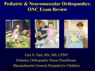
- Erin S. Hart, RN, MS, CPNP
- Pediatric Orthopaedic Nurse Practitioner
- Massachusetts General Hospital for Children
2
Pediatric Orthopaedics Lecture
Introduction Pediatric differences, Nursing
interventions Fractures/Trauma Pediatric
fractures, classification Common Conditions of
the Newborn/ Infant Developmental Hip
Dysplasia, Clubfoot Hip Legg-Calve-Perthes
Disease, Slipped Capital Femoral Epiphysis
3
Pediatric Orthopaedics Lecture
Spine Scoliosis (idiopathic, neuromuscular)
In-toeing other common rotational and angular
disorders Syndromes Osteogenesis imperfecta,
Achondroplasia Neuromuscular Nonprogressive
Cerebral palsy, myelo Neuromuscular
Progressive Muscular Dystrophy, NF
4
Objectives Pediatric Orthopaedics
- 11 of ONC Exam
- Identify signs and symptoms in selected pediatric
and neuromuscular disorders - Outline interventions for common pediatric
orthopaedic nursing protocols - List strategies to maximize function in patients
and families with neuromuscular disorders
5
Nursing Interventions
- Nursing see Table 1 from Core Curriculum
- Childs Developmental Level
- Parent and Childs Ability to Learn
- Amt Disorder Interferes With
- ADLs
- Growth
- Learning Ability
- Social Adjustment
6
Nursing Interventions
- Parents
- Realistic Expectations
- Understanding of the Disease / Disorder
- Follow-up With Treatment
- Response to public inquiry
7
Pediatric Orthopaedics
8
Developmental Stages
- Infancy 0-18 months trust vs mistrust
- Toddler 18mos 3 yrs autonomy vs shame/doubt
- Pre-school 3-5 yrs initiative vs guilt
- School age 6-12 yrs industry vs inferiority
- Adolescence 13 20 yrs identity vs role
confusion
9
Fractures in Children General
- Overall higher incidence of fractures in children
- Force required to break bone in child is less
than adult - Physes (growth plate) are the weak link (ligament
injury in adult, physeal fracture in child) - Bone healing much more rapid in children (femur
fracture in neonate heals in 2-3 weeks, early
childhood 4-6 weeks, and adult 16-20 weeks) - Much less likely to need operative fixation,
greater remodeling of bone - Physeal fractures unique to children 20-25 of
all fractures - Salter-Harris classification Based on fracture
pattern
10
X-ray taken at 2 weeks of age Note
extensive callus formation
Diaphyseal Humerus Fracture DOL 2
X-ray taken at 2 months of age Note callus and
remodeling
BIRTH TRAUMA HUMERUS FRACTURES IN THE NEWBORN
11
Fractures in Children General
- Closed reduction and casting Most common
treatment of pediatric fx - Thick periosteum of growing child aids in keeping
the fracture reduced - Most pediatric fractures referred to pedi ortho
specialist for definite management
12
Salter-Harris (SH) Classification
- Fractures of growth plate divided into five
categories based on pattern - I Fracture through the growth plate (very
difficult to see on x-ray) - II Fracture through metaphysis into growth plate
- III Fracture through epiphysis into growth plate
- IV Fracture though epiphysis through growth
plate and into metaphysis - V Crush injury to growth plate
13
ALWAYS RULE OUT NON-ACCIDENTAL TRAUMA!!
16 month old toddler with left diaphyseal femur
fracture, found to have proximal radius
fracture and right distal radius fracture
in advanced stage of healing
14
Trauma Child Abuse
- 3.14 million children reported abused/year
- Physical Abuse
- Greatest lt 3 years (66-78)
- 30 under 6 mos
15
Common Fractures Inflicted
- Rib Fx. seen in 5-20 of abused
- Scapular/ distal clavicle / ulna night stick fx
- Vertebral fx. or subluxation
- Bilateral, Multiple, or Different Stages of
Healing - Corner fracture of tibia/femur
16
Multiple Rib Fractures Corner fx of Distal Tibia
17
Developmental Hip Dysplasia Introduction
- No longer referred to congenital dislocation of
hip - DDH is developmental (ongoing) process
- Dysplasia refers to improper development or
formation of acetabulum and/or femoral head - Incidence 1-5 per 1,000 newborns
- From mild dysplasia to frank dislocation of hips
18
Developmental Hip Dysplasia Etiology
- Exact cause unknown multifactorial
- Risk factors for DDH
- 1.) First-born children
- 2.) Female sex (60-70)
- 3.) Breech presentation (30-50)
- 4.) Family History DDH
- 5.) Prematurity
- 6.) Other also associated with other ortho
conditions Torticollis, foot deformities
(calcaneovalgus), Trisomy 21 (increased laxity)
19
Developmental Hip Dysplasia Definitions
- 60 affect left hip, 20 right, 20 bilateral
- 25-30 present after newborn period (much harder
to manage!) - Complete dislocation femoral head completely
outside the acetabulum MUST be located prior to
PAVLIK harness - Subluxation femoral head partially dislocated
- Unstable femoral head can be pushed out from
acetabulum with stress
20
Developmental Hip Dysplasia Examination
- IMPERATIVE to assess hips during all well-child
visits during first year!! - DDH detected by different signs based on infants
age - Always examine each hip individually and then
together to compare - NEWBORN EXAM
- 1.) Barlows confirms instability. Attempt to
gently displace the hip out of the socket over
the posterior acetabulum
BARLOWs SIGN
21
Developmental Hip Dysplasia Examination
- 2.) Ortolonis Confirms joint is reducible.
Clunk sensation when thigh is abducted while
lifting up the greated trochanter with the finger
- Barlow and Ortoloni are much more reliable in
neonates and infants up to 3-5 months - Make sure infant is relaxed and comfortable
ORTOLONIS SIGN
22
Developmental Hip Dysplasia
- X-rays of pelvis not very useful until 4-6 months
of age (femoral head not fully ossified) - Generally use ultrasound of hips to follow
infants with DDH - Effectiveness depends on the skill and experience
of person performing U/S
23
Developmental Hip Dysplasia Examination
- Older Children Asymmetrical thigh/buttock skin
folds - Limited abduction compare hips
- Galeazzi Sign uneven knee heights when lying
supine with knees flexed and soles of feet on
table (indicates shortening) - Trendelenburg Gait often indicates weak hip
abductors
LIMITED ABDUCTION
GALEAZZI SIGN
24
Positive Galeazzi Sign on Physical Exam
25
Developmental Hip Dysplasia Management
- Refer to pediatric orthopaedics once diagnosis is
confirmed - Ideal age for management 0-6 months
- 60 of unstable hips will resolve spontaneously
in 1st month - First line of treatment in infants 0-6 months
PAVLIK harness - Harness should be properly fitted to avoid
pitfalls in management
26
Correct Position of Pavlik Harness
27
Developmental Hip Dysplasia Management
- Pavlik harness generally needed for 6-8 weeks
allows hip to become stable - Monitor with U/S imaging every 2-4 weeks
- If a dislocated hip has NOT reduced by 3-4 weeks
STOP Pavlik harness and proceed with closed/open
reduction with spica (body) cast - Toddlers and older children generally need
operative treatment open reduction/ osteotomy
28
9 month old female with Left DDH
(posterior/superior dislocation of femoral head)
29
Older Infant/ Young Child with DDH....
2 year old female with right hip dislocation
30
3 month old with DDH s/p Closed Reduction and
Spica Casting
- Nursing Care Spica Cast
- Neurovascular assessment
- Support for parent/family (often need cast for
prolonged time 3 months) - Keeping cast clean/dry
- (positioning, diaper care)
- Keeping the child a child (diversional activities
based on age of child)
31
Talipes Equinvarus Clubfoot Deformity
- Congenital deformity of the foot
- Approx 1 in 1,000 births in U.S.
- Multifactorial cause most often isolated and
idiopathic - 2X more common in males
- Bilateral in approx 50
- Higher incidence assoc. with neurogenic
conditions (spina bifida, cerebral palsy,
arthrogryposis)
32
Talipes Equinovarus Clubfoot
- Diagnosis usually obvious and made prenatally (18
week U/S) or at birth - Three classic signs Fixed Plantar flexion
(equinous of ankle) - Adduction (Varus), or turning in of
heel/hindfoot - Supination, or turning under of
forefoot/midfoot
33
Clubfoot Current Management
- Treatment usually begins immediately after birth
- Refer to pediatric orthopaedics as soon as
diagnosis suspected - Overall goal is to correct clubfoot, maintain
correction, facilitate normal growth and
development - Current shift away from early surgical correction
- Emphasis on early casting variable techniques
used
34
Clubfoot General Management
- Ponsetti approach Dr. Ignacio Ponsetti (U Iowa)
pioneered - Serial manipulation and plaster casting of
clubfoot - 4-5 Long Leg Plaster Casts applied weekly,
Percutaneous heel cord tenotomy (cut achilles
tendon) 85 patients - Splinting is essential part of management Denis
Brown Abduction Brace (prevents recurrence)
35
Ponsetti Technique Used in Our Clinic
4 week old female with Bilateral Clubfoot
36
Denis-Brown Abduction Brace
- Essential part of management in Ponsetti
Technique - Worn full time (23 hours/day) for 1st two-three
months after last cast removed, then at nighttime
for 2-3 years - Done to avoid high rate of recurrence
37
Need regular follow-up in orthopaedics until end
of growth Children with Clubfoot usually do well
with treatment, develop normally, and participate
fully in athletics
38
The Denis Browne Abduction Bar Ponsetti Technique
39
Rotational Angular Deformities in Children
Introduction
- Rotational and Angular Deformities are quite
common in pediatrics - Very diverse spectrum of diagnoses physiologic
to pathologic - In-toeing/Out-toeing, Genu varum (bowlegs)/valgum
(knock-knees)
40
Causes of In-toeing Gait in Children
- The most frequent causes of childhood in-toeing
- Femoral anteversion
- Medial Tibial Torsion
- Metatarsus adductus
41
The In-Toeing Toddler/Child Assessment
- Assess Femoral Version Measure external and
internal rotation of the hips with the child
prone and the knees flexed to 90 degrees. Assess
both sides simultaneously. Internal rotation
usually less than 65-70 degrees - If greater than 70 degrees in-toeing likely from
femoral anteversion/femoral torsion - If rotation is asymmetrical, evaluate with AP of
pelvis to r/o DDH or hip problem
42
Internal Femoral Torsion/Anteversion
- In standing position, patellae will point inwards
when feet are forward - Compensatory external rotation of tibia
43
Internal Femoral Torsion/Anteversion
- Usually first seen in the 3-5 year age group,
usually most severe b/w 4-6 years - Almost always symmetrical
- More common in females approx. 2 1 ratio, often
familial - Gait/running described as awkward/clumsy by
parents
44
Femoral Anteversion Management
- Gait is often worse when running or when fatigued
- Children prefer the W sitting position because
it is more comfortableshould not be discouraged
or avoided - Reassurance and Observation!!
- Special shoes, twister cables, etc avoided.no
difference in outcome!!
45
Internal (Medial) Tibial Torsion
- Toddler or young child often presents with c/o
bowing legs - Usually symmetric in-toeing, if
unilateral--usually worse on left - Often noticed when child is first starting to
walk - With patellae facing forwards
- (in neutral position), feet turn in
46
Measurement of Thigh Foot Angle Medial Tibial
Torsion
- Quantitate Tibial Version
- Thigh Foot Angle patient is pone, knees flexed
90 degrees TFA is the angular difference between
the axis of the foot and the axis of the thigh - Allow foot to fall into natural position, avoid
manual positioning of foot - Medial Tibial Torsion Negative Thigh Foot Angle
47
Tibial Torsion
- Resolves spontaneously in 95-98 of patients by
age 4-6 years - Stretching, special shoes are inefffectivedoes
not speed up resolution and makes no clinical
difference - Can occasionally have mild persistence with no
handicap or functional significance - Simple observation is best treatment and all that
is needed
48
Metatarsus Adductus in Infants
- Assess the foot for forefoot adductus
- Lateral border of foot should be straight
- Convexity of lateral border and forefoot
adduction are features of metatarsus adductus
49
14 month old with Metatarsus Adductus
50
Metatarsus Adductus Management
- Forefoot can gently be stretched passively with
each diaper change - Occasionally will use serial casting and
reverse/straight last shoes to correct deformity - Observation and Reassurance will resolve
spontaneously in 95 of patients (tends to
persist until age 12-18 months)
51
Lower Extremity Rotational Profile at Various Ages
- Normal alignment progresses from 10-15 degrees of
varus at birth to maximum valgus angulation of
10-15 degrees at 3-4 years of age
52
Genu Valgum Assessment
- Physiologic knock-knee deformity very common in
children aged 3-5 years - Screening evaluation normal height and body
proportions, symmetrical, localized or
generalized, limb lengths equal - Measure rotational profile, measure
intra-malleolar distance with the knees together - If generalized deformity, order metabolic
screening labs
53
Pathologic Causes of Genu Valgum
- Post-traumatic (most common)
- Dysplasias
- Primary tibial valga
- Tumor
- Infection
- Rickets
- Renal osteodystrophy
- Congenital deficiency of fibula (fibular
hemimelia)
54
Post-Traumatic Genu Valgum
- Usually results from overgrowth following
fracture of the proximal tibial metaphysis in
early childhood - Valgus deformity develops during the 1st 12-18
months post-injury due to tibial overgrowth - Management Most will correct spontaneously over
course of years without operative treatment - If deformity persists osteotomy or
hemiepiphyseodesis
55
Genu Valgum General Management
- Age 2-6 years 97-98 will resolve spontaneously
- If intermalleolar distance is gt 8-10 cm at age 10
- 1.) Hemiephiphyseodesis of distal femur and/or
proximal tibia - 2.) If skeletally mature a.) tibial varus
osteotomy - b.)
femoral osteotomy medial
56
Physiologic Genu Varum Assessment
- Parents will often note bow leg deformity,
usually recognized when child starts to walk
(12-18 months) - Commonly bilateral and symmetric bowing
- Seldom causes functional disability X-rays
unnecessary until at least 18 months of age - Physiologic bowing usually spontaneously resolves
by the age of two years
57
Infantile Blounts Disease Epidemiology
- Risk factors Obesity, African American,Walking
at early age, Family history - Differential Diagnosis Physiologic genu varum
(metaphyseal-diaphyseal angle less than 15
degrees) -
- Very difficult to differentiate from
physiologic varus/ - bowlegs in patients lt 2 years
58
Blounts Disease Radiographs
59
Adolescent Blounts Disease
- Definition Growth disorder involving the medial
portion of the proximal tibial growth plate that
produces a localized varus deformity - More often unilateral, usually seen in obese
individuals, slightly more males than females,
African American, certain geographic regions - Definite cause unkown biomechanical overload to
proximal tibia physis due to varus alignment and
excessive body weight
60
Adolescent Blounts Disease Clinical Assessment
61
Leg Length Discrepancy
- Congenital vs Acquired
- Sx Short limb, Limp, back pain
- Dx X-ray, CT, Bone Age
62
Leg Length Discrepancy Treatment
- Mosely graph calculate age for epiphyseodesis
- lt2cm LLD no treatment
- 2-6 cm lifts, epiphysiodesis
- gt6 - 15cm Shorten opp limb
- Limb lengthening, External fixation, Spatial
frames Ilizarov
63
Plan Management based on --Age of
Diagnosis --Severity --Projected Height at
Maturity Distal femur/prox tibia
physes Generally close at age 14 in Females and
16 in males
64
Leg Length Discrepancy External Fixation
- Nursing Care
- Pain Management
- Pin Care Infection common
- Compartment Syndrome
- Provide Emotional Support
65
Legg-Calve Perthes Disease
- Legg Calve Perthes disease (LCPD) is defined as
an idiopathic avascular necrosis of the femoral
head - Dr Legg, Calve, and Perthes independently
researched and described a vascular injury to
the hip in young children in 1910.
66
Early Concepts and Treatment
- Early treatment concepts for LCPD was often quite
severe - Involved bed rest, immobilization, long
hospitalizations, special carts, slings, and
braces
67
Epidemiology of LCPD
- Affects about 1 in 10,000 children
- Age of Onset age 2-12 with peak between age 4-8
- Boys affected more often than girls (51 ratio)
- More common among Asian and Central European
populations - Short stature and delayed bone age are also risk
factors
68
Etiology of LCPD
- Blood flow to the femoral head is temporarily
interrupted - Pathology is consistent with repeated bouts of
infarctions and subsequent pathologic fractures - Subchondral fracture leads to avascular necrosis
of the femoral head - Widening, flattening, and deformity---may
partially or completely affect the femoral head
69
Radiograph of child with LCPD
70
Early Crescent Sign of Subchondral Fracture in
LCPD
71
Clinical Features of LCPD
- Insidious onset
- Most frequent symptom is painless limp
- If pain is present, it is worse with activity and
relieved by rest - Hip pain can ALWAYS refer to the knee
- May also complain of anterior thigh or groin pain
72
Prognostic Factors of LCPD
- Age is the Key to Prognosis younger
patients have more growth remaining for
remodeling/reshaping of femoral head - Age less than 6 years usually good outcome
regardless of treatment - Greater than age 8-9 years often have poor
prognosis requiring surgical correction - Containment Maintenance of ROM
73
Slipped Capital Femoral Epiphysis
- SCFE is a displacement of the femoral head
relative to the femoral neck which occurs through
the physis (growth plate) of the femur - Considered one the few orthopaedic surgical
emergencies/urgency - THE most common orthopaedic hip disorder
affecting adolescents age range 9-16 years - Vast majority of patients are obese increased
shear stress across the physis - Mean age at diagnosis Females12.0 years
- Males13.5 years
74
Slipped Capital Femoral Epiphysis Etiology
- Local Trauma 26 report
- Mechanical forces obesity, decreased
anteversion, oblique physis - Inflammation/synovitis
- Endocrine imbalances
- Heredity 5
75
Slipped Capital Femoral Epiphysis
- Patients will often present with an antalgic limp
- Lower limb often kept in external rotation while
standing, decrease in internal rotation, flexion,
and abduction of the hip when supine - Involved hip will often abduct and externally
rotate with passive flexion
76
SCFE Management
- Objective is to stabilize the growth plate to
prevent further slippage and to avoid
complications - Mild and Moderate stable slips In situ Pin
Fixation (single screw) - Prevents further slippage and leads to fusion of
the growth plate - If child lt 8 years fix with smooth pins to allow
growth - Severe slips In situ Fixation, Osteotomy (neck,
base of nech, intertrochanteric, subtrochanteric
location of femur)
77
Surgical Pinning Technique for SCFE
78
(No Transcript)
79
OR Radiograph of In-Situ Fixation
80
Careful observation of contralateral hip
Note early L SCFE
81
The Pediatric Spine Scoliosis
82
Scoliosis Classification
- Idiopathic
- Infantile (lt3 years)
- Juvenile (3-10 years)
- Adolescent (gt10 years) MOST COMMON
- Congenital
- Failure of formation
- Failure of segmentation
- Neural tissue disorders
83
Scoliosis Classification
- Neuromuscular
- Upper neuron (cerebral palsy)
- Lower neuron (polio)
- Myopathic (muscular dystrophy)
- Secondary
- Muscle spasm
- Leg length discrepancy
- Functional disorders
84
Scoliosis Examination of Spine
- Note truncal symmetry
- Note tenderness, defects, and cutaneous abnl. of
the midline spine - Note difference in shoulder height, scapular
prominence, flank crease, and pelvic symmetry - Perform the Adams forward bend test measure with
scoliometer - Assess the sagitaal balance of the spine using
plumb line
85
Adams Forward Bend Test
86
Adolescent Idiopathic Scoliosis
- Prevalence 1-3 in general population
- Curves measuring gt25 degrees 0.5 population
- Female Male ratio 11 curves 6-10 degrees
- 1.41 curves
11-20 degrees - 5.4 1
curves exceeding 21 degrees (no tx) - 7.2 1
curves requiring orthopaedic intervention
87
Scoliosis Who Needs Treatment?
- Two biggest factors to determine
- 1.) Growth remaining
- 2.) Curve magnitude
- Treatment Options Observation, Bracing, Surgery
88
Remaining Growth Key to Management
- Risk for progression
- Risser Sign on Radiographs
- Females Menarche status (very important)
89
Scoliosis Observation
- Curves lt 25 degrees simple observation all that
is needed - If skeletally immature generally follow-up
radiograph and clinical exam every 6 months
90
Scoliosis Mangement Bracing
- Growing patients (Risser 0,1,2) with
- Curves that measure between 25-45 degrees
- Cosmetically acceptable deformity
- Compliance willing to wear the brace
- Low profile Boston Brace most commonly used
- Goals are the prevent curve progression and
prevent the need for surgery
91
Scoliosis Boston Brace
- Underarm TLSO, Low profile brace
- Recommended 16-23 hour day wear, continued until
growth is complete or if curve progresses to need
surgery (45-50 degrees) - Goal is to prevent curve progression..does not
eliminate curve that was there pre-bracing!
92
Scoliosis Surgical Management
- Surgical indications
- Curves that have progressed to 45-50 degrees
- Goals are to stabilize spine and prevent
continued progression - Gold standard posterior instrumented spinal
fusion with autogenous bone graft (iliac crest,
rib)
93
Posterior Instrumented Spinal Fusion
94
Osteogenesis Imperfecta OI
- Brittle Bone disease fracture with minimal stress
- Four types some fatal
- Etiol Autosomal dom vs recess depend on type
- Defect/ Mutation in Type I Collagen synthesis
- Rare 125,000 births
95
Osteogenesis Imperfecta OI
- Dx Clinical deformities
- Blue sclera, Shepard-crook of femur
- Dentinogenisis, deafness
- Progressive skeletal deformity
- Scoliosis, chest wall deformity
- Macrocephaly, short stature
- Bone density osteopenia
96
Osteogenesis Imperfecta OI
- Standing program, ambulation if possible
- IM rodding/osteotomy, spinal fusion
- Bisphosphonates Pamidronate treatment
- Brittle baby NO BPs signs Dont pull limbs
- Usually normal intelligence Physical NOT mental
handicap - Encourage independence of child
97
Achondroplasia
- Most common form of skeletal dysplasia, short
stature, very shortened limbs - Rare 1 of every 26,000 live births MgtF
- Disordered endochondral ossification
- genetic defect autosomal dominant
- 90 spontaneous mutations (75 born to normal
parents) - Typical adult height Approximately 4 feet
98
Achondroplasia Clinical Spectrum
- Hypotonia resulting in slow motor development
- Can have joint contractures most common at elbow
- Rhizomelia short proximal limbs with
preservation of trunk length - Low back pain 2 to spinal stenosis (short
pedicles) - Thoracolumbar kyphosis, Increased lumbar lordosis
- Difficulty performing ADLs Maximize function
99
Neuromuscular Disorders
- Non Progressive
- Cerebral Palsy Static encephalopathy
- Myelodysplasia Spina Bifida
- Arthrogryposis
100
Neuromuscular Disorders
- Nursing see Table 2 Core Curriculum
- Level of knowledge of the disease
- Realistic Expectations
- Activities with-in patient limits
- Psychological Functional levels School program
- Support systems
- Bowel / Bladder Function
- Skin, Nutrition, Immobility
101
Cerebral Palsy- CP
- Motor disorder following anoxia to cerebral
cortex - Single largest disability in children
- 1-41,000 births
- Trauma, infection, hypoxia, prematurity, neonatal
illness - Static, Non-Progressive CNS disorder
- Time of occurrence
- Prenatal 25
- Perinatal 33
- Postnatal 10
- No known etiologyapprox 30
102
Cerebral Palsy- CP
- Classification Based on type of movement
abnormality and affected region of body - Spastic 80
- Extrapyramidal 20
- athetosis, chorea, ataxia, hypotonic
- Diplegia 50
- Hemiplegia 30
- Quadriplegia (total body involvement) 20
103
Cerebral Palsy- CP
- Nursing management is challenging and extensive
- Mainstream as much as possible Maintain
communication, socialization, independence - Parent will initially grieve loss of typical
child - Multi-disciplinary clinics, multiple providers
- Splinting, Bracing, Mobility Aids, Physical and
Occupational therapy, Schooling, Botox, Baclofen - Skin problems, Pressure Sores, Nutritional
deficiency must be monitored
104
Myelodysplasia- Spina Bifida
- Spectrum of deformities resulting from neural
tube failure to close early in 1st trimester - Dramatic decrease secondary to folic acid
supplementation - Level of defect usually determines the
neurological level - Meningocele menigeal sac
- Myelomeningocele spinal cord too
- Has neuro deficits distal to lesion
- Hydrocephalus 90, Chiari malformation
105
Myelodysplasia- Spina Bifida
- F gt M 1 1,000 births
- Etiol Genetic , ? folic acid, Valproic acid
- Scheduled cesarean delivery
- Dx Clinical exam, X-ray, MRI, usually diagnosed
prenatally via AFP (blood) and U/S - Multi-disciplinary team Neuro- Ortho- Urologic
- Wide range of symptoms Hip/Knee deformities,
foot deformities, scoliosis, kyphosis,
106
Myelodysplasia- Spina Bifida
- Nursing Promote mobility, ADLs, Diet
- Walking most sacral, many lumbar, and a few
thoracic level pts walk - Note that ability to walk will often deteriorate
in late childhood/adolescence as wt increases
more than muscle mass - Latex Precautions for all pts
- Neuro status VP shunt problems
- Insensate skin, skin infections, water temp,
braces (KAFOs, AFOs, etc) - Multiple Ortho corrections, Multiple surgeries
107
Arthrogryposis
- Non-progressive disorder with multiple congenital
joint contractures - Decreased fetal movement due earlier loss of
movementmore severe deformities - Etiol Unknown approx 1 3,000 births
- Dx clinical exam x-ray, muscle bx.
- Amyoplasia classic form
- Multi-disciplinary team
108
Arthrogryposis
- Common features multiple contractures, severe
clubfeet, hip dysplasia, flexed knees, internally
rotated and abducted shoulders, elbow
flexed/extended with pronated forearms, often
scoliosis - Usually normal IQ/Intelligence
- Rx Aggressive Physical therapy
- Casting, Bracing, adaptive devices, Surgical
releases very common
109
Progressive Neuromuscular Conditions
- Muscular Dystrophy Duchenne MD
- Neurofibromatosis
- Peroneal Muscular Atrophy
- Freidricks Ataxia
- Polio
110
Muscular Dystrophy- Myopathy
- Progressive hereditary degenerative weakness and
wasting of skeletal muscles - Many types Duchennes and Beckers most common
- Duchennes MD absent/impaired dystrophin gene
- X-linked recessive, only males about 1 3,500
- Clinical findings onset usually in early
childhood, - delayed or wide based gait, loss of motor
skill, - calf hypertrophy
- Gowers sign climb up legs w/ hands
111
Duchennes Muscular Dystrophy
- Serum CPK elevated 200-300 times normal
- Muscle biopsy, EMG
- Progressive deterioration contractures of hips,
knees, loss of walking ability at about 10-12
years - Severe progressive scoliosis
- Cardiomyopathy, pulmonary compromise, Malignant
hyperthermia - Physical therapy, bracing, surgery
- Death often by age 20
112
Neurofibromatosis
- Autosomal dominant disorder tumors
(neurofibromas) in central and peripheral nervous
system - Type I (NF I) more common and Type II (NF II)
- Progressive over time affects development and
growth of neural tissues - Dx café au lait spots gt6, axillary freckling,
- Lisch nodules in iris, acoustic neuroma (Type II)
113
Neurofibromatosis
- Instigated by puberty
- Spinal deformities very common 20-25
- Scoliosis sharp angulation
- Pseudarthrosis of tibia, also bowing
- Malignant transformation of diseased tissues
neurofibrosarcoma - Nursing - Genetic counseling
- Multi system effected bone, nervous system, soft
tissue, skin - (See table 13 Core Curriculum)
114
Charcot-Marie-Tooth Polyneuropathy
- Charcot-Marie Tooth hereditary motor and sensory
neuropathy - Autosomal dominant often see high arch, weak
foot intrinsic muscles, weak foot peroneals
(eversion) - Pes cavovarus hindfoot varus, very high arch
(cavus) - Etiol unknown about 1 2,500
- Dx decrease sensation / function
- Rx Orthotics surgical releases
- Genetic counseling
115
Friedreich Ataxia
- Spinocerebellar degeneration unstable repeat of
genetic coding - Autosomal recessive
- Rare 1 50,000 births
- Slowly progressive ataxia, poor balance
- MF presents 5-20 yrs.
- Dx Unsteady gait (Ataxia) 1st symptom
- Usually absent deep tendon reflex
- Scoliosis, Pes cavovarus, Difficult
- walking by age 20, Fibromyopathic changes
- Heart muscle, death usually in 4th-5th decade
116
Polio
- Acute infection of nervous system by polio virus
- More common in developing countries where
immunization rare or scattered use - Sx fever malaise, muscle pain, paralysis, some
recover 4 mos-2yrs, others chronic weakness,
paralysis - Post-polio Syndrome
- Immunizations stress importance
117
Questions Case Study
- Anita is a 7 hour old Caucasian female born to a
32-year-old gravida 1, para 1 mother. She was
born by elective caesarean section at full term
due to breech presentation. Her birth weight is
8lb 1oz, with a length of 21 inches. Family and
prenatal histories are entirely negative. Upon
your initial observation, Anita is noted to be
quiet, alert, and moving all extremities. - Which of the following would be MOST
significantly increase Anitas risk of
developmental dysplasia of the hip (DDH)? - 1.) Elective caesarian section
- 2.) Breech presentation
- 3.) Birth weight and length
- 4.) Gravida 1, para 1 mother
118
Case Study DDH
- Ortolani and Barlow techniques reveal a clunk
on the left side and left hip abduction of 50
degrees. Examination of the right hip is
entirely normal. While prone, Anitas gluteal
skin folds are noted to be asymmetrical. - All the following statements regarding DDH are
true EXCEPT - 1.) Routine radiographic studies are
helpful in diagnosing DDH in - newborns
- 2.) DDH is more common in female infants
that in male infants - 3.) Diagnosis of DDH in the newborn is
made primarily by PE - 4.) The etiology of DDH is often
multifactorial
119
Case Study DDH
- Anita is referred to pediatric orthopaedics for
further evaluation and treatment. The goal of
early detection and treatment of DDH is - 1.) To shorten the necessary course of
treatment - 2.) To prevent avascular necrosis
- 3.) To restore the articulation of the
femur within the acetabulum - 4.) All of the above
- The MOST likely management of DDH in the newborn
would include - 1.) Observation of the newborns hip over
a three month period - 2.) Traction with surgical reduction
- 3.) Pavlik harness to maintain the hip in
flexion and abduction - 4.) Triple diapering for 3 months
120
Case Studies In-toeing
- Sarah is a 2 week old, full-term, healthy infant.
She presents today for a health supervision
visit with her mother and grandmother. Sarahs
grandmother expresses concern because Sarahs
foot turns in - The MOST common congenital foot deformity is
- A.) Metatarsus adductus
- B.) Pes planus
- C.) Equinovarus
- D.) Femoral anteversion
121
Case Study In-toeing
- On physical exam, you can passively correct the
forefoot to neutral position. Hip examination is
normal and there is no torticollis. You suspect
flexible metatarsus adductus - All of the following statements regarding supple
metatarsus adductus are true EXCEPT - A) It usually occurs secondary to
intrauterine positioning - B) The majority of infants do not
require treatment and have - resolution with growth
- C) Corrective shoes are required
- D) Internal tibial torsion is
frequently present
122
Questions
- The single largest cause of disability in
children is - A. Neurofibromatosis
- B. Myelodysplasia
- C. Arthrogryposis
- D. Cerebral Palsy
123
Questions
- John is 11 year old pre-pubertal male. He is
being seen today for scoliosis screening and you
notice has 6 large light brown spots on his trunk
and axillary freckling. You worry that he may
have - A. Neurofibromatosis
- B. Myasthenia Gravis
- C. Spinal Muscular Atrophy
- D. Osteogenesis Imperfecta.
124
Questions
- Mary is a 10 year old female who failed
school screening for scoliosis and was found to
have a 30 degree thoracic curve at her
orthopaedic appointment. You know that treatment
most likely will consist of - 1.) Simple observation with 6 month follow-up
- 2.) Intense physical therapy
- 3.) Full-time Boston Brace TLSO
- 4.) Surgery Posterior spinal fusion
125
Questions
- True/False Questions
- Scoliosis is common in many neuromuscular
conditions - DDH is more common in males
- Children heal bones slower than adults
- Leg leg discrepency gt2.5 cm is often treated with
an epiphyseodesis
Answer key 2,1,4,3,A,C,D,A,3,T,F,F,T
126
THANK YOU!!!
Recommended
