
SALIVARY GLAND IMAGING PowerPoint PPT Presentation
1 / 15
Title: SALIVARY GLAND IMAGING
1
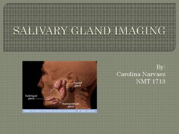
SALIVARY GLAND IMAGING
- By
- Carolina Narvaez
- NMT 1713
2
OBJECTIVES
- Learn about the Radiopharmaceutical,
localization, dosage, QC, dosage and method of
administration of salivary gland imaging
procedure. - Learn about the indication and contraindications.
- Learn about what patient preparation and history
to be taken.
3
RADIOPHARMACY
- Radionuclide
- Tc99m
- T1/2 6 hours
- Energy 140 keV
- Radiopharmaceutical
- Na 99mTcO4- (pertechnetate)
- Localization
- Parenchyma cells of the parotid gland
- QC
- Moly-Al breakthrough
- Adult Range Dose
- 8-12 mCi (10 mCi)
- Method of Administration
- IV Injection (good bolus for flow)
4
INDICATIONS CONTRAINDICATIONS
- Indications
- Tumors
- Salivary gland function
- Masses in parathyroid
- Salivary tissue
- Anatomic size position of salivary glands
- Contraindications
- NONE
5
PATIENT HISTORY PREPARATION
- Patient History
- Do you have history of family history of cancer,
salivary dysfunction? - Dry mouth?
- Pain or palpable lumps in or under jaw?
- Labial biopsy (minor salivary gland tissue)?
- Patient Preparation
- Withhold thyroid blocking meds (ex. iodine) for
48 hrs. - Administer patient gum to stimulate salivary
glands
6
PROCEDURE (60-90 minutes)
- Patient in supine position
- Pillow under shoulders
- Neck extended camera as close as possible
- Administer injection to patient Start camera
- Obtain one of the following
- Flow dynamic 30-60 min Anterior marker, left
right laterals - Flow with statics15 min PI3 min/view Anterior
marker, left right laterals - Statics only15 min PI3 min/view some images at
2,5,10,15,20, , 30 min Anterior marker, left
right laterals
7
PROCEDURE.continued
- Lemon Phase
- Place lemon extract or gauze soaked with lemon
juice on both sides of mouth - (some do this at 40 min dynamic or after
dynamics statics- use 2 ml of lemon juice
through straw) - Remove gauze after 5 min allow patient to
expectorate excess saliva rinse with water - Repeat anterior, left, right laterals at 15-30
min after lemon administration
8
(No Transcript)
9
PROCEDURES ..continued
- Salivary function evaluation
- Draw ROI around thyroid, submandibular, parotid
glands, backgrounds-lateral/anterior views - Record counts repeat for all sequential timed
images
10
(No Transcript)
11
RESULTS
- normal
- abnormal
- Symmetric uptake in the parotid submandibular
glands - Uptake in sublingual, thyroid glands nasal
cavity - Significant reduction of activity in salivary
glands after lemon admin
- Asymmetrical uptake on one/both sides of face
- Activity remains local after lemon admin
- Increased activity in mass (Warthins tumor)
- Patchy decreases in activity (Sjogrens
syndrome-dry mouth) - Inflammation of salivary glands
12
(No Transcript)
13
CONCLUSION
- With the lemon extract administration, the
salivary glands are evaluated to rule out tumors,
lesions, or infections.
14
QUESTIONS
- How much Na 99mTcO4- (pertechnetate) should be
administered to the patient? - What is the method of localization of the
salivary glands? - What views are acquired in the salivary gland
imaging?
15
ANSWERS
- 10 mCi Na 99mTcO4-
- Parenchyma cells of the parotid gland and
salivary glands - Anterior marker, left and right laterals
Recommended
