
Knee Joint PowerPoint PPT Presentation
1 / 54
Title: Knee Joint
1
(No Transcript)
2
(No Transcript)
3
Knee Joint
- The knee is primarily a hinge joint allowing
flexion extension these movements are
accompanied by gliding rolling in addition to
rotation about a vertical axis. The knee joint
is robust but given the complexity of its
movements which must occur during loading at or
above body weight its function is often at risk
particularly when it is hyperextended. Knee
injuries are consistently the most common cause
of disabling injuries in the NFL
4
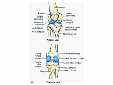
Articular surfaces
- The knee consists of three articulations
- 1. Two femorotibial articulations (lateral
medial) between the lateral medial femoral and
tibial condyles. - 2. One intermediate femoropatellar articulation
between the patella and femur - The fibula is not involved in the knee joint.
5
(No Transcript)
6
- The stability of the knee joint depends on
- 1. Strength and actions of surrounding muscles
and tendons, the most important muscles in
stabilizing the knee are the quadriceps femoris
particularly the fibers of the vastus lateralis. - 2. Ligaments connecting the femur tibia
7
Joint Capsule
- The joint capsule consists of an external fibrous
layer and an internal synovial membrane that
lines the internal surfaces of the articular
cavity not covered by articular cartilage. - The fibrous layer attaches to the femur
superiorly just proximal to the articular margins
of the condyles. - Posteriorly it encloses the condyles the
intercondylar fossa. - The fibrous layer has an opening posterior to the
lateral tibial condyle to allow the popliteus
tendon to pass out of the joint capsule and
attach to the tibia. - Inferiorly the fibrous layer attaches to the
margins of the articular surface of the tibia
(tibial plateau).
8
(No Transcript)
9
- The quadriceps tendon, patella patellar
ligament are intimately applied to the fibrous
capsule anteriorly.
10
- The synovial membrane lines the internal aspects
of the fibrous capsule and attaches to the
periphery of the patella and the edges of the
cartilaginous menisci. - The synovial membrane reflects from the posterior
aspect of the joint anteriorly into the
intercondylar region, covering the cruciate
ligaments and the infrapatellar fat pad
separating them for the articular cavity. This
creates a median infrapatellar synovial fold, a
vertical fold of synovial membrane that
approaches the posterior aspect of the patella
creating left right femorotibial articular
cavities. - Fat filled lateral medial alar folds cover the
inner surface of the fat pads that occupy the
space on each side of the patellar ligament
internal to the fibrous layer.
11
(No Transcript)
12
- Superior to the patella, the knee cavity extends
deep to the vastus lateralis as the suprapatellar
bursae. - Muscle fibers of the vastus intermedius form the
articular muscle of the knee which attaches to
the synovial membrane and retracts the bursa
during knee extension.
13
Ligaments
- The joint capsule is strengthened by 5
extracapsular (capsular) ligaments - 1. Patellar ligament The distal part of the
quadriceps tendon, it passes from the apex and
margins of the patella to the tibial tuberosity.
Laterally it receives aponeurotic expansions of
the vastus lateralis (medial lateral
retinacula) these are important for maintaining
the alignment of the patella. - The collateral ligaments are taut in extension
but become lax as the knee flexes which allows
rotation of the knee. - 2. Fibular (Lateral) collateral ligament Extends
inferiorly from the lateral epicondyle of the
femur to the lateral surface of the head of the
fibula. The tendon of the popliteus passes deep
to the FCL separating it from the lateral
meniscus. The FCL splits the tendon of the
biceps femoris.
14
(No Transcript)
15
Ligaments
- 3. Tibial (Medial) collateral ligament (TCL)
extends from the medial epicondyles of the femur
to the medial condyle and superior medial surface
of the tibia. The deep fibers of the TCL are
attached to the medial meniscus. - 4. Oblique popliteal ligament This is a
reflected expansion of the tendon of the
semimembranosus that strengthens the joint
capsule posteriorly. It arises from the medial
tibial condyle passes superolaterally to the
central part of the posterior aspect of the joint
capsule. - 5. Arcuate popliteal ligament strengthens the
capsule posterolateral. It arises from the
posterior aspect of the fibular head passes
supero-medially over the popliteal tendon and
attaches to the posterior surface of the knee
joint.
16
(No Transcript)
17
(No Transcript)
18
Intra Articular ligaments (cruciate menisci,
popliteus tendon)
- Cruciate ligaments These ligaments join the
femur to the tibia, cris-crossing within the
joint but outside the articular cavity. These
ligaments cross each other obliquely. During
medial rotation of the tibia on the femur the
cruciate ligaments wind around each other this
limits the amount of medial rotation to 10. With
lateral rotation the ligaments unwind thus
allowing 60 of lateral rotation when the knee
is flexed at gt90. The cross over point serves
as an axis for rotatory movements.
19
- 1. Anterior cruciate ligament the weaker of the
2 ligaments it arises from the anterior
intercondylar area of the tibia just posterior to
the attachment of the medial meniscus. - It extends superiorly posteriorly and laterally
to the posterior part of the medial side of the
lateral condyle of the femur. - The ACL limits the posterior rolling of the
femoral condyles on the tibial plateau during
flexion converting it into spin. - It also prevents posterior displacement of the
femur on the tibia and hyperextension of the knee
joint. - When the joint is flexed at a right angle the
tibia cannot be pulled anteriorly because it is
held by the ACL.
20
(No Transcript)
21
- 2. Posterior cruciate ligament (PCL) the stronger
of the 2 ligaments, it arises from the posterior
condylar area of the tibia. - The PCL passes superiorly and anteriorly on the
medial side of the ACL to attach to the anterior
part of the lateral surface of the medial condyle
of the femur. - The PCL limits the anterior rolling of the femur
on the tibial plateau during extension,
converting it to spin. - It also prevents anterior displacement of the
femur on the tibia or posterior displacement of
the tibia on the femur and helps prevent
hyperflexion of the knee joint. - In the weight-bearing flexed knee the PCL is the
main stabilizing factor for the femur
22
(No Transcript)
23
(No Transcript)
24
- ACL under tension when knee extended
- PCL under tension when knee flexed
25
Menisci of the knee
- These are cresentic plates of fibrocartilage on
the articular surface of the tibia, they serve to
deepen the joint surface and shock absorption. - They are thicker at their outer margins and
taper to thin inner margins. - The menisci are firmly attached at their ends to
the intercondylar area of the tibia. - Their external margins are attached to the
fibrous joint capsule. - The coronary ligaments are capsular fibers that
attach the margins of the menisci to the tibial
condyles. - The transverse ligament joins the anterior
margins of the menisci together.
26
- 1. Medial meniscus a C-shaped structure broader
posteriorly, the anterior horn attaches to the
anterior intercondylar area anterior to the
attachment of the ACL. The posterior end
attaches to the posterior intercondylar area
anterior to the attachment of the PCL. It is
firmly adherent to the deep surface of the tibial
collateral ligament.
27
- 2. Lateral meniscus Nearly circular, smaller and
more mobile than the medial meniscus. The tendon
of the popliteus separates the lateral meniscus
from the fibularr collateral ligament. The
posterior meniscofemoral ligament joins the
lateral meniscus to the PCL and the medial
femoral condyle
28
- Meniscus tears
- Meniscal repair
29
(No Transcript)
30
Bursae Around the Knee
- There are at least 12 bursae around the knee
joint because most tendons run parallel to the
bones and pull lengthwise across the joint during
knee movements. The subcutaneous prepatellar and
infrapatellar bursae are located at the convex
surface of the joint allowing the skin to be able
to move freely during knee movements. Four
bursae communicate with the articular cavity of
the knee joint suprapatellar bursa (deep to the
distal quadriceps), popliteus bursa, anserine
bursa and gastrocnemius bursa.
31
(No Transcript)
32
Arterial Supply to the Knee 5-62 63B
- The arteries supplying the knee joint are the 10
vessels that form the perigenicular anastomoses
around the knee the genicular branches of the
femoral, popliteal and anterior and posterior
recurrent branches of the anterior tibial
recurrent and circumflex fibular arteries. The
middle genicular branches of the popliteal artery
penetrate the fibrous layer of the joint capsule
and supply the cruciate ligaments, synovial
membrane and peripheral margins of the menisci.
33
(No Transcript)
34
Innervation of the Knee
- Hiltons law applies the nerves supplying the
muscles acting on the knee also innervate the
joint. Articular branches of the femoral nerve
supply the anterior knee, tibial nerve branches
go to the posterior knee and the common fibular
nerve supplies the lateral aspect of the knee.
The obturator and saphenous nerves also supply
genicular articular branches.
35
Movements of the Knee
- Flexion extension are the main movements of the
knee, some rotation can occur when the knee is
flexed. When the leg is extended with the foot on
the ground the knee passively locks because of
medial rotation of the femur on the tibia, this
allows weight bearing without muscular exertion
by the leg muscles. To unlock the knee the
popliteus contracts rotating the femur laterally
5on the tibial plateau allowing flexion to
occur.
36
(No Transcript)
37
(No Transcript)
38
(No Transcript)
39
(No Transcript)
40
(No Transcript)
41
(No Transcript)
42
(No Transcript)
43
(No Transcript)
44
(No Transcript)
45
(No Transcript)
46
(No Transcript)
47
(No Transcript)
48
(No Transcript)
49
(No Transcript)
50
(No Transcript)
51
(No Transcript)
52
(No Transcript)
53
(No Transcript)
54
(No Transcript)
Recommended
