
Muscle Structure and Function PowerPoint PPT Presentation
1 / 20
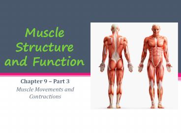
Title: Muscle Structure and Function
1
Muscle Structure and Function
- Chapter 9 Part 3
- Muscle Movements and Contractions
2
Structures of a skeletal muscle fiber
3
Neuromuscular Junction
- Connects the nervous system to the muscular
system through synapses between nerve and muscle
fibers - Directs action potentials basically a small
impulse from YO BRAIN that sets off a whole chain
of events that lead to muscle contractions - Acetylcholine is the neurotransmitter that motor
neurons use to control skeletal muscle
contraction - Synthesized in the cytoplasm of motor neuron
- Its to blame for muscle contractions, its
basically an instigator
4
Neuromuscular Junction
5
7 Steps of the Neuromuscular Junction
- An action potential travels the length of the
axon of a motor neuron to an axon terminal - Voltage-gated Calcium channels open and Ca ions
diffuse into the axon terminal - Ca ion entry causes vesicles to release their
contents, ACh, via exocytosis - ACh diffuses across the synaptic cleft and binds
to ACh receptors, in the sarcolemma, which
contain ligand-gated cation channels - ACh binding opens ion channels that allow
simultaneous passage of Sodium (Na) ions into the
muscle fiber and Potassium (K) ions out of the
muscle fiber. More Na ions enter than K ions
leave, produces a change in the membrane
potential - ACh effects are terminated by its enzymatic
breakdown in the synaptic cleft or its diffusion
away from the synaptic cleft - Once the membrane potential reaches a threshold
value, an action potential spreads along the
sarcolemma
6
Sarcomere
- Contractile unit of a muscle
7
Sarcomere Regions
- A Band middle area containing thin/thick
filaments - I Band Region containing only thin filaments
- Z Disc(Line) Separation line between sarcomeres
thin filaments attached - H Zone Region containing only thick filaments
- M Line runs through the center of H zone
8
Sarcomere Components
- Protein myofilaments
- Thick filaments myosin protein
- Thin filaments actin protein
9
Myofilaments Structures
- Thick Filaments
- Thin Filaments
- Twisted strands of actin
- Tropomyosin protein strand stabilizes actin
- Troponin protein strand affected by Ca2
- Myosin head forms cross bridges with thin
filaments to contract muscle cell - Opposite end of filament
10
- Sarcoplasmic Reticulum (SR) specialized smooth
endoplasmic reticulus, surrounds each myofibril - Stores and releases calcium
- Lots of calcium pumps!
- T Tubule Conducts nerve impulses to every
sarcomere - Triggers release of calcium from sarcoplasmic
reticulum
11
Sliding Filament Model
- During contractions thin filaments slide past
thick ones so they overlap more - Sarcomere shortens
12
Sliding Filament theory- method by which muscles
are though to contract
- Boat Myosin (thick filament)
- Oar Myosin side arm
- Water Actin (thin filament)
- Life ring Calcium
13
Resting State
- ATP is bound to myosin side arm.
- ATP splits into ADP P (high energy)
14
Step 1 Action Potential
- A nerve action potential releases acetylcholine
into the synaptic cleft opening the Na channels. - Action potential spreads across sarcolemma
releasing Calcium into sarcoplasma
15
Step 2 Myosin/Actin binding
- Calcium binds to troponin
- A shape change in troponin moves tropomyocin out
of the way of actin binding site - Actin and myosin bind using energy from cleaved
ATP.
16
Step 3 Powerstroke
- Side arm pivots so myosin and actin slide by each
other shortening the sarcomere. - ADP and P released (low energy)
17
Step 4 ATP Binding Release
- A different ATP molecule binds to active site.
- Actin released
18
Step 5 ATP cleavage
- Return to high energy state
- Cycle will repeat if calcium ions are still
available.
19
Think it over
- The boat (myosin) does not move far in one cycle,
a muscle contraction requires many cycles - If a muscle is contracted what happens if a new
molecule of ATP is not available? - Muscle stays contracted cramps!
- Why does rigor mortis occur? (Hint What
chemical is no longer available to the body?) - ATP is not available to control calcium release
so contractions are continuous 6-8 hours after
death. - Body relaxes 16-24 hours as enzymes break down
contractile structures.
20
- Review Questions
- What chemical exposes the binding site for actin
and myosin? - Calcium
- What is the source of energy for a contraction?
- ATP
- What is the name of the step in which the actin
filament is actively contracted? - Powerstroke
- What chemical must be present in order for the
actin and myosin filaments to separate? - ATP
Recommended
