
Chapter%206%20The%20Skeletal%20System:Bone%20Tissue PowerPoint PPT Presentation
Title: Chapter%206%20The%20Skeletal%20System:Bone%20Tissue
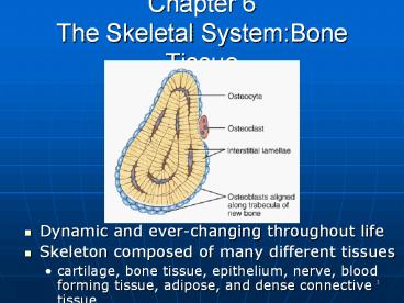
1
Chapter 6The Skeletal SystemBone Tissue
- Dynamic and ever-changing throughout life
- Skeleton composed of many different tissues
- cartilage, bone tissue, epithelium, nerve, blood
forming tissue, adipose, and dense connective
tissue
2
Functions of Bone
- Supporting protecting soft tissues
- Attachment site for muscles making movement
possible - Storage of the minerals, calcium phosphate --
mineral homeostasis - Blood cell production occurs in red bone marrow
(hemopoiesis) - Energy storage in yellow bone marrow
3
Anatomy of a Long Bone
- Diaphysis shaft
- Epiphysis one end of a long bone
- Metaphysis growth plate region
- Articular cartilage over joint surfaces acts as
friction shock absorber - Medullary cavity marrow cavity
- Endosteum lining of marrow cavity
- Periosteum tough membrane covering bone but not
the cartilage - fibrous layer dense irregular CT
- osteogenic layer bone cells blood vessels
that nourish or help with repairs
4
Histology of Bone
- A type of connective tissue as seen by widely
spaced cells separated by matrix - Matrix of 25 water, 25 collagen fibers 50
crystalized mineral salts - 4 types of cells in bone tissue
5
Cell Types of Bone
- Osteoprogenitor cells ---- undifferentiated cells
- can divide to replace themselves can become
osteoblasts - found in inner layer of periosteum and endosteum
- Osteoblasts--form matrix collagen fibers but
cant divide - Osteocytes ---mature cells that no longer secrete
matrix - Osteoclasts---- huge cells from fused monocytes
(WBC) - function in bone resorption at surfaces such as
endosteum
6
Matrix of Bone
- Inorganic mineral salts provide bones hardness
- Organic collagen fibers provide bones
flexibility - their tensile strength resists being stretched or
torn - Mineralization (calcification) is hardening of
tissue when mineral crystals deposit around
collagen fibers - Bone is not completely solid since it has small
spaces for vessels and red bone marrow - spongy bone has many such spaces
- compact bone has very few
7
Compact or Dense Bone
- Looks like solid hard layer of bone
- Makes up the shaft of long bones and the external
layer of all bones - Resists stresses produced by weight and movement
8
Histology of Compact Bone
- Osteon is concentric rings (lamellae) of
calcified matrix surrounding a vertically
oriented blood vessel - Osteocytes found in spaces called lacunae
- Osteocytes communicate through canaliculi filled
with extracellular fluid that connect one cell to
the next cell
9
The Trabeculae of Spongy Bone
- Latticework of thin plates of bone called
trabeculae oriented along lines of stress - Spaces in between these struts are filled with
red marrow where blood cells develop - Found in ends of long bones and inside flat bones
such as the hipbones, sternum, sides of skull,
and ribs.
No true Osteons.
10
Bone Scan
- Radioactive tracer is given intravenously
- Amount of uptake is related to amount of blood
flow to the bone - Hot spots are areas of increased metabolic
activity that may indicate cancer, abnormal
healing or growth - Cold spots indicate decreased metabolism of
decalcified bone, fracture or bone infection
11
Blood and Nerve Supply of Bone
- Periosteal arteries
- supply periosteum
- Nutrient arteries
- enter through nutrient foramen
- supplies compact bone of diaphysis red marrow
- Metaphyseal epiphyseal aa.
- supply red marrow bone tissue of epiphyses
12
Bone Formation or Ossification
- All embryonic connective tissue begins as
mesenchyme. - Intramembranous bone formation formation of
bone directly from mesenchymal cells. - Endochondral ossification formation of bone
within hyaline cartilage.
13
Intramembranous Bone Formation
- Mesenchymal cells become osteoprogenitor cells
then osteoblasts. - Osteoblasts surround themselves with matrix to
become osteocytes. - Matrix calcifies into trabeculae with spaces
holding red bone marrow. - Mesenchyme condenses as periosteum at the bone
surface. - Superficial layers of spongy bone are replaced
with compact bone.
14
Endochondral Bone Formation (1)
- Development of Cartilage model
- Mesenchymal cells form a cartilage model of the
bone during development - Growth of Cartilage model
- in length by chondrocyte cell division and
matrix formation ( interstitial growth) - in width by formation of new matrix on the
periphery by new chondroblasts from the
perichondrium (appositional growth)
15
Endochondral Bone Formation (2)
- Development of Primary Ossification Center
- nutrient artery penetrates center of cartilage
model - osteoblasts deposit bone matrix over calcified
cartilage forming spongy bone trabeculae - osteoclasts form medullary cavity
16
Endochondral Bone Formation (3)
- Development of Secondary Ossification Center
- blood vessels enter the epiphyses around time of
birth - spongy bone is formed but no medullary cavity
- Formation of Articular Cartilage
- cartilage on ends of bone remains as articular
cartilage.
17
Bone Growth in Length
- Epiphyseal plate or cartilage growth plate
- cartilage cells are produced by mitosis on
epiphyseal side of plate - cartilage cells are destroyed and replaced by
bone on diaphyseal side of plate - Between ages 18 to 25, epiphyseal plates close.
- cartilage cells stop dividing and bone replaces
the cartilage (epiphyseal line) - Growth in length stops at age 25
18
Zones of Growth in Epiphyseal Plate
- Zone of resting cartilage
- anchors growth plate to bone
- Zone of proliferating cartilage
- rapid cell division (stacked coins)
- Zone of hypertrophic cartilage
- cells enlarged remain in columns
- Zone of calcified cartilage
- thin zone, cells mostly dead since matrix
calcified - osteoclasts removing matrix
- osteoblasts capillaries move in to create bone
over calcified cartilage
19
Factors Affecting Bone Growth
- Nutrition
- adequate levels of minerals and vitamins
- calcium and phosphorus for bone growth
- vitamin C for collagen formation
- vitamins K and B12 for protein synthesis
- Sufficient levels of specific hormones
- during childhood need insulinlike growth factor
- promotes cell division at epiphyseal plate
- need hGH (growth), thyroid (T3 T4) and insulin
- sex steroids at puberty
- growth spurt and closure of the epiphyseal growth
plate - estrogens promote female changes -- wider pelvis
20
Hormonal Abnormalities
- Oversecretion of hGH during childhood produces
giantism - Undersecretion of hGH or thyroid hormone during
childhood produces short stature - Both men or women that lack estrogen receptors on
cells grow taller than normal - estrogen responsible for closure of growth plate
21
Bone Remodeling
- Ongoing since osteoclasts carve out small tunnels
and osteoblasts rebuild osteons. - release calcium and phosphorus into interstitial
fluid - Continual redistribution of bone matrix along
lines of mechanical stress - distal femur is fully remodeled every 4 months
22
Fracture Repair of Bone
- Fracture is break in a bone
- Healing is faster in bone than in cartilage due
to lack of blood vessels in cartilage - Healing of bone is still slow process due to
vessel damage - Clinical treatment
- closed reduction restore pieces to normal
position by manipulation - open reduction surgery
23
Fractures
- Named for shape or position of fracture line
- Common types of fracture
- closed -- no break in skin
- open fracture --skin broken
- comminuted -- broken ends of bones are
fragmented - greenstick -- partial fracture
- impacted -- one side of fracture driven into the
interior of other side - Potts -- distal fibular fracture
- Colless -- distal radial fracture
- stress fracture -- microscopic fissures from
repeated strenuous activities
24
Repair of a Fracture (1)
- Formation of fracture hematoma
- damaged blood vessels produce clot in 6-8 hours,
bone cells die - inflammation brings in phagocytic cells for
clean-up duty - new capillaries grow into damaged area
- Formation of fibrocartilagenous callus formation
- fibroblasts invade the procallus lay down
collagen fibers - chondroblasts produce fibrocartilage to span the
broken ends of the bone
25
Repair of a Fracture (2)
- Formation of bony callus
- osteoblasts secrete spongy bone that joins 2
broken ends of bone - lasts 3-4 months
- Bone remodeling
- compact bone replaces the spongy in the bony
callus - surface is remodeled back to normal shape
26
Calcium Homeostasis Bone Tissue
- Skeleton is reservoir of Calcium Phosphate
- Calcium ions involved with many body systems
- nerve muscle cell function
- blood clotting
- enzyme function in many biochemical reactions
- Small changes in blood levels of Ca2 can be
deadly (plasma level maintained 9-11mg/100mL) - cardiac arrest if too high
- respiratory arrest if too low
27
Exercise Bone Tissue
- Pull on bone by skeletal muscle and gravity is
mechanical stress . - Stress increases deposition of mineral salts
production of collagen (calcitonin prevents bone
loss) - Lack of mechanical stress results in bone loss
- reduced activity while in a cast
- astronauts in weightlessness
- bedridden person
- Weight-bearing exercises build bone mass (walking
or weight-lifting)
28
Aging Bone Tissue
- Bone is being built through adolescence, holds
its own in young adults, but is gradually lost in
aged. - Demineralization loss of minerals
- very rapid in women 40-45 as estrogens levels
decrease - in males, begins after age 60
- Decrease in protein synthesis
- decrease in growth hormone
- decrease in collagen production which gives bone
its tensile strength - bone becomes brittle susceptible to fracture
29
Osteoporosis
- Decreased bone mass resulting in porous bones
- Those at risk
- white, thin menopausal, smoking, drinking female
with family history - athletes who are not menstruating due to
decreased body fat decreased estrogen levels - people allergic to milk or with eating disorders
whose intake of calcium is too low - Prevention or decrease in severity
- adequate diet, weight-bearing exercise,
estrogen replacement therapy (for menopausal
women) - behavior when young may be most important factor
30
Disorders of Bone Ossification
- Rickets
- calcium salts are not deposited properly
- bones of growing children are soft
- bowed legs, skull, rib cage, and pelvic
deformities result - Osteomalacia
- new adult bone produced during remodeling fails
to ossify - hip fractures are common
Recommended
