
Virus and bullous dermatoses PowerPoint PPT Presentation
1 / 48
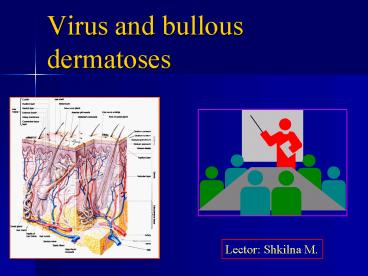
Title: Virus and bullous dermatoses
1
Virus and bullous dermatoses
Lector Shkilna M.
2
CONENT
- 1.Clinical types of pemphigus
- Pemphigus vulgaris
- Pemphigus foliaceus
- Pemphigus vegetans
- Pemphigus erythematous
- 2. Classification
- 3.Diagnosis of HSV Infections
- 4. Epidemiology
- 5. Disease caused by Herpes Simplex Viruses
- 6. Disease caused by Herpes Zoster
- 7. Other human Herpes Viruses Disease
- 8. Diagnosis and treatment
3
Skin layers
4
Bulla formed due to fluid in the skin and fluid
collection occurs at sites where the cohesion on
the skin is weak
- subcorneal
- intra epidermal, due to individual
keratinocytes - dermo epidermal junction
A circumscribed collection of free fluid more
than 0,5 sm in diameter
5
Pemphigus ( from the Greek pemphix) -
- meaning blister is a rare, of autoimmune,
intraepidermal blistering diseases involving the
skin and mucous membranes. - It is a particular group of bullous dermatoses
presenting with a distinct histopathology
characterized by intraepidermal bulla and
acantholysis.
6
Acantholysis
- Normally the cells of the spinous cell layer are
kept together by the of desmosomes and a series
extracellular proteins known as cadherins. - Autoantibodies, (IgG) are directed against the
extracellular protein desmoglein 3 which is one
of the cadherins. Desmoglein 3 is treated as an
antigen and this process produces the separation
of the cells of the spinous cell layer with
consequent formation of vesicles and bullae. The
process of destruction (lysis) of the
intercellular connections (desmosomes) of the
epithelial cells is known as acantholysis.
7
Acantholysis
- Acantholytic cells
- is (which are present both in the blister cavity
and at the edge of the blister) are rounded
keratinocytes. The cytoplasm - is condensed in the periphery resulting in a
perinuclear pale halo.
8
Four clinical types of pemphigus
- 1. Pemphigus vulgaris cleft is deeply situated
between the basal layer and the rest of epidermis
and there is sufficient fluid to produce the
characteristic bulla. - 2. Pemphigus vegetans superficial cleft and
proliferate changes producing papillomatous
masses. - 3. Pemphigus foliaceus subcorneal cleft and
little fluid. - 4. Pemphigus erythematous abortive phase of
Pemphigus foliaceus.
9
Pemphigus vulgaris
- It is an autoimmune disease caused by drugs,
chemicals and infections. - Pathology.
- 1. The bulla of Pemphigus vulgaris are
intra-dermal and irregular in shape with acute
lateral margins . - 2. They are formed by the separation of
acantholytic epidermal cells( Tzanck cells ). - 3. Acantholytic cells may be in the bulla cavity.
- 4. Dermis beneath the bulla shows number of
inflammatory cells including a few lymphocytes
and plasma cells.
10
Skin lesions predominantly present on
Axillae
Trunk
11
Skin lesions
- Tense of flaccid bulla appear on normal skin.
- The lesions may be few and sparse, or extensive.
- The eruption is usually symmetrical.
- They are usually irregular in shape.
- On rupturing, form painful erosions which have a
tendency to spread . - Positive Nikolskys sign.
12
Nikolsky's sign
- application of tangential pressure on normal
skin results in formation of anew bulla or if
applied to pre-existing bulla results in the
spread of bulla (Nikolskys sign). - where the epidermis is detached and slipping free
from the dermis with slight pressure
13
Mucosal lesions
- Eventually present in all patients oral mucosa
moat frequently involved. - The mouth is often involved, but denuded areas
may be seen on conjunctive, vagina, nose. - Patients have painful raw areas with detachable
shreds of epithelium in the mouth, these may
extend to the pharynx and larynx resulting in
dysphagia and hoarseness.
14
Pemphigus foliaceus
- Pathology
- It is superficial pemphigus (in granular cell
layer or under the stratum corneum).
15
Skin lesions
- Flaccid bulla and exfoliating scales.
- Usually flaccid bulla develop first on the face.
- Slowly the disease spreads symmetrically till the
whole of the integument is covered with bulla
(when it looks like erythroderma). - Bulla rapture rapidly and produce a moist, red,
raw, and oedematous surface and flake-like
plaques of imperfectly keratinized, horny cells. - The conjunctivae and mucosa may be affected.
- The scalp may also be involved it is covered
with moist, yellowish scales. The hair may fall.
16
Pemphigus vegetans
- It is the rarest variety of pemphigus.
- Individuals of any
- group may be
- affected.
- It is more common in females than in males.
17
Skin lesions
- The initial lesions, in the form of broken
bullae, appear on the mucosa of the lips, angle
of mouth or nose. - Later, they develop in the axillae, groins and
some-times on the other parts of the body. - When ruptured, the bulla develop into moist,
superficial ulcers. - The ulcers undergo proliferative changes
producing fungoid vegetations with malodorous
discharge. - The vegetations may also seem to arise de novo on
the normal skin. - Nikolskys sign is often positive.
18
Pemphigus erythematous
- Skin lesions
- The early lesions which are erythematous and
crusted, appear on the nose and ears, resembling
lupus erythematosus both in their location and
appearance. - However, the lesions exhibit a moist, raw surface
when the crust is removed. - The greasy crust may indicate seborrhoeic
dermatitis. - These lesions may appear along with bullae on the
chest and extremities. - The eruption is symmetrical in distribution.
19
Diagnosis
- Laboratory diagnosis of pemphigus is based on
- Tzanck Smear.
- Histology.
- Immunopathology.
20
Preparation of Tzanck smear
- The vesicle should be unroofed or the crust
removed, and the base scraped with a scalpel or
the edge of a spatula. - The material is transferred to a glass slide by
touching the spatula to the glass slide
repeatedly but gently. - The slide should be clean, since cells will not
adhere to a slide marred by fingerprints. - In the case of blistering disorders
- The intact roof of a blister is opened along one
side, folded back and the floor gently scraped. - The material thus obtained is smeared onto a
microscopic slide, allowed to air dry, and
stained with Giemsa or any of the Romanowskys
stains.
21
Tzanck Smear findings in bullous disorders
- Pemphigus (Acantholytic cells)
- Bullous pemphigoid (Predominantly eosinophils)
- Chronic bullous disease of childhood
(Predominantly polymorphs) - Varicella zoster infection (Multinucleated giant
cells) - Herpes simplex infection (Multinucleated giant
cells) - Toxic epidrmal necrolysis (Necrotic cells).
22
ImmunopathologyTwo classes of tests
areavailable
- 1. Direct immunofluorescence (DIF) Done on the
skin of the patient, shows intercellular deposits
of Ig G and C3 giving a fish net appearance. - 2. Indirect immunofluorescence (IIF) Done on
patient serum to detect autoantibody titers
correlate with the clinical activity and may be a
useful guide to the dose of oral steroids needed.
23
Treatment
- Supportive treatment
- Local hygiene of mucosal and skin lesions.
- Therapeuticas well as prophylactic use of
antibiotics (forcoetaneous infection) and
anticandidal agents (formucosal lesions). - Maintenance of water and electrolyte balance.
- Specific treatment
- Specific treatment depends on the judicious use
of corticosteroids and immunosuppressive drugs
since pemphigus - is an autoimmune disorder.
24
Treatment
- Corticosteroids
- Two regimes are commonly used
- Daily dose of 1 -2 mg / kg body weight of
prednisoloneequivalent is used to suppress
disease activity andsteroids are tapered when
the disease is controlledthis form of steroid
therapy is associated withsubstantial adverse
events. - Monthly steroid therapy.
- Monthly 1-2 mg / kg of betamethasone orally
/dexamethasone intravenous is given. - Usually combined with immunosuppressivetherapy.
- gt May induce remissions with less side effects.
- Immunosuppressive therapy
- Drug regimes
- Azathioprine Usually along with oral
steroidtherapy. 2-3 mg/kg of body weight till
clearing ofdisease maintain on 1 mg / kg. - Methotrexate Usually along with oral
steroidtherapy given as weekly 20-25 mg. - Cyclophosphamide Usually along with oral
steroidtherapy. As daily dose (50-200 mg) or
monthlybolus dose (500-1000 mg) intravenously.
25
Human Herpes Viruses
- Latent Viruses
26
Vesicle
- Description
- Circumscribed collection of free fluid
- Up to 0.5 cm in diameter
Herpes zoster
27
EROSION
- Description
- A focal loss of epidermis
- erosions do not penetrate
- below the dermoepidermal
- junction
- and therefore heal without scarring
Toxic epidermal necrolysis
28
CRUST
- Description
- Is a collection of dried serum and cellular
debris- a scab - Examples
- Acute eczematious inflammation
- Atopic on the face
- Impetigo- golden or honey colored
- Tinea capitis
Impetigo. A thick, honey-yellow adherent crust
covers the entire eroded surface.
29
Classification
- There are 25 families in the Herpeotoviridae but
only 6 of them infect man with any regularity. - Herpes Simplex virus Type 1 (HSV-1)
- Herpes Simplex virus Type 2 (HSV-2)
- Epstein Barr virus (EBV)
- Cytomegalovirus (CMV)
- Varicella Zoster virus (VZV)
- Human Herpes virus 6
- Human Herpes virus 8
30
Herpes Simplex Virus (HSV)
- These are very large viruses and their genome
encodes at least 80 proteins. - Half are not directly involved in the virus
structure. - Almost any human cell type can be affected by
HSV.
31
Epidemiology
- HSV-1 and 2 infections are life-long.
- The virus is found in the lesions on the skin but
can be present in body fluids including saliva
and vaginal secretions. - As a result of poor hygiene in underdeveloped
countries, HSV-1 antibodies are found in more
than 90 of children.
32
Epidemiology 2
- HSV-2 is normally spread sexually and is found in
the anus, rectum and upper alimentary tract as
well as the genital area. - An infant can be infected at birth by a
genitally-infected mother. - The infant can also be infected in utero if the
mothers infection spreads. - Because of the infants underdeveloped immune
system, the resulting infection can be very
severe and sometimes be deadly.
33
Disease caused by Herpes Simplex Viruses
- Oral Herpes - Cold sores
- Herpetic gingiovostomatitis, the infection, often
initially on the lips spreads to all parts of the
mouth and pharynx.
34
Disease caused by Herpes Simplex Viruses
- Eczema Herpeticum
- This is found in children with active eczema.
- The virus can spread to other organs such as the
liver and adrenals.
35
Disease caused by Herpes Simplex Viruses
- Genital Herpes
- Is usually the result of HSV-2.
- Primary infection is often asymptomatic but many
painful lesions can be developed on the shaft of
the penis and vulva, vagina, cervix and perianal
region of women.
36
Genital Herpes
37
Genital Herpes
- In both sexes, the urethra can be involved.
- Genital Herpes infections can be accompanied by a
variety of symptoms including fever, myalgia,
glandular inflammation of the groin area
(inguinal). - Some patients have only infrequent recurrences
but others experience recurrences as often as
every 14-21 days.
- Is usually the result of HSV-2.
- Primary infection is often asymptomatic but many
painful lesions can be developed on the shaft of
the penis and vulva, vagina, cervix and perianal
region of women.
38
(No Transcript)
39
Diagnosis of HSV Infections
- Cells may be obtained from the base of the lesion
(called a Tzank smear) and histochemistry
performed. - These can be seen in the smears as multinucleated
giant cells and contain Cowdry type A inclusion
bodies.
- The cells can also be stained with specific
antibodies in an immunofluorescence test. - It can also be detected by viral DNA by in situ
hybridization. - Type-specific antibodies can distinguish between
HSV-1 and HSV-2.
40
Diagnosis of HSV Infections
41
HERPES ZOSTER
- Reactivation of HVZ
- dermatomal distribution
- may recur
- can disseminate in immunocompromised patients
- complications
- post herpetic pain
- ophthalmic zoster -corneal scarring and loss of
vision
DIAGNOSIS
CLINICAL EM of vesicle fluid SEROLOGY IgM
detection
42
Pain and hyperaesthesia
43
Pain and hyperaesthesia
44
Pain and hyperaesthesia
45
OTHER HUMAN HERPES VIRUSES
- HHV6
- virus replicates in T and B cells
- infection occurs in first 3 years of life
- Clinical Exanthem subitum (roseola infantosum)
- mild acute febrile illness
- incubation period of 2 weeks
- fever lasts several days
- macular papular rash appears within 2 days of
fever - 85 of adults carry virus in saliva
- HHV7
- isolated from CD4 positive cells
- virus present in saliva of gt75 of adults
- role in disease unclear
- Evidence of infection present (seroconversion)
- HHV8
- detected in epithelial cells of Kaposi sarcoma
- also present in semen
- postulated as cause of Kaposi sarcoma
46
Exanthem subitum (roseola infantosum)
47
Treatment
- Acyclovir
- A Safe and extremely well-tolerated drug.
- More than 35 million patients have been
consistent and reassuring. - Some authorities have proposed making acyclovir
available as a non-prescription drug. - Adverse effects, usually mild, include nausea,
vomiting, rash and headache.
- Valacyclovir
- New antiviral agent
- Is the 1-valine ester prodrug of acyclovir.
- It has an oral bioavailability three to five
times greater than that of acyclovir. - Several large trials have shown that it is safe
and well tolerated.
48
THANK YOU !
Recommended
