
The cell cycle PowerPoint PPT Presentation
1 / 72
Title: The cell cycle
1
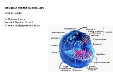
Molecules and the Human Body Module Leader Dr
Graham Ladds Warwick Medical School Graham.ladds_at_w
arwick.ac.uk
2
Topics to cover today
- Signalling
- Neuromuscular junction events
- G proteins pathways
- Alkalosis and acidosis
- Genetics group work in lecture theater
- Preparation for next few weeks
- The bolics
- Endocrinology
- Mitosis/meiosis
- Cell cycle
- Oncogenes/tumour suppressor genes
- ESA style questions
3
For once size does matter
Size
Name
Units
Example
10-3 10-6 10-9 10-12 10-15 10-18
milli micro nano pico femto atto
m µ n p f a
mM µL ng pM fL ag
Drug levels
moles mass(g) e.g. 1 mole of carbon 12g
MW
12
1 mole 6.022x10²³ molecules Avogadros number
4
Signal gt Translator gt Response
- The translator detects the signal - it is a
receptor. - The translator converts the signal into the
response - it is an effector. - The translator is a protein (or series of
proteins). - Effectors use three mechanisms to change cell
behaviour. - 1. Alter gene transcription.
- 2. Alter ion balance across the plasma membrane.
- Alter the activity level of existing enzymes.
- There are five types of receptor
- 1. Intracellular receptors.
- 2. Receptors that are ion channels.
- 3. Receptors with intrinsic enzyme activity.
- 4. Receptors linked to protein kinases.
- 5. Receptors coupled to target proteins via a G
protein.
5
Alter gene transcription Changing the protein
composition changes cell behaviour. Some
genes are turned on. Some genes are turned
off. Not suitable for rapid, short-term
changes. Common mechanism for development and
differentiation.
6
Alter ion balance across the plasma
membrane Changing ion balance changes cell
behaviour. Transport of Na, K or Cl- changes
membrane potential. Transport of Ca2 changes
intracellular concentration. Ca2 is a second
messenger (affects activity of target proteins).
7
Many signals alter the activity level of
enzymes Many enzymes affect protein
phosphorylation. Protein kinases
(phosphorylate target proteins). Protein
phosphatases (dephosphorylate target
proteins). Many enzymes affect second messenger
levels. Phospholipase C (hydrolyses PIP2 to
IP3 and DAG). Adenylate cyclase (converts ATP
to cAMP). Guanylate cyclase (converts GTP to
cGMP). cGMP phosphodiesterase (converts cGMP
to GMP). Phosphoinositide 3-kinase
(phosphorylates phosphoinositides).
8
Second messengers Concentration of second
messenger changes after stimulation. Second
messengers regulate the activity of target
proteins.
Exoplasm
O
O
Cytoplasm
CH
CH2
CH2OH
Diacylglycerol
9
Signal gt Translator gt Response
- The translator detects the signal - it is a
receptor. - The translator converts the signal into the
response - it is an effector. - The translator is a protein (or series of
proteins). - Effectors use three mechanisms to change cell
behaviour. - 1. Alter gene transcription.
- 2. Alter ion balance across the plasma membrane.
- Alter the activity level of existing enzymes.
- There are five types of receptor
- 1. Intracellular receptors.
- 2. Receptors that are ion channels.
- 3. Receptors with intrinsic enzyme activity.
- 4. Receptors linked to protein kinases.
- 5. Receptors coupled to target proteins via a G
protein.
10
Intracellular receptors that are
enzymes Activating receptor changes their enzyme
activity. Some enzymes become more active.
Some enzymes become less active. Changing
enzyme activity changes cell behaviour.
Nitric oxide and intracellular guanylate
cyclase. NO diffuses across the membrane and
binds to guanylate cyclase. Guanylate cyclase
converts GTP to cGMP (a second messenger). cGMP
affects the activity of target proteins (protein
kinase G). NO is used in many signalling
pathways. Controls blood vessel dilation
(amyl nitrate spray). Allows peristaltic
movement through the gut.
11
Nitric oxide and intracellular guanylate cyclase
12
Signal gt Translator gt Response
- The translator detects the signal - it is a
receptor. - The translator converts the signal into the
response - it is an effector. - The translator is a protein (or series of
proteins). - Effectors use three mechanisms to change cell
behaviour. - 1. Alter gene transcription.
- 2. Alter ion balance across the plasma membrane.
- Alter the activity level of existing enzymes.
- There are five types of receptor
- 1. Intracellular receptors.
- 2. Receptors that are ion channels.
- 3. Receptors with intrinsic enzyme activity.
- 4. Receptors linked to protein kinases.
- 5. Receptors coupled to target proteins via a G
protein.
13
The opening of ion channels Voltage-gated
channels (changes in membrane potential).
Channels for Na, K and Ca2. Ligand-gated
channels (extracellular ligands -
neurotransmitters). Excitatory transmitters
open Na/K-channels (depolarisation). Acetylchol
ine (nicotinic receptor) sympathetic NS
. Glutamate. Serotonin (5HT-3 receptor).
14
(No Transcript)
15
The 6 events that occur when a nerve impulse
reaches the NMJ resulting in neurotransmitter
release
1.Voltage-regulated calcium channels in the axon
membrane open. 2. Allows Ca2 to enter the
axon. 3. Ca2 inside the axon terminal causes
some of the synaptic vesicles to fuse with the
axon membrane. 4. Release of acetylcholine into
the synaptic cleft (exocytosis). 5. acetylcholine
diffuses across the synaptic cleft and attaches
to acetylcholine receptors on the sarcolemma. 6.
Binding of acetylcholine to receptors on the
sarcolemma initiates an action potential in the
muscle.
16
(No Transcript)
17
The opening of ion channels Voltage-gated
channels (changes in membrane potential).
Channels for Na, K and Ca2. Ligand-gated
channels (extracellular ligands -
neurotransmitters). Excitatory transmitters
open Na/K-channels (depolarisation). Acetylchol
ine (nicotinic receptor) sympathetic NS
. Glutamate. Serotonin (5HT-3 receptor).
Inhibitory transmitters open Cl- channels
(hyperpolarisation). g-aminobutyric acid
(GABA). Glycine.
18
The opening of ion channels Voltage-gated
channels (changes in membrane potential).
Channels for Na, K and Ca2. Ligand-gated
channels (extracellular ligands -
neurotransmitters). Excitatory transmitters
open Na/K-channels (depolarisation). Acetylchol
ine (nicotinic receptor) sympathetic NS
. Glutamate. Serotonin (5HT-3 receptor).
Inhibitory transmitters open Cl- channels
(hyperpolarisation). g-aminobutyric acid
(GABA). Glycine. Ligand-gated channels
(intracellular ligands - second messengers).
cAMP (olfaction), cGMP (phototransduction), Ca2.
19
The opening of ion channels Voltage-gated
channels (changes in membrane potential).
Channels for Na, K and Ca2. Ligand-gated
channels (extracellular ligands -
neurotransmitters). Excitatory transmitters
open Na/K-channels (depolarisation). Acetylchol
ine (nicotinic receptor) sympathetic NS
. Glutamate. Serotonin (5HT-3 receptor).
Inhibitory transmitters open Cl- channels
(hyperpolarisation). g-aminobutyric acid
(GABA). Glycine. Ligand-gated channels
(intracellular ligands - second messengers).
cAMP (olfaction), cGMP (phototransduction),
Ca2. Mechanically-gated channels (sound, touch,
stretch).
20
Signal gt Translator gt Response
- The translator detects the signal - it is a
receptor. - The translator converts the signal into the
response - it is an effector. - The translator is a protein (or series of
proteins). - Effectors use three mechanisms to change cell
behaviour. - 1. Alter gene transcription.
- 2. Alter ion balance across the plasma membrane.
- Alter the activity level of existing enzymes.
- There are five types of receptor
- 1. Intracellular receptors.
- 2. Receptors that are ion channels.
- 3. Receptors with intrinsic enzyme activity.
cancer stuff. - 4. Receptors linked to protein kinases.
- 5. Receptors coupled to target proteins via a G
protein.
21
Signal gt Translator gt Response
- The translator detects the signal - it is a
receptor. - The translator converts the signal into the
response - it is an effector. - The translator is a protein (or series of
proteins). - Effectors use three mechanisms to change cell
behaviour. - 1. Alter gene transcription.
- 2. Alter ion balance across the plasma membrane.
- Alter the activity level of existing enzymes.
- There are five types of receptor
- 1. Intracellular receptors.
- 2. Receptors that are ion channels.
- 3. Receptors with intrinsic enzyme activity.
- 4. Receptors linked to protein kinases. EPO.
- 5. Receptors coupled to target proteins via a G
protein.
22
(No Transcript)
23
Signal gt Translator gt Response
- The translator detects the signal - it is a
receptor. - The translator converts the signal into the
response - it is an effector. - The translator is a protein (or series of
proteins). - Effectors use three mechanisms to change cell
behaviour. - 1. Alter gene transcription.
- 2. Alter ion balance across the plasma membrane.
- Alter the activity level of existing enzymes.
- There are five types of receptor
- 1. Intracellular receptors.
- 2. Receptors that are ion channels.
- 3. Receptors with intrinsic enzyme activity.
- 4. Receptors linked to protein kinases. EPO.
- 5. Receptors coupled to target proteins via a G
protein.
24
G protein-coupled receptors (GPCRs)
g
g
g
g
a
a
a
a
and
b
b
b
b
b
b
b
b
GTP
GDP
GDP
GTP
Nucleotide exchange
Activate target proteins Transcription
factors Ion channels Protein kinases and
phosphatases Phospholipase C Phosphoinositide
3-kinase Cyclases and phosphodiesterases
GDP
GTP
25
Variations on a very common theme Human cells
are estimated to have at least 1000 GPCRs.
Neurotransmitters, hormones, lipids, chemokines,
odours. Human cells contain many different types
of G proteins. There are at least 20 Ga
subunits. There are at least 5 Gb subunits.
There are at least 12 Gg subunits.
Some ligands bind to more than one GPCR. Some
GPCRs activate more than one G protein. Dissociate
d subunits can regulate more than one target
protein. Some target proteins are regulated by
more than one G protein.
26
If you really want a simple version Gas
stimulates adenylate cyclase. Glucagon,
ACTH. Gai inhibits adenylate cyclase.
Prostaglandin PGE1, adenosine. Gat stimulates
cGMP phosphodiesterase. Photons
(rhodopsin). Gaq stimulates phospholipase C.
Bombesin, vasopressin. Ga13 activates ion
channels (Na/H exchange). Thrombin.
27
Dopamine There are D1-like and D2-like
receptors. D1 and D5 couple through Gs to
stimulate adenylate cyclase. D2, D3 and D4
couple through Gi to inhibit adenylate cyclase.
Acetylcholine parasymathetic ns There are five
muscarinic acetylcholine receptor subtypes.
M1, M3 and M5 couple through Gq to stimulate
phospholipase C. M2 couples through Gi to
open a K-channel. M4 couples through Gi to
inhibit adenylate cyclase.
.and dont forget the nicotinic acetylcholine
receptor. This is a Na/K-channel.
28
Serotonin There are 15 serotonin receptor
subtypes. 5HT-1 couple through Gi to inhibit
adenylate cyclase. 5HT-2 couple through Gq to
stimulate phospholipase C. 5HT-4, 5, 6 and 7
couple through Gs to stimulate adenylate cyclase.
.and dont forget the 5HT-3 receptor. This
is a Na/K-channel. 5HT 5-hydroxytryptamine.
Adrenergic receptors Multiple receptors for
adrenaline (epinephrine) and noradrenaline.
a1 receptors couple through Gq to stimulate
phospholipase C. a2 receptors couple through
Gi to inhibit adenylate cyclase. b receptors
couple through Gs to stimulate adenylate cyclase.
29
Signal integration in cardiomyocytes Contraction
is regulated by stimulatory and inhibitory
signals. b-adrenergic receptors stimulate
adenylate cyclase. a-adrenergic receptors
inhibit adenylate cyclase Both receptors work
through G proteins. Adenylate cyclase converts
ATP to cAMP (a second messenger).
b-adrenergic receptor
a-adrenergic receptor
Stimulate
Inhibit
Adenylate cyclase
Gs protein
Gi protein
cAMP
ATP
30
Topics to cover today
- Signalling
- Neuromuscular junction events
- G proteins pathways
- Alkalosis and acidosis
- Genetics group work in lecture theater
- Preparation for next few weeks
- The bolics
- Endocrinology
- Mitosis/meiosis
- Cell cycle
- Oncogenes/tumour suppressor genes
- ESA style questions
31
The Bohr effect Metabolically active tissues
generate H. The pH of the blood can be reduced
from 7.4 to 7.2. The presence of H lowers the O2
affinity of Hb (O2 is released). Hb release 10
more O2 at pH 7.2 than pH 7.4. This helps to
deliver more O2 to active muscles.
32
Molecular explanation of the Bohr effect The
protons (H) bind to particular residues in Hb.
N-terminal amino group of the a-chains, Hisa122
and Hisb146. Positively charged groups form new
electrostatic bonds. For example, Hisb146
interacts with Aspb94. The additional
interactions stabilise deoxy-Hb (the
T-form). Deoxy-Hb has a lower O2 affinity than
oxy-Hb. Thus, protonation reduces the O2 affinity
of Hb.
33
(No Transcript)
34
Transport of CO2 from the tissuesto the Lungs
- 70-80 is transported back to the lungs
- dissolved in the blood as bicarbonate (HCO3-)
- 20-30 is transported back to lungs attached to Hb
Erythrocyte Carbonic anhydrase
35
Conditions in the lungs promote addition of O2 to
Hb and release of CO2
1. Hb(H) O2 HbO2 H
2. Hb(H) CO2 HbCO2
3. CO2H2O H2CO3 HCO3-
H Lowering Hb(H) forces eqn 2 to the left
promoting unloading of CO2 from carbaminoHb Incre
asing H forces eqn 3 to the left forcing CO2
out of solution
36
O-Hb Exchange in the Blood
37
Acid-base Balance
6.8
acidosis
- Normal blood pH is from 7.35 - 7.45
- Going outside of this range can be very dangerous
- even deadly.
pH
7.0
7.35
7.45
alkalosis
7.8
38
Acid-base Balance
- pH balance is maintained by buffers in the blood.
The primary buffers are - Bicarbonate
- CO2 H2O H2CO3 HCO3- H
- Phosphate
- H2PO4- HPO42- H
- Plasma proteins - various
39
The carbonate system contributes the most to
Blood pH control
- HH eqn
- pH (of blood) pKa Log A- ( base HCO3)
- HA
( acid CO2) - the pKa of the CO2/HCO3 system is 6.1
- Normal pCO2 blood 40mmHg 1.2mM (0.03 Ksol)
- Normal HCO3 blood 24mM
- therefore Log 24mM 1.3 6.1 7.40
- 1.2mM
40
Respiratory Control of Blood pH
- Blood pH can be maintained by controlling the
level of CO2 exhaled. - CO2 H2O H2CO3 HCO3-
H - Hyperventilation
- Increase in rate and depth of breathing.
- Reduces the CO2 in blood, increasing pH.
- Hypoventilation
- Reduced rate and depth of breathing.
- Increases CO2 in blood, decreasing pH.
41
Respiratory Acidosis and Alkalosis
- When breathing patterns are improper, the
following can occur. - Respiratory alkalosis (exam stress?)
- Caused by hyperventilation.
- Treatment - rebreathe air or administer CO2.
- Respiratory acidosis
- Caused by inadequate breathing.
- Treatment - administer HCO3- via IV.
42
Metabolic Acidosis and Alkalosis
- Various metabolic conditions can also create
improper pH levels in the blood. - Metabolic acidosis
- Can be caused by uncontrolled diabetes mellitus,
diarrhea, aspirin overdose and after heavy
exercise. - Note that hyperventilation, while a cause of
respiratory alkalosis, can be a response to
metabolic acidosis. - Metabolic alkalosis
- Caused by prolonged vomiting, excessive use of
bicarbonate for treating an upset stomach.
43
Allosteric regulation of Hb - a summary O2
affinity is determined by electrostatic
interactions at a-b interface. Deoxy-Hb has
more interactions and lower O2 affinity.
Oxy-Hb has fewer interactions and higher O2
affinity. O2 increases the O2 affinity of Hb
(cooperative binding). Structural changes
reduce interactions at the a-b interface. H,
CO2 and BPG lower the O2 affinity of Hb. They all
increase electrostatic interactions at the a-b
interface. Protonation (H) creates
electrostatic bonds between chains.
Carbamation (CO2) creates electrostatic bonds
between chains. BPG forms electrostatic
interactions with the polypeptide chains.
44
15 minute breakGenetic problems worksheet
45
Topics to cover today
- Signalling
- Neuromuscular junction events
- G proteins pathways
- Alkalosis and acidosis
- Genetics group work in lecture theater
- Preparation for next few weeks
- The bolics
- Endocrinology
- Mitosis/meiosis
- Cell cycle
- Oncogenes/tumour suppressor genes
- ESA style questions
46
Metabolism terms and phrases
47
The bolics (processes)
- Ana-bolic
- To build up
- (think anabolic steroids-these are the sex
steroids) - The synthesis of muscle proteins
- The synthesis of adipose tissue
- Storage of glucose
- Cata-bolic
- To break down
- The breakdown of muscle
- The breakdown of adipose
- Breakdown of glycogen
48
Carbohydrate terms/process
- Glycolysis
- The lysis of glucose (usage thereof)
- Gluco(se)neogenesis
- The synthesis of new glucose
- Glycogenolysis
- The lysis of glycogen
- Glycogen synthesis
- The synthesis of glycogen
49
Fat/protein terms/processes
- Lipolysis
- Breakdown of fats
- Lipogenesis
- Synthesis of fats
- Proteolysis
- Breakdown of proteins
- Protein synthesis
- synthesis of proteins
We cover little on this please look up yourself
50
Major organ/systems involved
- Liver
- Brain
- Muscle
- Stomach/GI tract
- Pancreas
- Hypothalamus Pituitary Adrenals (HPA) axis
- Cf Repro HPO-vary axis
51
Different types of signals
Paracrine
Odours
Gap junction
Taste
Autocrine
Endocrine
Light
Direct contact
"Electrical"
Gas
52
Brain
Pituitary
Thyroid
Endocrine glands
Thymus
Kidney
Testes
Ovary
Uterus
53
Endocrine system
- System of tissues/organs/glands that secrete and
respond to hormones to control (all?) body
processes - Including metabolism
- Mitosis
- etc
- Combined with neuronal control of process
- Neuro-endocrine
- Example is adrenaline
54
How is metabolism controlled
- Via endocrine control-this is global control
- Endocrine control between organs and tissues
frequently involves negative feedback mechanisms
(sometimes positive feed forward) - Local control is via substrate (or pathway
intermediates) availability/concentration - Therefore involves enzymic parameters such as Km
- Local (intracellular) control of metabolism
mainly involves negative feedback mechanisms
(sometimes positive feed forward)
55
Key endocrine hormones
- Insulin (which bolic? And why?)
- Glucagon
- Adrenaline
- Cortisol
- Growth hormone
56
Key endocrine hormones
- Insulin ana
- Glucagon cata
- Adrenaline cata
- Cortisol cata
- Growth hormone ana
57
Topics to cover today
- Signalling
- Neuromuscular junction events
- G proteins pathways
- Alkalosis and acidosis
- Genetics group work in lecture theater
- Preparation for next few weeks
- The bolics
- Endocrinology
- Mitosis/meiosis
- Cell cycle
- Oncogenes/tumour suppressor genes
- ESA style questions
58
Names
- Chromosomes 23 pairs per cells therefore 46
chromosomes. - Chromatin DNA histones.
- Chromatids one arm of a chromosome.
- Centromeres point where 2 chromatids touch.
- Telomere region of repetitive DNA at chromosome
tip to prevent destruction.
Short arm
Long arm
59
Mitosis
- The stages
- Interphase
- Prophase
- Prometaphase
- Metaphase
- Anaphase
- Telophase
- Interphase
Clip of mitosis
60
Meiosis
- Clip of Meiosis
61
Differentiation
- The specialisation of cells.
- Cell differentiation causes its size, shape,
metabolic activity, and responsiveness to signals
to change dramatically. - A cell that is able to differentiate into many
cell types is known as pluripotent. These cells
are called stem cells in animals. - A cell that is able to differentiate into all
cell types is known as totipotent. In mammals,
only the zygote and early embryonic cells are
totipotent. - Cells that are fully differentiated reside in
what is called a Go arrest.
62
Cell cycle
63
Cell cycle
Timings
M 1 hr G1 10-12 hr S 8 hr G2 4-6 hr
64
Cell cycle checkpoints Molecular processes that
monitor progress around the cycle.
- G2 checkpoint
- Is all the DNA replicated ?
- Triggers entry into mitosis.
- G1 checkpoint (restriction point)
- Is environment favourable ?
- Triggers entry into S phase.
- DNA damage checkpoint
- prevents progression through the cell cycle until
the damage is repaired. Arrests at G1,S and G2.
65
Factors that regulate cell cycle progression
- Passage through specific checkpoints
- G1 checkpoint point of no return
- M phase checkpoint cell will divide.
- DNA damage checkpoint.
- Growth factors
- Inhibitory growth factors
66
Growth factors Growth factors stimulate
division. Growth-inhibiting factors inhibit
division.
Growth factor
Growth-inhibiting factor
67
Growth factor signalling
GTP
GDP
Ras
adaptor
Raf
Raf
MEK
MEK
P
MAPK
P
MAPK
Activation of TCN factors
68
Oncogenes
- Oncogenes are mutations in DNA that promote cell
division. - Mutations activate the signalling cascade that
promotes cell division. - Mutations render the proteins active all the time
or increase cellular content. - Originally identified in chickens. Now many human
genes identified. - Oncogenes have normal cell equivalent.
- These normal cell equivalents are called
- Proto-oncogenes.
- prefixed with c- e.g. c-ras, c-erb, c-myc.
- Only requires one of the cells two alleles to be
mutated.
69
Signalling cascade for inhibitory growth factors
Inhibitory growth factor
Binding of inhibitory growth factor induces
receptor dimerisation
Receptor dimers phosphorylate SMAD protein
SMAD-P forms complex with coSMAD (SMAD4) protein
SMAD-coSMAD complex migrates to nucleus. Complex
activates transcription factors. Transcription of
target genes.
70
Tumour suppressor genes
- Tumour suppressor genes are genes that protects a
cell from one step on the path to cancer. - Mutations result in reduction or loss of function
of proteins. - 3 mains types
- Repression of genes that are essential for the
continuing of the cell cycle. Inhibitory growth
factors. - DNA damage checkpoint. As long as there is
damaged DNA in the cell, it should not divide. If
the damage can be repaired, the cell cycle can
continue. - If the damage cannot be repaired, the cell should
initiate apoptosis to remove the threat it poses
for the greater good of the organism. - Follows the two-hit hypothesis.
- BOTH ALLELES MUST BE AFFECTED BEFORE EFFECT IS
OBSERVED.
71
Cancer
- Requires many different oncogenes to become
activated. - Many different tumour suppressor genes to become
deactivated. - However, certain chemicals, radiation, etc can
speed up this process. - Treatments?
72
(No Transcript)
Recommended
