
Molecular Spectroscopy Identification of Molecular Structure PowerPoint PPT Presentation
1 / 88
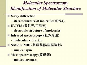
Title: Molecular Spectroscopy Identification of Molecular Structure
1
Molecular SpectroscopyIdentification of
Molecular Structure
- X-ray diffraction
- -stereostructure of molecules (DNA)
- UV/VIS (???/???)
- -electronic structure of molecules
- Infrared spectroscopy (????)
- -molecular vibration
- NMR or MRI (????/????)
- -nuclear spin
- Mass spectroscopy (???)
- -molecular mass
2
Internal Energy of Molecules
- EtotalEtransEelecEvibErotEnucl
- Eelec electronic transitions (UV, X-ray)
- Evib vibrational transitions (Infrared)
- Erot rotational transitions (Microwave)
- Enucl nucleus spin (nuclear magnetic
- resonance) or (MRI magnetic resonance
- imaging)
3
(No Transcript)
4
(No Transcript)
5
The molecular orbital diagram for the ground
state of NO
6
The molecular structure of beta-carotene
7
The electronic absorption spectrum of
beta-carotene.
8
- Ultraviolet 190400nm
- Violet 400 - 420 nm
- Indigo 420 - 440 nm
- Blue 440 - 490 nm
- Green 490 - 570 nm
- Yellow 570 - 585 nm
- Orange 585 - 620 nm
- Red 620 - 780 nm
9
(No Transcript)
10
(No Transcript)
11
Electronic Spectroscopy
- Ultraviolet (UV) and visible (VIS) spectroscopy
- This is the earliest method of molecular
spectroscopy. - A phenomenon of interaction of molecules with
ultraviolet and visible lights. - Absorption of photon results in electronic
transition of a molecule, and electrons are
promoted from ground state to higher electronic
states.
12
UV and Visible Spectroscopy
- In structure determination UV-VIS spectroscopy
is used to detect the presence of chromophores
like dienes, aromatics, polyenes, and conjugated
ketones, etc.
13
Electronic transitions
- There are three types of electronic transition
- which can be considered
- Transitions involving p, s, and n electrons
- Transitions involving charge-transfer electrons
- Transitions involving d and f electrons
14
Absorbing species containing p, s, and n electrons
- Absorption of ultraviolet and visible radiation
in organic molecules is restricted to certain
functional groups (chromophores) that contain
valence electrons of low excitation energy.
15
Vacuum UV or Far UV (?lt190 nm )
UV/VIS
16
(No Transcript)
17
s s Transitions
- An electron in a bonding s orbital is excited to
the corresponding antibonding orbital. The energy
required is large. For example, methane (which
has only C-H bonds, and can only undergo s s
transitions) shows an absorbance maximum at 125
nm. Absorption maxima due to s s transitions
are not seen in typical UV-VIS spectra (200 - 700
nm)
18
n s Transitions
- Saturated compounds containing atoms with lone
pairs (non-bonding electrons) are capable of n
s transitions. These transitions usually need
less energy than s s transitions. They can be
initiated by light whose wavelength is in the
range 150 - 250 nm. The number of organic
functional groups with n s peaks in the UV
region is small.
19
n p and p p Transitions
- Most absorption spectroscopy of organic compounds
is based on transitions of n or p electrons to
the p excited state. - These transitions fall in an experimentally
convenient region of the spectrum (200 - 700 nm).
These transitions need an unsaturated group in
the molecule to provide the p electrons.
20
Chromophore Excitation lmax, nm Solvent
CC p?p 171 hexane
CO n?pp?p 290180 hexanehexane
NO n?pp?p 275200 ethanolethanol
C-X XBr, I n?sn?s 205255 hexanehexane
21
(No Transcript)
22
Orbital Spin States
- Singlet state (S)Most molecules have ground
state with all electron spin paired and most
excited state also have electron spin all paired,
even though they may be one electron each lying
in two different orbital. Such states have zero
total spin and spin multiplicities of 1, are
called singlet (S) states.
Total Spin
Multiplicities
23
Orbital Spin States
- For some of the excited states, there are states
with a pair of electrons having their spins
parallel (in two orbitals), leading to total spin
of 1 and multiplicities of 3.
Total Spin
Multiplicities
24
Orbital Spin States
- For triplet state Under the influence of
external field, there are three values (i.e. 3
energy states) of 1, 0, -1 times the angular
momentum. Such states are called triplet states
(T). - According to the selection rule, S?S, T?T, are
allowed transitions, but S?T, T?S, are forbidden
transitions.
25
Selection Rules of electronic transition
- Electronic transitions may be classed as intense
or weak according to the magnitude of emax that
corresponds to allowed or forbidden transition as
governed by the following selection rules of
electronic transition - Spin selection rule there should be no change in
spin orientation or no spin inversion during
these transitions. Thus, S?S, T?T, are allowed,
but S?T, T?S, are forbidden. (?S0 transition
allowed)
26
(No Transcript)
27
p?p
28
Instrumentation
?? ??? ?? ??? ???
29
Components of a SpectrophotometerLight Source
- Deuterium Lamps-a truly continuous spectrum in
the ultraviolet region is produced by electrical
excitation of deuterium at low pressure.
(160nm375nm) - Tungsten Filament Lamps-the most common source of
visible and near infrared radiation.
30
Components of a SpectrophotometerMonochromator
(???/???)
- Used as a filter the monochromator will select a
narrow portion of the spectrum (the bandpass) of
a given source - Used in analysis the monochromator will
sequentially select for the detector to record
the different components (spectrum) of any source
or sample emitting light.
31
MonochromatorCzerny-Turner design
32
Grating
33
Photomultiplier Detector
34
Principle of Photomultiplier Detector
- The type is commonly used.
- The detector consists of a photoemissive cathode
coupled with a series of electron-multiplying
dynode stages, and usually called a
photomultiplier. - The primary electrons ejected from the
photo-cathode are accelerated by an electric
field so as to strike a small area on the first
dynode.
35
Principle of Photomultiplier Detector
- The impinging electrons strike with enough energy
to eject two to five secondary electrons, which
are accelerated to the second dynode to eject
still more electrons. - A photomultiplier may have 9 to 16 stages, and
overall gain of 106109 electrons per incident
photon.
36
Single and Double Beam Spectrometer
- Single-Beam There is only one light beam or
optical path from the source through to the
detector. - Double-Beam The light from the source, after
passing through the monochromator, is split into
two separate beams-one for the sample and the
other for the reference.
37
(No Transcript)
38
Quantitative AnalysisBeers Law
- Aebc
- e the molar absorptivity (L mol-1 cm-1)
- b the path length of the sample
- c the concentration of the compound in solution,
expressed in mol L-1
39
Transmittance
I0
I
b
40
(No Transcript)
41
External Standard and the Calibration Curve
42
Vibrational Spectroscopy
43
The potential curve for a diatomic molecule
44
Morse energy curve for a diatomic molecule.
45
IR spectrum
46
Selection Rule of Infrared Spectrum
- There must be a change in the dipole moment
during a vibrational cycle. - Homonuclear diatomic molecules will have no IR
spectrum.
47
Molecules with permanent dipole moments (µ) are
IR active
48
Types of Molecular VibrationsStretch-change in
bond length
- symmetric stretching
- asymmetric stretching
49
(No Transcript)
50
Types of Molecular Vibrations Bend-change in
bond angle
scissoring
wagging
rocking
twisting/torsion
51
(No Transcript)
52
Normal Modes of Vibration
- Linear molecule of N atoms normal modes 3N -
5 - Nonlinear molecule of N atoms normal modes 3N
- 6
53
Only some modes may be IR active
54
The three fundamental vibrations for sulfur
dioxide
55
How many vibrational modes?
- 2 atoms (H2) - 1 vibration
- 3 atoms (H2O) - 3 vibrations
- 3 atoms (CO2) - 4 vibrations
- 4 atoms (H2CO) - 6 vibrations
56
(No Transcript)
57
Rotational Spectroscopy
Selection Rule A molecule must have a permanent
dipole moment
58
(No Transcript)
59
(No Transcript)
60
Vibrational-Rotational Spectrum
61
(No Transcript)
62
(No Transcript)
63
(No Transcript)
64
Calculate Bond Length of Heteronuclear Diatomic
Molecule
65
Nuclear Magnetic Resonance Spectroscopy
- The rules for determining the net spin of a
nucleus - 1. If the number of neutrons and the number of
protons are both even, then the nucleus has NO
spin. - 2. If the number of neutrons plus the number of
protons is odd, then the nucleus has a
half-integer spin (i.e. 1/2, 3/2, 5/2) - 3. If the number of neutrons and the number of
protons are both odd, then the nucleus has an
integer spin (i.e. 1, 2, 3)
66
Nuclei Unpaired Protons Unpaired Neutrons Net Spin
1H 1 0 1/2
2H 1 1 1
31P 1 0 1/2
23Na 1 2 3/2
14N 1 1 1
13C 0 1 1/2
19F 1 0 1/2
- A nucleus of spin I will have 2I 1 possible
orientations.
67
Larmor Precession
68
- In the absence of an external magnetic field,
these orientations are of equal energy. - If a magnetic field is applied, then the energy
levels split. Each level is given a magnetic
quantum number, m.
69
Nucleus in a Magnetic Field
- The lower energy level will contain slightly more
nuclei than the higher level. - It is possible to excite these nuclei into the
higher level with electromagnetic radiation. - The frequency of radiation needed is determined
by the difference in energy between the energy
levels.
70
Calculating transition energy
g magnetogyric ratio and is a fundamental
nuclear constant which has a different value for
every nucleus. B the strength of the magnetic
field at the nucleus ?E?B?
71
The Absorption of Radiation by a Nucleus in a
Magnetic Field
- If energy is absorbed by the nucleus, then the
angle of precession, q, will change. - For a nucleus of spin 1/2
- , absorption of radiation "flips" the
magnetic moment so that it opposes the applied
field.
72
Chemical Shift
- The magnetic field at the nucleus is not equal to
the applied magnetic field electrons around the
nucleus shield it from the applied field. - The difference between the applied magnetic field
and the field at the nucleus is termed the
nuclear shielding.
73
Electrons in s-orbitals
- Spherical symmetry and circulate in the applied
field - A magnetic field which opposes the applied field.
- Applied field strength must be increased for the
nucleus to absorb at its transition frequency. - This upfield shift is also termed diamagnetic
shift.
74
Electrons in p-orbitals
- No spherical symmetry.
- They produce comparatively large magnetic fields
at the nucleus, which give a low field shift. - This "deshielding" is termed paramagnetic shift.
75
Proton Chemical Shift Ranges
76
Spin - Spin coupling
- The protons on neighboring carbons will generate
magnetic fields whose magnetic moments will
interact with the magnetic moment of the external
magnetic field. - This results in the splitting of the NMR signal.
77
NMR of Ethanol
-CH3
-CH2-
78
Methyl peak splitting into a triplet
the ratio of areas 121
79
Methylene peak splitting into a quartet
the ratio of areas 1331
80
The molecular structure of bromoethane
81
The NMR spectrum of CH3CH2Br (bromoethane) with
TMS reference
82
The molecule (2-butanone)
83
(C)
(A)
(B)
84
(No Transcript)
85
(D)
(A)
(B)
(C)
86
(C)
(B)
(A)
87
A technician speaks to a patient before heis
moved intot eh cavity of a magnetic resonance
imaging (MRI).
88
A colored Magnetic Resonance Imaging (MRI) scan
through a human head, showing a healthy brain in
side view.
Recommended
