
Table 1. Patients who underwent DSAEK from 10/06 to 11/08 PowerPoint PPT Presentation
Title: Table 1. Patients who underwent DSAEK from 10/06 to 11/08
1
Outcomes of Descemet Stripping Automated
Endothelial Keratoplasty in patients with a
Pre-Existing Anterior Chamber Intraocular Lens
S. Elderkin1A, E. Tu1A, J. Sugar1A, S. Reddy1A,
A. Kadakia1B, R. Ramaswamy1B, Djalilian1A AOphtha
lmology, BMedical School, 1Univ of Illinois Eye
and Ear Infirmary, Chicago, IL
Abstract
Results (cond)
Purpose This study was performed to evaluate
the outcome of Descemet Stripping Automated
Endothelial Keratoplasty (DSAEK) in patients with
a retained anterior chamber intraocular lens
(ACIOL). Methods Retrospective review of 11
patients with corneal decompensation an ACIOL who
underwent DSAEK in the 2 year period. All
patients except one had an open loop style
ACIOLs, and in all cases, there was adequate
anterior chamber depth. At the time of surgery, 6
patients had a temporary suture to secure their
graft, 2 of which were retained from the suture
method and the other 4 were placed additionally.
Postoperatively, the rate of donor detachment,
graft clarity, corneal pachymetry and visual
acuity were noted.
This was one of the 5 patients who did not have a
suture holding the graft. The dislocated graft
was successfully reattached with an air
bubble. The mean follow up period was 12 months
(range 6 - 25 mos). By 6 months, all patients
had experienced at least a 2 line improvement in
their visual acuity except for patient 6 who had
limited visual potential and patient 3, whose
graft never completely cleared (primary graft
failure). Patient 3 had required excessive
manipulation intraoperatively to unfold the graft
and therefore was considered iatrogenic failure.
This patient has subsequently undergone a repeat
DSAEK and at the 4 month follow-up was found to
have a clear graft with UCVA of 20/70 and a CCT
of 675µm (data not included). The final visual
acuities in most patients were limited because of
comorbidities (listed below). None of the 11
patients experienced acute graft rejection. In
all patients except patient 3, the central
corneal thickness (CCT) after DSAEK was lower
compared to before surgery. The mean thickness
pre-operatively was 910 µm (range 777 -1000µm).
The average CCT at the last recorded follow-up
for the 10 successful cases was 668µm (range 595
- 785µm). (Fig 1).
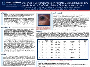
A
Fig 1 Patient 10 below after DSAEK
demonstrating existing ACIOL and a clear graft
Introduction
Endothelial keratoplasty (EK) is now the
preferred treatment for patients with symptomatic
corneal edema whose pathology is limited to the
endothelium.1, 2 It has several advantages over
penetrating keratoplasty, most notably, a more
rapid visual recovery, less induced astigmatism,
and greater resistance to trauma
post-operatively. Currently, the most popular
technique for EK is the Descemet's Stripping
Automated Endothelial Keratoplasty (DSAEK).1, 2
Pseudophakic corneal edema is one of the most
common indications for DSAEK. However, most
reported cases involve patients with posterior
chamber lenses.1, 2 Patients with an ACIOL and
corneal endothelial decompensation, however, may
also be candidates for DSAEK. Typically, if the
ACIOL is felt to be directly responsible for the
endothelial failure, an IOL exchange is indicated
either prior to or at the time of DSAEK. However,
in cases where the ACIOL is not considered the
primary reason for the endothelial decompensation
and there is adequate anterior chamber depth, it
may be more desirable to leave the ACIOL in
place. The presence of the ACIOL, however,
can pose additional challenges during DSAEK.
Specifically, it can interfere with the surgical
placement of the donor graft while making it
difficult to maintain an air bubble in the
anterior chamber. Given these challenges some
surgeons may lean towards exchanging the IOL
prior to DSAEK in patients with an ACIOL.3 This
series examines the early outcomes of DSAEK in
patients with adequate anterior chamber where the
ACIOL was left in place.
Summary
This series confirms previous smaller reports
that for select patients with corneal
decompensation and an ACIOL, DSAEK without IOL
exchange may be a viable alternative in order to
minimize the risks of an IOL exchange. These
patients include those with adequate anterior
chamber depth whose risk of subsequent damage by
the ACIOL is minimal. Moreover, this series
demonstrates that the use of a suture, either as
part of the insertion technique or as a safety
suture at the end of the case may help reduce
the risk of graft detachment without any
significant adverse effects on the short term
results. Further long term studies, including
endothelial cell counts, are necessary to further
confirm these results.
Patient Age Detached Day1? Clear or Failed? VA Pre, Post U or BCVA Comorbidities Pachy (6mos) Follow-up (mos.)
1 55 N Clear CF _at_ 3, 20/100 UCVA PDR, Fuchs 715 25
2 81 N Clear CF _at_ 2, 20/50 BCVA none 670 12
3 76 N Edema 20/200, 20/100 BCVA ACG 973 12
4 77 Y Clear 20/400, 20/70 UCVA RD 603 21
5 65 N Clear CF, 20/200 UCVA Acantham, RD, Prior PK 785 6
6 64 N Clear 20/400, 20/300 UCVA Uveitic Glaucoma 676 17
7 78 N Clear 20/300, 20/50 BCVA Glaucoma 725 8
8 85 N Clear 20/400, 20/200 BCVA ARMD 660 13
9 70 N Clear 20/200, 20/50 BCVA none 623 6
10 73 N Clear 20/200, 20/50 BCVA Chronic CME 595 6
11 65 N Clear CF, 20/60 BCVA Prior PK 623 6
Results
A total of 11 eyes were identified and the
clinical features of each patient are summarized
in Table 1. The mean age at the time of DSAEK was
72 years (range 55 to 85 years). The time from
ACIOL to DSAEK ranged from 6 months to 30 years.
All patients except for one had an open loop
style ACIOLs, patient 7, who had a closed loop
ACIOL which had led to corneal decompensation
over the course of 30 years. The primary etiology
of the corneal decompensation in the other
patients included complicated cataract surgery,
multiple intraocular procedures, graft failure,
and Fuchs dystrophy. There were no major
intraoperative complications. There was one
complete graft dislocation into the anterior
chamber on POD 1 (4).
Table 1. Patients who underwent DSAEK from 10/06
to 11/08
References
1. Price MO, Price FW. Descemet's stripping
endothelial keratoplasty. Curr Opin Ophthalmol
2007 Jul18(4)290-4. 2. Terry MA, Shamie N, Chen
ES, Hoar KL, Friend DJ. Endothelial keratoplasty
a simplified technique to minimize graft
dislocation, iatrogenic graft failure, and
pupillary block. Ophthalmology 2008
Jul115(7)1179-86. 3. Wylegala E, Tarnawska D.
Management of pseudophakic bullous keratopathy by
combined descemet-stripping endothelial
keratoplasty and intraocular lens exchange. J
Cataract Refract Surg 2008
Oct34(10)1708-14.
Recommended
