
Conditions of the Lymph System PowerPoint PPT Presentation
1 / 10
Title: Conditions of the Lymph System
1
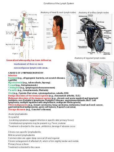
Conditions of the Lymph System
Anatomy of axillary lymph nodes
Anatomy of head neck lymph nodes
Anatomy of inguinal lymph nodes
- Generalized adenopathy has been defined as
- involvement of three or more
- noncontiguous lymph node areas.
CAUSES OF LYMPHADENOPATHY Infection Bacterial
(e.g., all pyogenic bacteria, cat-scratch
disease, syphilis) Mycobacterial (e.g.,
tuberculosis, leprosy) Fungal (e.g.,
histoplasmosis ) Chlamydial (e.g.,
lymphogranuloma venereum) Parasitic (e.g.,
toxoplasmosis, filariasis)
Viral (e.g., Epstein-Barr virus, cytomegalovirus,
rubella, HIV) Benign disorders of the immune
system (e.g., rheumatoid arthritis,
SLE) Malignant disorders of the immune system
(e.g., chronic and acute myeloid and lymphoid
leukemia, non-Hodgkin's lymphoma, Hodgkin's
disease, angioimmunoblastic-like T-cell lymphoma,
multiple myeloma with amyloidosis, malignant
histiocytosis) Other malignancies (e.g., breast
carcinoma, lung carcinoma, melanoma, head and
neck cancer, gastrointestinal malignancies, germ
cell tumors, Kaposi's sarcoma) Storage diseases
(e.g., Gaucher's disease)
Acute lymphadenitis It is painful Localizing
symptoms suggest infection in specific site(
primary focus ) Constitutional symptoms may be
present e.g. Fever, malaise Treatment is
directed to the cause ,antibiotics, derange if
abscess occur
Chronic non specific lymphadenitis Mild
recurrent lymphadenitis Common sites are upper
deep cervical LN and inguinal Chronic
enlargement of affected LN, which is firm
slightly tender and mobile Primary focus is
there Treatment is directed to the cause
2
- Specific lymphadenitis
- Bacterial (cat-scratch disease, syphilis)
- Mycobacterial (e.g., tuberculosis, leprosy)
- Fungal (e.g., histoplasmosis, coccidioidomycosis)
- Chlamydial (e.g., lymphogranuloma venereum)
- Parasitic (e.g., toxoplasmosis, filariasis)
- Viral (e.g., Epstein-Barr virus, cytomegalovirus,
HIV) - HIV- related.Persistent Generalized
Lymphadenopathy (PGL) - Lymph nodes larger than 1.5 cm in diameter in 2
or more extrainguinal sites of 3 or more months
duration - Nodes are non-tender, symmetrical, and often
involve the posterior cervical, axillary,
occipital, and epitrochlear nodes - Develops in up to 50 of HIV-infected individuals
- Up to one-third do not have any other symptom on
presentation (stage 1) - In HIV-positive patients, PGL is a clinical
diagnosis. - PGL may slowly regress during the course of HIV
infection and may disappear before the onset of
AIDS - Tuberculosis lymphadenopathy
- Cervical nodes most commonly involved
- Organism
- Root lymph Vs blood
3
- syphilis
- Clinical Symptoms may evolve
- Generalized painless lymphadenopathy
- Maculo-papular, papular, or pustular rash on
entire body, especially on palms and soles - Highly infectious lesions on mucous membranes
(lips, mouth, pharynx, vulva, glans penis) which
are silvery grey superficial erosions with a red
halo and not painful unless there is a secondary
infection. - 40 of these patients will have CNS involvement
with headache and meningismus - 1-2 will develop acute aseptic meningitis
- benzathine penicillin 2.4 million units IM single
dose
- Lymphoma
- Lymphoma is a malignant disease that affects
blood cells called lymphocytes immune cells
that normally protect you from illness. - Damage to genes in these cells can sometimes lead
to abnormal cell behavior which makes the cells
immortal unable to die when they should or
causes sustained rapid cell division.. - These malignant cells then may accumulate to form
tumors that enlarge the lymph nodes or spread to
other areas of the lymphatic system, such as the
spleen or bone marrow, or outside the lymphatic
system to the skin, or mucosal linings of the
stomach. - They arise as the result of abnormal
proliferation of the lymphoid system, and hence
occur at any site where lymphoid tissue is found.
Most commonly they are manifest by the
development of lymphadenopathy at single or
multiple sites, although primary extranodal
presentations account for up to 20 of
non-Hodgkin's lymphoma. - The prognosis is determined by the specific
subtype of lymphoma and the anatomical extent of
disease and its bulk, the clinical course ranging
from months to years. - Lymphomas are currently classified on the basis
of histological appearance into - Hodgkin's
lymphoma - non-Hodgkin's
lymphoma. - The two types not only have different morphologic
characteristics but differ also in their clinical
behavior and their response to various
therapeutic regimens.
- HODGKIN'S LYMPHOMA (HL)
- Aetiology
- There is epidemiological evidence linking
previous infective mononucleosis with HL and up
to 40 of patients with HL have increased EBV
antibody titres at the time of diagnosis and
several years prior to the clinical development
of HL. - Other environmental and occupational exposure to
pathogens have been postulated. - ?? Chronic infection,?? Depressed immunity,??
Chemical exposure pesticides,cancer therapies,
herbicides ,?? Viral exposures - Pathology
- The hallmark of HL is the Reed-Sternberg cell)
which is usually derived from germinal centre B
cells or, rarely, peripheral T cells. - Pathological classification of Hodgkin's lymphoma
- Nodular lymphocyte-predominant Hodgkin's
lymphoma - Classical Hodgkin's lymphoma
- Nodular sclerosis HL(young females,
involving particularly lymph nodes in the
mediastinum and neck ). - Lymphocyte-rich HL(It often occurs in
peripheral lymph nodes. It is often an indolent
disease(. - Mixed cellularity HL) more common in
men and is associated with B symptoms ( - Lymphocyte-depleted HL) It is seen in
HL associated with HIV (
4
- Clinical features
- Lymph node enlargement, most often of the
cervical nodes (other causes are shown in, these
are usually painless and with a rubbery
consistency. - Enlargement of the spleen/liver.
- 'B' symptoms fever, (25) drenching night
sweats, weight loss of gt 10 bodyweight - Other constitutional symptoms, such as pruritus,
fatigue, anorexia and, occasionally,
alcohol-induced pain at the site of enlarged
lymph nodes. - Symptoms due to involvement of other organs (e.g.
lung - cough and breathlessness ( - Investigations
- Blood count may be normal, or there can be a
normochromic, normocytic anaemia. Lymphopenia and
occasionally eosinophilia are present. - Erythrocyte sedimentation rate )ESR) is usually
raised and is an indicator of disease activity. - Liver biochemistry is often abnormal, with or
without liver involvement. - Serum lactate dehydrogenase raised level is
adverse prognostic factor. - Chest X-ray may show mediastinal widening, with
or without lung involvement. - CT scans show involvement of intrathoracic nodes
in 70 of cases. Abdominal or pelvic lymph nodes
are also found. It is the investigation of choice
for staging although PET scanning is increasingly
being used. - Lymph node biopsy is required for a definitive
diagnosis - Cotswolds modification of Ann Arbor staging
classification - Stage I Involvement of a single lymph-node region
or lymphoid structure (e.g. spleen, thymus,
Waldeyer's ring) or involvement of a single
extralymphatic site - Stage II Involvement of two or more lymph-node
regions on the same side of the diaphragm (hilar
nodes, when involved on both sides, constitute
stage II disease) localized contiguous
involvement of only one extranodal organ or site
and lymph-node region(s) on the same side of the
diaphragm (IIE). The number of anatomic regions
involved should be indicated by a subscript (e.g.
II3)
5
- NON-HODGKIN'S LYMPHOMA (NHL)
- These are malignant tumours of the lymphoid
system classified separately from Hodgkin's
lymphoma. Most (70) are of B cell origin
although T cell tumours are increasingly being
recognized. - NHL is associated with the EBV virus (Burkitt's
lymphoma) and the human T cell lymphotropic virus
which is prevalent in Japan, Africa, South
America and the Caribbean. Herpes virus 8 is
associated with primary effusion lymphomas and
Castleman's disease there is an increase in
lymphoma in patients with AIDS. Helicobacter
pylori is an aetiological factor in gastric
lymphoma. - Lymphomas also occur in congenital
immunodeficiency, post-transplantation and in
autosomal family cancer syndromes . - Other causes, e.g. occupation, dietary and
exposure to chemicals, have been linked to the
increasing incidence but the evidence is
unconfirmed - Types
- Follicular
- Lymphoplasmacytict
- Mantle cell
- Diffuse large B cell
- Burkitts
- Anaplastic
- MALT (mucosal associated lymphoid tissue)
- WHO classification of lymphoid neoplasms
- B cell lymphomas
- Precursor B cell lymphoma Precursor B
lymphoblastic lymphoma/leukaemia (highly
(aggressive) - Mature B cell lymphoma
6
The Lymphatics
- Treatment options
- Aggressive combination chemotherapy gives high
complete remission rates and molecular remission.
- Antibody therapy. The monoclonal antibody
rituximab induces remission (partial) in 30-70
of patients, almost without toxicity. Molecular
remissions are observed. Complications include
the cytokine release syndrome, with fever,
vomiting and allergic reactions (angio-oedema,
bronchospasm and dyspnoea). - Rituximab/chemotherapy combination. These have
now been reported to improve the complete
remission rate (with disappearance of Bcl-2
positive cells from the bone marrow in 100 of
patients), freedom from progression and
event-free survival, even though there is (as
yet) no effect on overall survival. This may
become the standard therapy for CD20 positive
lymphoma. - Antibody-targeted irradiation
- Clinical approach
- A careful history
- , -Age of the patient.
- -The occurrence of fever, sweats, or
weight loss - Site of infection, a particular medication, a
travel history, or a previous malignancy. - physical examination
- localized or generalized
- size of nodes
- Texture
- Mobility
- presence or absence of nodal tenderness
- signs of inflammation over the node
- skin lesions
7
- Clinical picture
- The edematous limb has a firm and hardened
consistency. - There is loss of the normal perimalleolar shape,
resulting in a tree trunk pattern. - The dorsum of the foot is characteristically
swollen, resulting in the appearance of the
buffalo hump, and the toes become thick and
squared . - In advanced lymphedema, the skin undergoes
characteristic changes, such as lichenification,
development of peau dorange, and
hyperkeratosis.Additionally, the patients give a
history of recurrent episodes of cellulitis and
lymphangitis after trivial trauma and frequently
present with fungal infections affecting the
forefoot and toes. - Patients with isolated lymphedema usually do not
have the hyperpigmentation or ulceration one
typically sees in patients with chronic venous
insufficiency. - Lymphedema does not respond significantly to
overnight elevation, whereas edema secondary to
central organ failure or venous insufficiency
does.
8
- Differential Diagnosis
- The most common causes of bilateral extremity
edema are of systemic origin. The most common
etiology is cardiac failure, followed by renal
failure. Hypoproteinemia secondary to cirrhosis,
nephrotic syndrome, and malnutrition can also
produce bilateral lower extremity edema. - Another important cause to consider with
bilateral leg enlargement is lipedema. Lipedema
is not true edema but rather excessive
subcutaneous fat found in obese women. It is
bilateral, nonpitting, and greatest at the ankle
and legs, with characteristic sparing of the
feet. There are no skin changes,and the
enlargement is not affected by elevation. The
history usually indicates that this has been a
lifelong problem that runs in the family. - Once the systemic causes of edema are excluded,
in the patient with unilateral extremity
involvement, edema secondary to venous and
lymphatic pathology should be entertained. The
edema responds promptly to overnight leg
elevation. In the later stages, the skin is
atrophic with brawny pigmentation. Ulceration
associated with venous insufficiency occurs above
or posterior and beneath the malleoli. - CLASSIFICATION of lymph- edema
- Lymphedema is generally classified as primary
when there is no known etiology and secondary
when its cause is a known disease or disorder. - Primary lymphedema has generally been classified
on the basis of the age at onset and presence of
familial clustering. - Primary lymphedema with onset before the first
year of life is called congenital. The familial
version of congenital lymphedema is known as
Milroys disease and is inherited as a dominant
trait. - Primary lymphedema with onset between the ages of
1 and 35 years is called lymphedema praecox. The
familial version of lymphedema praecox is known
as Meiges disease. - Finally, primary lymphedema with onset after the
age of 35 is called lymphedema tarda. - Worldwide the most common cause of secondary
lymphedema is infestation of the lymph nodes by
the parasite Wuchereria bancrofti in the disease
state called filariasis. In the developed
countries the most common causes of secondary
lymphedema involve resection or ablation of
regional lymph nodes by surgery, radiation
therapy, tumor invasion, direct trauma, or, less
commonly, an infectious process. - Investigation
- The diagnosis of lymphedema is relatively easy in
the patient who presents in the second and third
stages of the disease. It can however, be a
difficult diagnosis to make in the first stage,
particularly when the edema is mild, pitting, and
relieved with simple maneuvers such as elevation.
For patients with suspected secondary forms of
lymphedema, computed tomography (CT) and magnetic
resonance imaging (MRI) are valuable and indeed
essential for exclusion of underlying oncologic
disease - In patients with known lymph node excision and
radiation treatment as the underlying problem of
their lymphedema, additional diagnostic studies
are rarely needed except as these studies relate
to follow-up of an underlying malignancy. - For patients with edema of unknown etiology and a
suspicion for lymphedema, lymphoscintigraphy is
the diagnostic test of choice. - When lymphoscintigraphy confirms that lymphatic
drainage is delayed, the diagnosis of primary
lymphedema should never be made until neoplasia
involving the regional and central lymphatic
drainage of the limb has been excluded through CT
or MRI. - If a more detailed diagnostic interpretation of
lymphatic channels is needed for operative
planning, then contrast lymphangiography may be
considered.
9
pleomorphic adenomaof parroted gland
Neck lump
Ludwigs angina
Multiple cervical metastases visible in the nodal
basins that drain the site of the primary
malignancy
Virchows lymph node
Ranula
Branchial cyst
Thyroglossal cyst
10
Neck lumpThyroid, lymph node ,carotid ,salivary
gland , sternomastoid muscle
Cystic Hygroma
- Goitre.
- Neoplasm
- Thyroid neoplasms
- Metastatic carcinoma.
- Primary lymphoma.
- Salivary gland tumour.
- Sternocleidomastoid tumour.
- Carotid body tumour.
- Inflammatory
- Acute infective adenopathy.
- Collar stud abscess.
- Parotitis.,submandibular sialadinitis
- Congenital
- Thyroglossal duct cyst.
- Cystic hygroma.
- Branchial cyst.
- Dermoid cyst.
- Torticollis.
- Vascular
- .Caroted body tumors
- Subclavian aneurysm.
- Subclavian ectasia.
Children
Congenital and inflammatory lesions are common.
Cystic hygroma in infants, base of the neck,
brilliant transillumination. Thyroglossal or
dermoid cyst midline, discrete, elevates
with tongue protrusion. Torticollis rock-hard
mass, more prominent with head flexed, associated
with fixed rotation (a fibrous mass in the
sternocleidomastoid muscle). Branchial cyst
anterior to the upper third of the
sternocleidomastoid. Viral/bacterial adenitis
usually affects jugular nodes, multiple, tender
masses. Neoplasms (lymphoma most
common). Young adults Inflammatory neck masses
and thyroid malignancy are common. Viral (e.g.
infectious mononucleosis) or bacterial
(tonsillitis/pharyngitis) adenitis. Papillary
thyroid cancer isolated, non-tender, thyroid
mass, possible lymphadenopathy. Older age group
Neck lumps are malignant until proven
otherwise. Metastatic lymphadenopathy
multiple, hard, nontender, tendency to be
fixed. 75 in primary head and neck (thyroid,
nasopharynx, tonsils, larynx, pharynx), 25 from
infraclavicular primary (stomach, pancreas,
lung). Primary lymphadenopathy (thyroid,
lymphoma) fleshy,rubbery, large size. Primary
neoplasm (thyroid, salivary tumour) firm,
nontender, fixed to tissue of origin.
Recommended
