Endocrine System PowerPoint PPT Presentation
1 / 35
Title: Endocrine System
1
Endocrine System
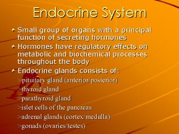
- Small group of organs with a principal function
of secreting hormones - Hormones have regulatory effects on metabolic and
biochemical processes throughout the body - Endocrine glands consists of
- gtpituitary gland (anterior/posterior)
- gtthyroid gland
- gtparathyroid gland
- gtislet cells of the pancreas
- gtadrenal glands (cortex/medulla)
- gtgonads (ovaries/testes)
2
Endocrine System
- In-vivo imaging and non-imaging applications in
NM has a significant role in understanding the
function and disorders of the endocrine system - Thyroid, parathyroid, and adrenal glands most
common. - No other imaging or non-imaging procedures exists
for other glands
3
Adrenal Gland Imaging
- Located at superior poles of the kidneys.
- Consist of outer cortex and inner medulla
- Cortex produces steroid hormones
(aldosterone,cortisol) - Medulla manufactures catecholamines
(epinephrine/norepinephrine) hormones that
control bodies response to stress
4
Adrenal Gland Imaging
- Tumors of the adrenal medulla sre called
Pheochromocytomas - Symptoms include increased levels of
epinephrine/non-epinephrine in blood and urine. - NM used to identify sites of increased
epinephrine/norepinephrine within adrenal bed and
outside metastatic sites.
5
Adrenal Gland Imaging
- Radiopharmaceutical used to image the adrenal
medulla is I-131 methyliodobenzylguanidine (MIBG) - Compound is structurally the same as
norepinephrine, does not exert same effect.
6
Adrenal Gland Imaging
- 1 day prior to injection, patient prep receives
Lugols solution (potassium iodide) - Solution saturates the thyroid gland preventing
uptake of I-131 (minimize unnecessary exposure to
thyroid) - 0.5mCi I-131 administered IV.
7
Adrenal Gland Imaging
- Anterior and Posterior images acquired at 1,3,
and 7 days PI. (skull to pelvis) - Patient is asked to void prior to study.
- Uptake normally in liver, spleen, and heart.
- Salivary glands and bladder may also be
visualized.
8
Adrenal Gland Imaging
- Metastases from Pheochromocytomas may be
visualized in liver, bone, lymph nodes, heart,
and lungs.
9
Thyroid Imaging
- Located in the anterior neck between suprasternal
notch and thyroid cartilage. - Consists of 2 lobes approx 3-4 cm long.
- Isthmus connects lobes. Thyroid overlies trachea
- Highly vascular (superior/inferior thyroid
arteries)
10
Thyroid Imaging
- Thyroid imaging is one of the earliest NM
procedures developed - Based on physiological process of thyroid hormone
production T-3(triiodothyronine) T- - 4(tetraiodothyronine)
11
Thyroid Imaging
- T-3 and T-4 are products of iodine absorbed into
the blood from digestion. - Blood transports iodine in the form of iodide to
the thyroid gland. - Iodide is trapped by thyroid follicular cells.
- Process is called iodide pump.
12
Thyroid Imaging
- Hormones produced in the thyroid are stored there
until they are required by body. - Thyroid controls many metabolic processes growth
and development, body temperature regulation,
metabolism of proteins, lipids, carbohydrates,
vitamins, and minerals.
13
Thyroid Imaging
- Thyroid hormone production and secretion
controlled by negative feedback. - TSH (thyroid stimulating hormone) released by
pituitary gland, regulates thyroid iodide uptake
and release.
14
Clinical Indications
- Evaluate gland structure to function
- Evaluate gland size and palpable nodules or
masses. - Identify ectopic thyroid tissue located from base
of tongue to below sternum.
15
Thyroid Imaging procedure
- Prior to dose administration patient should be
questioned previous thyroid surgery,thyroid
symptoms, thyroid medications, recent
radiographic procedures. - Many medications, iodine containing foods, and
radiographic procedures using iodinated contrast
can affect radioiodine uptake.
16
Thyroid Imaging procedure
- All females of chidbearing age should be
questioned - Lab values for thyroid hormones will aid in
interpretation. - Radiopharmaceuticals administered orally or
intravenously - Tc99m is tracer administered IV and imaging
begins 15-30min PI.
17
Thyroid Imaging Procedure
- I-123 sodium iodide is administered orally and
imaged 3-4hrs or 16-24hrs.after administration - I-131 sodium iodide is administered orally and
imaged 6-24hrs after administration - Patient placed supine with neck hyperextended.
18
Thyroid Imaging procedure
- Pinhole or LEHR collimator used for imaging
- Images are acquired in the anterior and oblique
projections - Marker images acquired anteriorly at supersternal
notch and over thyroid cartlidge. - Ectopic thyroid tissue suspected an anterior view
of mediastinum is obtained
19
(No Transcript)
20
Thyroid Imaging Procedure
- Normal findings
- gtbutterfly shaped structure with a uniform
symmetric distribution of activity - gtright lobe slightly larger than left
- gtisthmus not well defined
- gtpyramidal (third lobe) may be visualized.
21
Thyroid Imaging Procedure
- Abnormal findings
- gtenlarged thyroid gland
- gtvisualization of functioning and
non-functioning thyroid nodules - gtfunctioning HOT nodules represent benign
cysts or tumors - gtnon-functioning COLD nodules represent
carcinoma, benign adenoma, cysts, hematoma,
inflammatory condotions.
22
Thyroid Uptake Procedure
- If using I-123 or I-131 capsules a thyroid uptake
is obtained - Measures amount of radioactive iodide that is
taken and retained within thyroid gland. - Uptake at 2-6hrs measures iodine trapping
- Uptake at 24-48hrs measures rate of iodine is
lost from gland
23
Thyroid Uptake Procedure
- I-123 and I-131 in capsule or liquid form (most
common capsule) - Preferred thyroid uptake, possible with Tc99m.
- Patient preparation same as radioiodine thyroid
imaging - NPO and fasting 2hrs post administration
- Data (uptake) collected with thyroid uptake probe
(sodium iodide crystal flat field collimator)
24
Thyroid Uptake Procedure
- Thyroid gland counts are collected with patient
in supine or seated (erect) position - Neck is hyperextended, probe over centered
between suprasternal notch and thyroid cartlidge.
25
Thyroid Uptake Procedure
- Thyroid uptake calculations are obtained by
Patient counts from the neck and patient
background. - Counts from standard (capsule) and room
background - 1min counts are obtained for each.
- Normal values 4hrs 6-18
- 24hrs 10-35
26
Radioiodine Whole-body Imaging
- Post thyroid-ectomy (carcinoma) whole body
radioiodine imaging is acquired - To identify remaining residual thyroid tissue or
metastasis. - I-131 sodium iodide most commonly used.
- 1-10mCi I-131 administered orally
27
Radioiodine whole body Imaging
- Patient preparation similar to thyroid imaging
- Anterior and posterior images of body from head
to mid-femur acquired. - 24-48 hrs post administration
- Anatomical landmarks placed on images, activity
seen in salivary glands, thyroid tissue remnants,
stomach, esophagus, functioning metastasis.
28
Parathyroid Imaging
- 4 parathyroid glands located on posterior aspect
of poles of thyroid gland - Produce and secrete parathyroid hormone (PTH)
- Responsible for regulating level and distribution
of calcium and phosphorus.
29
Parathyroid Imaging
- Imaging useful when primary hyperthyroidism is
suspected. - Tumor in one of the parathyroid glands or
hyperplasia of all four glands lead to excess PTH
to be secreted. - PTH stimulates the removal of calcium from bones,
affects nervous system, muscle contraction, etc. - Treatment is surgical removal of hyperplastic
gland or tumor
30
Parathyroid Imaging
- Parathyroid imaging acquired with either dual
phase or dual tracer technique - dual phase Tc99m sestamibi 5-25 mCi is
administered - localizes in both thyroid/parathyroid tissue
- tracer washes out of normal thyroid tissue
quicker than abnormal parathyroid tissue
31
Parathyroid Imaging
- Imaging 10min PI and again 1.5hr-2.5hr PI is
obtained - Abnormal parathyroid demonstrates retention of
tracer, becomes better visualized on delay
images.
32
Parathyroid Imaging
- Dual tracer technique
- Tc99m or I-123 sodium iodide (distinguish normal
thyroid tissue) - Tc99m-sestamibi or thallium-201 (localizes in
both thyroid and abnormal parathyroid tissue) - Advantages/Disadvantages
- scatter high energy to low energy
- length of images (pt.cooperation)
- activity of each tracer/time required for
I123 to localize.
33
Parathyroid Imaging
- Both images are normalized, so counts per pixel
are the same for both images. - Tc99m/I-123 images are subtracted from Tc99m
sestamibi/thallium-201 images. - Final images reveal area of abnormal parathyroid.
34
Parathyroid Imaging
- Dual phase techniques has several advantages
- One injection and tracer (Tc99m sestemibi)
- No computer subtraction needed
- Radiation dose decreased to patient.
35
TURN ON THE LIGHTS
- ANY QUESTIONS???????

