Respiratory System II: PowerPoint PPT Presentation
1 / 32
Title: Respiratory System II:
1
Respiratory System II
2
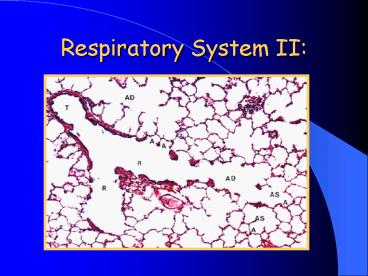
Bronchioles
- Function- same as bronchi with added involvement
in gas exchange - Types
- Bronchioles
- Terminal bronchioles
- Respiratory bronchioles
3
Bronchioles
- Bronchioles-
- less than one millimeter in diameter
- Mucosa
- Ciliated simple columnar w/Goblet cells
- Lamina propria
- Thin elastic fibroconnective tissue.
- Submucosa
- smooth muscle
- No mucosal glands
4
5
Bronchioles
- Terminal bronchioles
- Mucosa
- ciliated simple columnar to ciliated cuboidal to
non-ciliated cuboidal - Lacks goblet cells
- Clara cells
- Lamina propria
- Very thin elastic fibroconnective tissue
- Submucosa - same as bronchioles
6
7
Bronchioles
- Respiratory bronchioles
- Mucosa
- Cuboidal to flattened squamous epithelium.
- discontinuous smooth muscle in the lamina propria
- Terminate in the alveolar ducts
8
Terminal airways
- Alveolar ducts
- Small alveoli open up from thin walls of these
ducts - Terminate in alveolar sacs
- Smooth muscle at opening into the alveolar sacs
9
Lungs
- Alveoli- constitute the majority of the lung
parenchyma - Air exchange
- Squamous epithelium
- Type I alveolar cell (pneumocyte)
- Forms wall of the alveoli
- Basement membranes fused with basement membrane
of capillaries in septa
10
11
Lungs
- Type II alveolar cell (pneumocyte)
- Cuboidal rather than squamous
- Primary function is the production of surfactant
- Act as reserve cells- can differentiate into type
I alveolar cell if needed
12
13
Lungs
- Alveolar macrophages (dust cells)- derived from
blood monocytes, clean up debris
14
(No Transcript)
15
Lungs
- Septa
- Composed of capillaries supported by a fine
fibrous stroma - No smooth muscle
- Alveolar pores- equalize pressure and provide
collateral air circulation when bronchi leading
to the alveoli are blocked
16
17
Pleura
- Visceral pleura
- Thick in large animals (cattle, horses)
- Thin in carnivores
- Consists
- Mesothelium superficial layer of squamous
epithelial cells - Elastic fibrous connective tissue and some smooth
muscle - Divides lungs into lobules
- prominent in ruminants.
- Sparse in carnivores
18
19
Circulatory system
- Vasculature
- Dual system
- Pulmonary arterial system (primary blood supply)
- Bronchial system
20
Pulmonary Circulation
- Pulmonary system
- Relatively thin walled and large caliber
- Usually about the same size as accompanying
airway - Are elastic arteries not muscular
- Maintain pressure at a constant through out the
cardiac cycle, (expand and recoil)
21
(No Transcript)
22
Bronchial Circulation
- Bronchial system.
- Small branches off the aorta.
- Supply the walls of the airways and the pleura
- Venous return
- Majority returns to the left atrium via the
pulmonary veins - A portion from the bronchial system returns to
the right heart via the azygos system
23
Pulmonary Blood Flow
- Control of blood flow in capillaries.
- Matches blood flow (perfusion) to oxygenation
- Controlled by partial pressures of gases in
alveoli - High pO2 and low pCO2- vasodilation- increases
blood flow to alveolar system - Low pO2 and high pCO2- vasoconstriction-
decreases blood flow to alveolar system
24
Respiratory System
- Lymphatics
- Associated with pulmonary blood vessels and
airways. - Drain into thoracic duct at lung hilus
- No lymphatics are found in the alveolar septa
25
Respiratory System
- Avian Respiratory System
- Trachea
- Overlapping complete cartilaginous rings
- Epithelium of anterior segment- many mucus glands
- Epithelium of the posterior segment- glands are
replaced by goblet cells - Submucosa has very high elastic fiber component
26
Respiratory System
- Syrinx- phonation, located at tracheal
bifurcation - Bronchi
- Extrapulmonary primary bronchi (mesobronchus)
- Ciliated pseudostratified columnar epithelium
with goblet cells and mucous glands - Hyaline C-shaped cartilage.
- Smooth muscle
- Elastic fibers
27
Respiratory System
- Intrapulmonary primary bronchus (mesobronchus)
- Ciliated pseudostratified columnar epithelium
with goblet cells and mucous glands - Cartilage plates- number decreases as the
diameter of the bronchus decreases - Smooth muscle
- Elastic fibers
- Connects to the caudal air sacs
28
Respiratory System
- Secondary bronchi
- Ciliated columnar epithelium with mucus cells
- Numerous parabronchi branch from wall
29
Respiratory System
- Tertiary bronchi (parabronchi)- form anastomoses
with air capillaries - Cuboidal epithelium
- Smooth muscle
- Lead to atrium - cuboidal to squamous epithelium
30
Respiratory System
- Atrium
- opens into multiple air capillaries lined by
squamous epithelium - Vascular capillaries for gas exchange surround
air capillaries - Flow of air and blood is a counter-current system
31
Respiratory System
- Air Sacs
- Connect to lungs by mesobronchi
- May extend into hollow bones
- Variable epithelial lining- squamous, ciliated
columnar, ciliated cuboidal - Elastic fibers in stroma
- Do not participate in gas exchange
- Act as air pumps to move air by distension and
compression
32
Respiratory System
- Respiration is circular (one-way) verses the
in-out pattern of mammalian respiration - Much more efficient- extracts more Oxygen per
respiratory cycle

