PowerPointpresentasjon PowerPoint PPT Presentation
1 / 58
Title: PowerPointpresentasjon
1
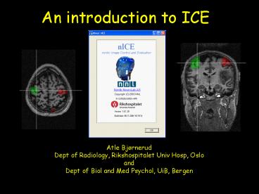
An introduction to ICE
Atle Bjørnerud Dept of Radiology, Rikshospitalet
Univ Hosp, Oslo and Dept of Biol and Med Psychol,
UiB, Bergen
2
Contents
- Overview of functionality
- Image loading/saving/viewing
- Image conversion (ICE-gtSPM-gtICE)
- Working with image overlays
- ROI functionality
- Image segmentation
- Image processing
- Q A
3
What is ICE?
- Image Control and Evaluation
- Viewing and processing of medical images
- Converting between image formats
- Performing image measurements
- Special emphasis on functional data
- fMRI
- Dynamic imaging
- Perfusion analysis
4
What isnt ICE?
- A complete fMRI analysis package (like SPM, FSL,
Brain Voyager, AFNI) - A 3D volume rendering tool
- A computer game
5
Feature overview (I)
Image loading and saving
- Compatibility with a variety of image formats
- DICOM 1.0-3.0, SPM/Analyze, RAW, BMP, JPEG, TIFF
- Ability to render most DICOM formats including
lossless jpeg compressed, Siemens MOSAIC, RGB and
multi-frame formats. - Image loading through many different interfaces,
depending on image type and user preferences. - Easy transfer of image- and measurement data to
other Windows applications - Dicom header modification prior to saving (e.g.
patient anonymization, etc.) - Easy batch conversion between image formats
6
Feature overview (II)
Image viewing
- Display of medical images from a variety of image
sources - Dicom reader
- SPM reader
- Drag Drop
- Dicom server
- Easy scrolling through 3- or 4-dimensional
datasets - Multiple viewing formats
- Cine / movie loop display
- Display /copy relevant image information (e.g.
from Dicom header)
7
Feature overview (III)
Image manipulation
- Flexible and intuitive tools for basic image
manipulation - Image size, Zoom, Pan
- Smoothing
- Image intensity / contrast
- Change colour palette
8
Feature overview (IV)
Image analysis
- Region of interest (ROI) analysis
- Multiple ROI types
- Easy and reproducible ROI analysis of large
datasets - Easy transfer of ROI data to other Windows
applications (e.g. Excel) - Dynamic ROI analysis (time-intensity curves,
etc.) - Advanced curve fitting of dynamic ROI data to
relevant algorithms - relaxation analysis
- dynamic contrast analysis
- other advanced MR analysis models
9
Feature overview (V)
Image analysis
- ROI Histogram analysis
- Volume of interest (VOI) analysis
- Intensity line profile analysis
- Pixel segmentation analysis
- Image masking
10
Feature overview (VI)
Image processing
- Multi-planar reformatting (MPR)
- Maximum/ minimum intensity projections (MIP, mIP)
- Comprehensive dynamic image analysis package
- Perfusion module including multiple deconvolution
techniques and easy identification/ specification
of arterial input function (AIF) - Kinetic modelling of dynamic contrast agent
effects - Diffusion analysis (including DTI)
- Image arithmetics (subtraction, addition, etc.)
- Quantitative T1/T2 relaxation analysis
11
Feature overview (VII)
Image processing
- Image overlays
- Any number of overlays on a given dataset
- Individual adjustments of each overlay (colour
palette, transparency, etc.) - Compatibility with SPM /Analyze image formats
- Direct co-registration of overlays based on Dicom
geometry information - Manual adjustment of overlay position, if needed
- fMRI pre-processing steps (compatible with SPM)
- Gaussian smoothing
- Re-slicing of SPM-coregistered data
12
Loading images
- DICOM images
- Dicom Reader
- Dicom Server (optional)
- SPM / Analyze images
- SPM Reader
- All image types
- File gt Open
- Drag Drop from Explorer or WinZip
DICOM universally used digital medical image
format Analyze image format used by SPM
13
Loading imagesexamples
14
Image Viewing
- Multiple datasets can be loaded in a single
session - Each dataset is contained in a single window with
a scroll-bar on the left side (if gt 1 image in
set) - Multi-dimensional datasets can be sorted
according to specified attributes using the
scroll bar on the top left of the window
15
Image Viewing
Image scroll bar only visible for 4-dimensional
datasets ltAllgt ltSlicegt ltDyn.scangt or ltEcho no.gt
16
Multi-planar reformatting (MPR)
- A quick method to view 3D data in three
orthogonal projections - Can be used to convert from one image orientation
to another (e.g. to convert sagittal to axial 3D
images required by SPM) - Can be used to co-register one image set to
another by manual repositioning - ICE is usually able to automatically determine
the correct orientation of the input images
17
MPR mode
18
MPR mode
19
Converting DICOM to SPM images Some important
issues
20
Some critical parameters when converting DICOM to
SPM
- Slice thickness and slice spacing
- Pixel dimensions
- Slice order (ascending vs descending)
- For Siemens MOSAIC images
- Matrix size and voxel dimensions
- Numaris 3. X vs 4. X variations
21
Siemens DICOM issues
- Siemens have made life hard for us by changing
many important DICOM attributes between Numaris
3.x and 4.x (Vision -gt Symphony/ Sonata) - Different definition of SLICE SPACING rel. to
SLICE THICKNESS - Different definition of image size for MOSAIC
images - Swapped defitition of ASCENDING and DECENDING
slice order
22
Siemens MOSAIC from Vision vs Symphony
Vision
Symphony
23
DICOM dump
Vision
Symphony
Series description (0008,103E)
ep2d_mosaic_functional Study description
(0008,1030) Standard ProtocolsfMRI Image type
(0008,0008) ORIGINAL\PRIMARY\M\ND\MOSAIC Manufac
ure's model name (0008,1090) SonataVision Repeti
tion time, TR (ms)(0018,0080) 3000.0 Pixel
spacing X (mm) (0028,0030) 3.1250 Pixel
spacing Y (mm) (0028,0030) 3.1250 Slice
thickness (mm) (0018,0050) 4.0000 Slice
spacing or gap (mm) (0018,0088) 4.4000 Recon
resolution (Rows) (0028,0010) 64 Recon
resolution (Columns) (0028,0011) 64 Image
matrix size (X Y pixels) 384 384 Slice
acquisition order (Siemens private) ASCENDING
(Siemens Syngo) Number of slices (Siemens
private) 29
Series description (0008,103E)
fMRI/ep2d_fid_ov_64_mosaic Image type
(0008,0008) ORIGINAL\PRIMARY\OTHER Manufacure's
model name (0008,1090) MAGNETOM VISION
plus Pixel spacing X (mm) (0028,0030)
0.4297 Pixel spacing Y (mm) (0028,0030)
0.4297 Slice thickness (mm) (0018,0050)
4.0000 Slice spacing or gap (mm) (0018,0088)
0.4000 FoV (freqphase, mm) 27.5000
27.5000 .. Recon resolution (Rows) (0028,0010)
64 Recon resolution (Columns) (0028,0011)
64 Image matrix size (X Y pixels) 512
512 Parameter filename (Siemens private)
/usr/appl/proto/016/c/head/fMRI/ep2d_fid_ov_64_mos
aic_pfizer.prg Sequence filename (Siemens
private) /usr/appl/sequence/ep2d_fid_60b2080_62_6
4.ekc Slice acquisition order (Siemens private)
ASCENDING Total measurement time (sec)(Siemens
private) 2.9038 Number of slices (Siemens
private) 28 No. of Rows in slice (Siemens
private) 64 No. of Columns in slice (Siemens
private) 64
24
How does ICE solve this mess?
- First, determine if image is MOSAIC from Numa 3.x
or Numa 4.x platform - Numa 4.x Image type xxx\MOSAIC
- Numa 3.x Use unique string in Parameter File
Name tag NB string is user defined! - Then interpret the appropriate DICOM tags in
order to extract correct image parameters needed
for conversion
25
Siemens Numaris 4. x (Symphony, Sonata)
Pixel spacing X (mm) (0028,0030) 3.1250 Pixel
spacing Y (mm) (0028,0030) 3.1250 Slice
thickness (mm) (0018,0050) 4.0000 Slice
spacing or gap (mm) (0018,0088) 4.4000 FoV
(freqphase, mm) 200.0000 200.0000 Image
type (0008,0008) ORIGINAL\PRIMARY\M\ND\MOSAIC
26
NB for Numa 3.x MOSAIC images
- Displayed pixel dimensions (Xdim, Ydim) true
pixel dimension / rows in MOSAIC image - ICE takes care of this conversion when image is
recognized as Numa 3.X MOSAIC
e.g. DICOM Pixel spacing X (mm) (0028,0030)
0.4297 cols/rows in MOSAIC image 8 TRUE
pixel spacing 0.4297 mm x 8 3.4376 mm
27
Siemens Numaris 3.x (Vision)
Pixel spacing X (mm) (0028,0030) 0.4297 Pixel
spacing Y (mm) (0028,0030) 0.4297 Slice
thickness (mm) (0018,0050) 4.0000 Slice
spacing or gap (mm) (0018,0088) 0.4000 FoV
(freqphase, mm) 27.5000 27.5000 Image
type (0008,0008) ORIGINAL\PRIMARY\OTHER Parameter
filename (Siemens private) /usr/appl/proto/016/c
/head/fMRI/ep2d_fid_ov_64_mosaic_pfizer.prg
28
Slice order SAME definition for both platforms
in ICE Feet-head DESCENDING
29
MOSAIC -gt SPM
Resulting AXIAL SPM dataset should always be
FEET-HEAD
Vision
Symphony
Increasing slice number --------------------------
-----?
30
MOSAIC -gt SPM
NB slice thickess in SPM Slice spacing in
DICOM
DICOM SPM ST 4.0 mm 4.4 mm SP 4.4 mm
(Numa 4.x) NA SP 0.4 mm (Numa 3.x) NA
31
Always a good idea to check the SPM-converted raw
data in MPR mode to ensure that ICE displays them
correctly
32
Image trasferICE-gtSPM SPM-gtICE
33
DICOM -gt SPM conversionImportant issues
- SPMs method for reading and displaying images is
not compatible with the DICOM standard - DICOM image read TOP -gtDOWN
- SPM image read from BOTTOM -gtUP
- DICOM left (image) right (patient)
- SPM (default) left (image) left (patient)
34
DICOM -gt SPM conversionImportant issues
- SPM expects all images to be AXIAL
- NOTE Patient sensitive data may be present in
SPM images!
35
ICE vs SPM image display
ICE
SPM
36
ICE-generated SPM files
SPM ( ice-generated .mat file)
SPM (- .mat file)
37
Reformatting non-axial images for use in SPM
Sagittal input images MPR display in ICE
38
Reformatting non-axial images for use in SPM
39
Reformatting non-axial images for use in SPM
REMEMBER when converting non-axial images to
AXIAL projection for use in SPM turn OFF option
for saving .mat file
This is because the reformatted .mat file cannot
be correct for both SPM and nICE (due to the
difference in image origin)
40
(No Transcript)
41
mat-file
- mat-file
42
DICOM -gt SPM conversionImportant issues
- Note that .mat files SHOULD be generated for
AXIAL images (e.g. raw BOLD images) when to be
used by SPM for further analysis - SPM will modify the .mat files upon realignment
- The modified .mat files can be read by ICE so
that result images (SPM_T, CON, filter result
images, etc) can be used as overlays in ICE.
43
Displaying ICE-generated SPM files in SPM
- If data is not normalised or coregistered (in
SPM), activation will not appear in the right
location in SPM. - HOWEVER, as long as .mat files are generated, the
results images WILL be correctly displayed in ICE - This is not an issue if data is normalized in SPM!
44
Displaying ICE-generated SPM files in SPM
SPM-example
45
Displaying SPM-generated files in ICE
Any of the results images generated by SPM can
now be displayed directly as overlays in ICE
- spmT_xxx
- Con_xxx
- Beta_xxx
- Mask_
- filtered results files
46
filtered SPM output overlayd on raw BOLD data
47
filtered SPM output overlayd on 3D data
48
Displaying SPM-generated files in ICE
Any of the results images generated by SPM can
now be used directly as overlays in ICE, provided
- .mat files were genrated when saving to SPM
- There was no significant movement between the
time of acquisiton of the raw BOLD data and the
underlay data (e.g. 3D series)
49
Displaying SPM-generated files in ICE
Hint to minimise effect of inter-scan motion on
coregistration in ICE
- Always use the BOLD image set acquired closest in
time to the 3D series as the reference
realignment image set in SPM! - All other datasets results images will then be
realigned according to this referece
50
Some words on displaying SPM-generated results
files in ICE
- What is the most appropriate results file to use?
- spmT_xxx
- Con_xxx
- Beta_xxx
- Mask_
- filtered results files
- How to modify the displayed threshold values
51
Working with image overlays
- Adding overlays to a dataset
- Issues with co-registration and image
compatibility - Modifying overlays
- Saving reformatted overlays
- Overlay thresholding
- ROI measurements in overlays
52
(No Transcript)
53
Image overlays
- nICE co-registers datasets by using the
geometrical information in the DICOM header - Voxel size
- Image position
- Image orientation
- From this info, a 4 x 4 transformation matrix is
created which can be used to co-register
different datasets - nICE saves this matrix as a .mat file when images
are converted to SPM format. This .mat file can
be used and modified by SPM.
54
Image overlays
- For this method to work the datasets to be
co-registered must be acquired in the same
imaging session with minimal movement between
acquisitions! - If this is not the case there are two options
- Perform a manual adjustement of the overlay
position in ICE - Do image co-registration in SPM
- Remember SPM requires all datasets to be AXIAL
so may need to reformat the data in ICE first.
55
Image overlays
- Any combination of SPM and DICOM images can be
used as overlay / underlays - Overlays can be added by
- Menu option ltOverlaygtltLoadgt
- Drag Drop file from explorer (ONLY for SPM
files) - Drag one image window onto another
56
Examples of image overlay functions
57
Image Measurements
- Region of Interest (ROI) analysis
- Time-intensity (dynamic) analysis
- Saving and loading single or multiple ROIs
- Creating binary masks from ROIs
- Creating ROIs from image segmentation by
thresholding
58
Examples of image mesurements

