
Descending Control of Movement PowerPoint PPT Presentation
1 / 16
Title: Descending Control of Movement
1
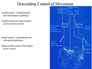
Descending Control of Movement
medial system vestibulospinal and
reticulospinal pathways medial system provides
postural and locomotor control lateral
system corticospinal and rubrospinal
pathways lateral system controls fine finger/
motor control
2
Medial Postural System
damage to more anterior brain regions results in
more and more posture related control of body
muscles- suggests this is located in
brainstem raphe nucleus and locus ceruleus send
serotinergic and noradrenergic axons to
modulate motor neuron function in the spinal
cord vision, proprioception and vestibular
senses are most important for
posture semicircular canals measure
rotational accelleration by increased of
action potentials otolithic maculae measure
linear accelleration
3
Medial Postural System
utricule and saccule are bony regions of the
cochlea that measure linear accelleration
using hair cells moved by dense crystals
(otoconia) that lie on hair cells using
gravity (lack of gravity causes
dizziness) different hair cells have different
orientations that measure accelleration in
various directions lesions in VIIIth nerve
(to canals) cause falling toward the side of
the lesion vestibulospinal tract mediates most
head/neck positioning
4
Medial Postural System
excitatory, lateral vestibulospinal afferents to
ipsilateral extensors counter tilt to the same
side-- straight negative feedback works through
head/neck tilt, but can go to legs to stabilize
body posture vestibular-cervical processing
deals with low and high frequency head
motions low frequency straight
repositioning high frequency anticipatory
changes to predict future head
location mechanisms of control are uncertain
timing and specific motor unit activation is
integrated from all inputs
5
Medial Postural System
proprioceptive responses of muscles is more
complicated, and only really addressed for
the neck-- arms, etc do have counteracting
movements, but which motor units are activated
could vary hugely in neck, rotation of the body
causes ipsilateral extensor activation as in
the vestibular system-- suggests a common motor
unit/regulation, but the specific muscle
groups used counteract motions are
complex posture reactions require the brainstem
ie. placing a limb on the ground stiffens
that limb-- positive supporting reaction others
require cerebral cortex- optical righting
reflexes hopping reaction is to try and find a
stable footing if a surface moves underneath
6
Medial Postural System
basic goal of posture is to keep the center of
mass of the body over the base of support,
otherwise a person falls or must change their
support when vision or vestibular or
proprioceptive inputs are altered, swaying
increases ie. body tries harder to balance the
center of mass if a platform changes, leg
muscles contract in sequence from ankle to
hip for other displacements, hips followed by
trunk and head alter position for
balance posture reflexes are also mediated by
negative feedback seem to work as additive
units depending upon situation/experience
7
Medial Postural System
damage to many neural areas can disrupt
posture basal ganglia lesions can cause
rigidity and reduced movement anterior
cerebellar lesions increase rigidity and
decrease adaptation unilateral vestibular
lesions cause pronounced leaning toward
side compensation occurs within weeks vestibular
lesions cause vision and proprioceptive inputs
to be more important for posture
8
Lateral Voluntary System
frontal lobe of the cortex can be stimulated to
cause motor responses has direct connections
through the central sulcus to spinal
cord corticospinal axons descend through the
contralateral lateral column as well as
synapsing on pontine neurons in the
cerebellum corticospinal axons synapse in the
ventral horn onto motor neurons, some crossing
the midline again allowing contra and ipsilateral
synapses
9
Lateral Voluntary System
different cortical neurons connect to the red
nucleus, which integrates information with the
cerebellum and enters the spinal cord through
the lateral column and synapses in the
dorsolateral ventral horn rubrospinal neurons
work synergistically with corticospinal neurons
and plays largest role in arms, hands, and
fingers medial reticular formation in the
pons/medulla also controls voluntary
movements projects through the ventral column
to ventromedial ventral horn and called the
reticulospinal tract
10
Lateral Voluntary System
the 'motor cortex' is, as usual, subdivided into
multiple motor areas generally layer 5
pyramidal cells are larger layer 4 receives
more sparse thalamic input than other cortical
areas 3 mediolaterally oriented strips of
cortex primary motor cortex M1 is most
posterior strip 'premotor' areas are anterior
to M1, each with 3 subdivisions cingulate sulcus
and cingulate gyrus also contain motor areas
11
Lateral Voluntary System
motor cortex also receives input from sensory and
association cortex including somatosensory and
visual cortex somatosensory connections
generally relate to the motor areas
activated visual and somatosensory inputs
also tend to have related fields muscle
intention also alters how somatosensory inputs
affect motor cortex
12
Lateral Voluntary System
in M1 where most studies occur, face is lateral,
lower extremities medial, and upper
torso/extremities in between, making a
somatotopic map areas with finer voluntary
motions (fingers, tongue, etc) are larger size
mapping is not 11, with several areas having
overlapping fields M1 cortical connections over
large areas can converge on small motor
units and territory overlaps single cortical
neurons can drive multiple muscle motor
pools horizontal cortical connections can
cover large regions of M1 cortex M1 has lots of
distributed activity regulating voluntary
movements
13
Lateral Voluntary System
plasticity is also maintained in M1 after
denervation use can also enlarge that area of
M1- practicing the piano really does
change the brain's layout damage to M1 can be
slowly and/or partly restored after
damage microelectrodes measure 1 to a few
neurons, while MRI measures 1000's of neurons,
but both have their place in understanding
voluntary motion planned movements of hands, for
example, activates S1 as well as premotor
areas, supplementary motor area, and several
others planning involves brain activation up
to a second before motion visualizing an action
really does happen before performing it
14
Lateral Voluntary System
M1 contains flexion/extension neurons- ie. move
that muscle group with firing rate depending
upon force required responds generally to
flexion or extension, not both other premotor
and motor areas also depend upon same
parameters M1 activity relies upon an ensemble
activity- population determines the specific
speed/direction/force very accurately modern
electrode nets can use this to actively
predict/generate moves
15
Lateral Voluntary System
cortical areas outside of M1 select and guide
movement using senses dorsal premotor cortex
(PMd) and suplementary motor area (SMA) fire
most while waiting to make a particular movement
areas store information on force/direction
if 'orders' change, often the wrong 'prepared'
action is taken! using mirrors and perceived
cues, some areas respond best to 'planned'
movements, while others respond to 'actual'
movement M1 is most correllated with actual
movement
16
Lateral Voluntary System
brain areas also differ based on remembered cues
or external instructions M1 fired equally for
remembered and external cues SMA is more active
for remembered cues PMv is more active during
external cues
internal external
Recommended
