
Topic 1: Cells PowerPoint PPT Presentation
1 / 63
Title: Topic 1: Cells
1
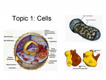
Topic 1 Cells
2
1.1 Cell Theory
- 1.1.1
- Discuss the theory that living organisms are
composed of cells. (3) - Skeletal muscle and some fungal hyphae are not
divided into cells but have a
multinucleate cytoplasm. Some biologists consider
unicellular organisms to be acellular.
3
1.1 Cell Theory
- 1.1.2
- State that a virus is a non-cellular structure
consisting of DNA or RNA surrounded by a protein
coat. (1)
4
1.1 Cell Theory
5
1.1 Cell Theory
- 1.1.4
- Explain three advantages of using light
microscopes. (3) - Advantages include
- colour images instead of monochrome,
- a larger field of view,
- easily prepared sample material,
- the possibility of examining living material and
observing movement.
6
1.1 Cell Theory
- 1.1.5
- Outline the advantages of using electron
microscopes. (2) - Greater
- Resolution the ability to distinguish between
two points on an image. Like pixels in a digital
camera. - Magnification how much bigger a sample appears
to be under the microscope than it is in real
life.
7
1.1 Cell Theory
- Transmission electron microscopes pass a beam of
electrons through the specimen. The electrons
that pass through the specimen are detected on a
fluorescent screen on which the image is
displayed. - Thin sections of specimen are needed for
transmission electron microscopy as the electrons
have to pass through the specimen for the image
to be produced.
8
1.1 Cell Theory
- Scanning electron microscopes pass a beam of
electrons over the surface of the specimen in the
form of a scanning beam. - Electrons are reflected off the surface of the
specimen as it has been previously coated in
heavy metals. - It is these reflected electron beams that are
focused on the fluorescent screen in order to
make up the image. - Larger, thicker structures can thus be seen under
the scanning electron microscope as the electrons
do not have to pass through the sample in order
to form the image. - However the resolution of the scanning electron
microscope is lower than that of the transmission
electron microscope.
9
1.1 Cell Theory
10
1.1 Cell Theory
- 1.1.6
- Define organelle. (1)
- Literally little organ
- An organelle is a discrete structure within a
cell, and has a specific function. - i.e. nucleus, cell membrane, mitochondria
11
1.1 Cell Theory
- 1.1.7
- Compare the relative sizes of molecules, cell
membrane thickness, viruses, bacteria, organelles
and cells, using appropriate SI units (2) - Appreciation of relative size is required,
- molecules (1 nm),
- thickness of membranes (10 nm), xref. 1.4
- viruses (100 nm),
- bacteria (1 µm), xref. 1.33
- organelles (up to 10 µm), xref. 6.4.2, 7.1.3,
7.2.1 - most cells (up to 100 µm).
- Dont forget all of these structures are in 3D
space
12
1.1 Cell Theory
- 1 nm 1/1,000,000,000 of a meter, or . . .
- 0.000000001m, or . . .
- 1 billionth of a meter
- 1 µm 1/1,000,000 of a meter, or . . .
- 0.000001m, or . . .
- 1 millionth of a meter
- 1 nm 1/1,000 of a µm, or
- 1 µm 1,000 nanometers
- Therefore. . .
13
1.1 Cell Theory
- A 100 µm cell 10x larger than a. . .
- A 10 µm organelle 10x larger than a. . .
- A 1 µm bacteria 10x larger than a. . .
- A 100 nm virus 10x larger than a. . .
- A 10 nm membrane 10x larger than a. . .
- A 1 nm molecule
14
1.1 Cell Theory
15
1.1 Cell Theory
- 1.1.8
- Calculate linear magnification of drawings. (2)
- Drawings should show cells and cell
ultra-structure with scale bars - Magnification could also be stated, eg x250.
16
1.1 Cell Theory
- 1.1.9
- Explain the importance of the surface area to
volume ratio as a factor limiting cell size. (3) - The rate of metabolism of a cell is a function of
its massvolume ratio, - Whereas the rate of exchange of materials and
energy (heat) is a function of its surface area. - Simple mathematical models involving cubes and
the changes in the ratio that occur as the sides
increase by one unit could be compared.
17
Assume we have 3 cubes
3
With sizes
2
1
1 cm
100 cm
10 cm
What will happen to ratio between V and S.A. as
their size increases?
18
Ratio of VS.A.
1 cm3
6 cm2
6
600 cm2
1 000 cm3
0.6
1 000 000 cm3
60 000 cm2
0.06
19
1.1 Cell Theory
- 1.1.10
- State that unicellular organisms carry out all
the functions of life. (1) - MOVEMENT Intracellular and/or extracellular
- RESPIRATION Gas exchange. Not always O2 and
CO2 - NUTRITION Need raw materials, i.e.- food,
water, minerals - EXCRETION Get rid of waste materials
- REPRODUCTION Ability to produce like organisms
- IRRATIBILITY Respond to external stimuli
- GROWTH Cells grow larger . . . and dont forget
. . . - MR. NERIG also carries out the functions of
life!
20
1.1 Cell Theory
- 1.1.11
- Explain that cells in multicellular organisms
differentiate to carry out specialized functions
by expressing some of their genes but not others.
(3)
21
1.1 Cell Theory
- 1.1.12
- Define (1)
- Tissue A group of cells working together to
perform a common function - Organ A group of tissues working together to
perform a common function - Organ System A group of organs working together
to perform a common function
22
Prokaryotic cell
- 1.2.1
- Draw a generalized prokaryotic cell as seen in
electron micrographs. (1) - Use images of bacteria as seen in electron
micrographs to show the structure. - The diagram should show the cell wall, plasma
membrane, mesosome, cytoplasm, ribosomes and
nucleoid (region containing naked DNA).
23
(No Transcript)
24
1.2 Prokaryotic Cells
- 1.2.2
- State one function for each of the following
(1) - cell wall forms a protective outer layer that
prevents damage from outside and bursting if
internal pressure is too high - plasma membrane controls entry and exit of
substances, pumping some of them in by active
transport
25
1.2 Prokaryotic Cells
- mesosome increases the area of membrane for ATP
production. May move the DNA to the poles during
cell division - cytoplasm contains enzymes that catalyse the
chemical reactions of meabolism and DNA in a
region call the nucleoid
26
1.2 Prokaryotic Cells
- ribosomes synthesize proteins by translating
messenger RNA. Some proteins stay in the cell
and others are secreted - naked DNA stores the genetic information that
controls the cell and is passed on to daughter
cells
27
1.2 Prokaryotic Cells
- 1.2.3
- State that prokaryotes show a wide range of
metabolic activity including fermentation,
photosynthesis and nitrogen fixation. (1)
28
1.3 Eukaryotic Cells
- 1.3.1
- Draw a diagram to show the ultrastructure of a
generalized animal cell as seen in electron
micrographs. (1) - The diagram should show ribosomes, rough
endoplasmic reticulum (rER), lysosome, Golgi
apparatus, mitochondrion and nucleus.
29
electron micrograph
30
1.3 Eukaryotic Cells
- 1.3.2
- State one function of each of these organelles
(1) - ribosomes protein synthesis
- rough endoplasmic reticulum (rER) synthesis of
proteins to be secreted - lysosome holds digestive enzymes
- Golgi apparatus for processing of proteins
- mitochondrion for aerobic respiration
- nucleus holds the chromosomes
31
1.3 Eukaryotic Cells
- 1.3.4
- Describe three differences between plant and
animal cells. (2)
Plant Cells
Structure
Animal Cells
Can produce its own food.
?
Chloroplast
X
Cannot produce its own food
Rigid, cannot easily change shape.
?
Cell Wall
X
Flexible, can easily change shape.
Stores large amounts of liquid (juice). Larger
size of cell.
?
Central Vacuole
X
Does not store large amounts of liquid. Smaller
size of cell.
Carbohydrates stored as starch.
Carbohydrates stored as glycogen.
32
1.3 Eukaryotic Cells
- 1.3.5
- State the composition and function of the plant
cell wall. (1) - The composition of the plant cell wall should be
considered only in terms of cellulose
microfibrils.
33
1.4 Membranes
- 1.4.1
- Draw a diagram to show the fluid mosaic model of
a biological membrane. (1) - The diagram should show the phospholipid bilayer,
cholesterol, glycoproteins and integral and
peripheral proteins. - Use the term plasma membrane not cell surface
membrane for the membrane surrounding the
cytoplasm. - Integral proteins are embedded in the
phospholipid of the membrane whereas peripheral
proteins are attached to its surface.
34
1.4 Membranes
- 1.4.2
- Explain how the hydrophobic and hydrophilic
properties of phospholipids help to maintain the
structure of cell membranes. (3) - Hydrophobic afraid of water
- Hydrophilic loves water
35
1.4 Membranes
36
1.4 Membranes
37
1.4 Membranes
38
1.4 Membranes
39
1.4 Membranes
- 1.4.3
- List the functions of membrane proteins including
(1) - hormone binding sites
- enzymes
- electron carriers
- channels for passive transport
- pumps for active transport.
40
(No Transcript)
41
1.4 Membranes
- 1.4.4
- Define diffusion (1)
- Diffusion is the passive movement of particles
from a region of high concentration to a region
of low concentration (down a concentration
gradient), until there is an equal distribution. - Define osmosis
- Osmosis is the passive movement of water
molecules, across a partially permeable membrane,
from a region of lower solute concentration (high
water concentration) to a region of higher solute
concentration (low water concentration).
42
1.4 Membranes
Diffusion moves down the concentration gradient
just like a ball rolling down a hill. It cannot
roll uphill without energy.
High Concentration
Low Concentration
43
1.4 Membranes
- 1.4.5
- Explain passive transport across membranes in
terms of diffusion. (3) - Requires no energy
- Moves from down the concentration gradient
- Some molecules pass through the membrane
- Some molecules use channels for facilitated
diffusion
44
1.4 Membranes
45
1.4 Membranes
- 1.4.6
- Explain the role of protein pumps and ATP in
active transport across membranes. (3) - Requires energy, in the form of ATP, or adenosine
triphosphate - Molecules are pumped across the membrane UP the
concentration gradient - Pumps fit specific molecules
- The pump changes shape when ATP activates it,
this moves the molecule across the membrane
46
Active Transport
47
1.4 Membranes
- 1.4.7
- Explain how vesicles are used to transport
materials within a cell between the rough
endoplasmic reticulum, Golgi apparatus and plasma
membrane. (3) - 1.4.8 Describe how the fluidity of the membrane
allows it to change shape, break and reform
during endocytosis and exocytosis. (2)
48
(No Transcript)
49
1.4 Membranes
- Endocytosis the mass movement INTO the cell by
the membrane pinching into a vacuole - Exocytosis the mass movement OUT of the cell by
the fusion of a vacuole and the membrane - This is possible because the of the fluid
properties of the membrane (able to break and
reform easily, phospholipids not attached just
attracted)
50
Exocytosis
51
Endocytosis
endo- and exo- -cytosis
52
1.5 Cell Division
- 1.5.1
- State that the cell-division cycle involves
interphase, mitosis, and cytokinesis. (1)
Mitosis
Cytokinesis
Interphase
53
1.5 Cell Division
- 1.5.2
- State that interphase is an active period in the
life of a cell when many biochemical reactions
occur, as well as DNA transcription and DNA
replication. (1)
54
1.5 Cell Division
- 1.5.3
- Describe the events that occur in the four phases
of mitosis (2)
55
1.5 Cell Division
- PROPHASE - breakage of nuclear membranes and
supercoiling of DNA to form visible chromosomes
56
1.5 Cell Division
- METAPHASE - chromosomes line up along equatorial
region of cell, attachment of spindle
microtubules to centromeres
57
1.5 Cell Division
- ANAPHASE - splitting of centromeres, movement of
sister chromosomes to opposite poles as spindle
microtubules shorten
58
1.5 Cell Division
- TELOPHASE - uncoiling of chromosomes and
reformation of nuclear membranes
59
1.5 Cell Division
P rophase M etaphase A naphase T elophase P-MAT
60
1.5 Cell Division
- 1.5.4 Explain how mitosis produces two
genetically identical nuclei. (3) - Synthesis of identical chromosomes in interphase
- Lining up during mitosis ensures that each new
cell gets a copy of each chromosome
61
1.5 Cell Division
- 1.5.5
- Outline the differences in mitosis and
cytokinesis between animal and plant cells. (2) - No centrioles in plant cells
- Cell plate formed in plants, membrane pinching
in animal cells
Cytokinesis
62
1.5 Cell Division
63
1.5 Cell Division
- 1.5.6
- State that growth, tissue repair and asexual
reproduction involve mitosis. (1) - 1.5.7
- State that tumours (cancers) are the result of
uncontrolled cell division and that these can
occur in any organ. (1)
Recommended
