
Poxviridae Orthopoxvirus (image) PowerPoint PPT Presentation
1 / 30
Title: Poxviridae Orthopoxvirus (image)
1
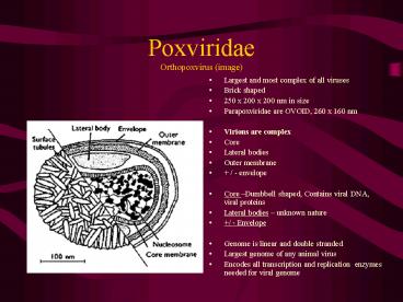
PoxviridaeOrthopoxvirus (image)
- Largest and most complex of all viruses
- Brick shaped
- 250 x 200 x 200 nm in size
- Parapoxviridae are OVOID, 260 x 160 nm
- Virions are complex
- Core
- Lateral bodies
- Outer membrane
- / - envelope
- Core Dumbbell shaped, Contains viral DNA, viral
proteins - Lateral bodies unknown nature
- / - Envelope
- Genome is linear and double stranded
- Largest genome of any animal virus
- Encodes all transcription and replication
enzymes needed for viral genome
2
PoxviridaeOrthopoxvirus, Parapoxvirus
- Several poxviruses encode virokines that affect
the response of the host to infection - Epidermal growth factor homologue
- Protein that down regulates complement proteins
- Virokines conferring resistance to INF etc.
- Cytoplasmic replication
- Eosinophilic intracytoplasmic inclusions
- All poxviruses share group specific nucleoprotein
NP antigen that is exposed after being
digested with alkaline - Each species in a genus is characterized by
specific polypeptides - Cross neutralization and cross- protection occurs
among members of the same genus none between
different genera - Only the Orthopoxviruses produce Hemagglutinin
3
PoxviridaeParapoxviridae Pseudopox virus
- Parapoxviridae causes Bovine Papular Stomatitis
as well as Milkers nodule lesions - Serologic Assays
- In the assays, a 2-fold serial dilution of
suspect serum is used - A constant amount of virus is added to each of
the dilutions - The titer is the highest dilution giving the
desired result - Antibodies can be detected and/or quantitated
using any of the assays below - Hemagglutination- inhibition test see next
slide - Complement fixation test
- Immunodiffusion test
- Indirect ELISA test
- Indirect Immunofluorescent test IFA
- Virus neutralization test
4
(No Transcript)
5
Poxviridae Subfamilies and Genera
- Orthopoxvirus vaccinia
- Parapoxvirus pseudocowpox virus
- Avipoxvirus fowlpox virus
- Capripoxvirus sheeppox virus
- Leporipoxvirus Leooripoxvirus
- Suiposvirus Swinepox virus
- Molluscipoxvirus Myxoma virus
- Yabapoxvirus Yaba monkey tumor virus
- Subfamily Entomopoxvirinae contains viruses of
insects - Viral replication cytoplasm
6
Poxviridae - Replication
- Occurs in the cytoplasm
- Following the release of the viral core into the
cytoplasm, the following occur - Adsorption and penetration via endocytosis for
non-enveloped viruses - Fusion with the plasma membrane are used with
enveloped viruses - Uncoating 2 step process
- Host cell enzymes partially uncoat the viral
particle, exposing some viral DNA and polymerase - Early mRNA are transcribed resulting in several
polypeptides, including an uncoating protein
which completes the uncoating of the core. - DNA replication using enzyme pools
7
Poxviridae Replication
- Late mRNA codes for structural proteins and
proteins to switch off early mRNA activity - Especially to block uncoating proteins in order
to protect the progeny virions that are being
assembled - Following assembly, mature enveloped virions are
released by budding or by exocytosis - Naked virions are released by cell lysis
- Most poxviruses are naked
- Both enveloped and naked are infectious
8
Poxviridae Epidemiology, Pathogenesis and
Immunity
- Epidemiology
- Poxviruses are resistant to ambient temperatures
and can survive for many months or years in dried
scabs - Poxviruses are transmitted between animals by
skin abrasions, aerosol to the URT, mechanical
transmission by arthropods - Pathogenesis and Immunity
- Highly epitheliotropic causing cutaneous and
systemic disease in birds and wild and domestic
mammals - Many are host specific, but orthopoxviruses
infect a wide range of hosts - After cutaneous introduction or inhalation the
virus gains access to the systemic circulation
through the lymphatics. - Multiplication of the virus at the skin wound
may lead to direct access to the blood and
primary viremia. - Secondary viremia disseminates the virus back to
the skin and other target organs
9
PoxviridaePathogenesis, Immunity
- Poxviruses induce lesions by a variety of
mechanisms - Degenerative changes in the epithelium
- Lesions start as erytheamatous macules, become
papular and then vesicular - Vesicles develop into umbilicated pustules POCK
LESION - Pustules rupture and for a crust/scab
- Lesions heal and leave a scar
- Rupture of the pustule can lead to secondary
bacterial infection - Proliferative lesions
- Poxviruses replicating in epidermis may result in
virus induced (encoded epidermal growth factor)
cellular hyperplasia - Poxviruses encode proteins which may counteract
host defenses - Immunity varies from short lived like in
parapoxvirus infections to prolonged in others
10
PoxviridaeDiagnosis
- Virus isolation
- Scrapings from skin lesions, vesicular lesions
and crusts - Chorioallantoic membrane
- Pock lesions not parapoxviruses DO NOT
replicate in embryonated eggs - Cell culture
- Poxviruse grow in a variety of cell cultures
- Virus identification
- Negative stain electron microscopy FAT etc.
- Histopathology
11
PoxviridaeOrthopoxvirus Vaccinia - Cowpox
- Distribution identified only in Europe
- Hosts cattle, wild and domestic cats, zoo
animals, elephants, rhinos etc., humans - Rodents serve as reservoir hosts
- Etiologic agent Orthopoxvirus One serotype
- Transmission infection of cattle and domestic
cats occurs through intact with rodents. In
dairy herds, the virus spreads by the process of
milking - Clinical features - Disease in cows
- IP 3-7 days. Lesions seen on teats and udder
- Ulcerated pustules give rise to thick red scabs
- Secondary bacterial infection of teat lesions are
very common - Uncomplicated teat lesion heal in 3-4 weeks
12
Orthopoxvirus DiseasesClinical Features, Immunity
- Disease in cats More severe than in cattle or
humans - Scarbs are widespread
- Secondary bacterial infection may result in
pneumonia VERY COMMOM - Cats usually recover in 6-8 weeks
- Disease in humans
- Maculopapular leisons on the hands and face
- Nausea, fever, lymphadenopathy
- Immunity
- Recovering animals have long lasting immunity
13
PoxviridaeParapoxvirus Diseases
- Range of species - most important are cattle,
sheep, goats and camels - Parapoxviruses of domestic animals are zoonotic
- Pseudocowpox
- Common endemic infection in cattle worldwide
- Chronic infection in many dairy herds and beef
herds - Etiologic agent Bovine parapoxvirus - one
serotype - Clinical features
- Infections are mild morbidity approaches 100
only 10-15 of cows are affected at one time - Lesions are dark red ring or horseshoe scabs
- Horseshoe scabs are pathognomonic for the disease
- Lesions affect teats, udder and perineum
- Major lesions desquamate by 6 weeks no scars
- Similar lesions on muzzle and in mouths of
nursing calves ulcers and vesicles are rare - Immunity short-lived 4-6 months duration
- Humans mild skin lesion milkers nodule
14
PoxviridaeParapoxvirus Contagious Ecthyma
- Worldwide disease resulting in an acute infection
of all breeds of sheep and goats - Mostly affecting lambs and kids
- Synonyms scabby mouth, contagious pustular
dermatitis, sore mouth and orf the human dz. - Etiologic agent Parapoxvirus one serotype
- Transmission
- Survival in the environment is indefinite in
scabs - Infection is through cutaneous abrasions
- Oral lesions in lambs or kids result from nursing
dams with teat and udder lesions - Pathogenesis
- Lesions are vesiculopapular eruptions followed by
pustules with yellow- brown scabs - Dermal tissue may proliferate resulting in
verrucose mass under the scabs
15
Contagious Ecthyma
- Clinical features
- IP 4-7 days
- High morbidity 90, but low mortality
- Primary lesion develops on skin of the lips and
extends to the mucosa of the mouth - Severe cases lesions on genitals, perineum and
feet lameness can ensue - Oral and facial lesions are painful and may lead
to anorexia and weight loss - Scabs drop off in 1-4 weeks no scars
- Mortality can result when primary lesions are
invaded with screwworm larvae or Fusobacterium
necrophorum (gram negative rod - post op wound
infections, navel ill, soft tissue abscesses,
metritis, aspiration pneumonia, necrotic lesions
like FOOT ROT, liver abscesses common) - Immunity sheep are susceptible to reinfection
and chronic infections can occur
16
Contagious Ecthyma
- Vaccination
- Ewes are vaccinated several weeks before lambing
- Non- attenuated virus vaccines from infected
scabs or from cell cultured virus - Vaccine is brushed over scarified areas of skin
inside of thigh where a localized lesion develops - Vaccine short term immunity
- Lambs and kids
- Vaccinated at 1 month, revaccinated at 2-3 months
later, followed by annual revaccination - Humans Public Health Significance
- Maculopapular lesions and nodular lesions are
observed 2-4 days following infection - Lesions last for 4-9 weeks and heal without scars
17
Poxviridae Capripoxvirus - Sheeppox
- Diseases include sheeppox, goatpox and lumpy skin
disease - Sheeppox is the most important disease of
domestic animals - Viruses causing the three diseases may represent
strains of a single virus. This results in
cross- infection and cross- protection, however,
most strains show definite host preferences - Sheeppox
- Distribution endemic in Africa, Asia and Europe
- Etiologic agent Ovine capripoxvirus. One
serotype - Transmission Aerosolization and direct contact
with saliva, nasal secretions, scabs shed by sick
animals - Scabs are infective for up to 6 months
18
PoxviridaeSheeppox
- Mechanical transmission by biting arthropods?
- Stomoxys calcitrans stable fly
- Pathogenesis systemic disease
- Inhalation is followed by a leukocyte associated
viremia - Leading to a virus localization in skin, and to a
lesser extent the internal organs - Severe necrotizing vasculitis developing in
arterioles and postcapillary venules in the skin
may be due to immune complex deposition - TYPE III hypersensitivity virus does not
multiply in endothelium - This results in ischemic necrosis of the dermis
and overlying epidermis
- Clinical features
- IP 4-8 days
- Morbidity up to 80
- Malignant form seen in lambs and susceptible
nonnative breeds, e.g. merino - Signs are
- Fever, salivation, lacrimation, hyperpnea, edema
of the eyelids and serous nasal discharge later
becoming mucopurulent - 1-2 days later cutaneous nodules develop
- Can be widely distributed throughout the body
- Nodules scab and persist for 3-4 weeks, leaving a
permanent depressed scar - Mortality rate up to 50, Fatality up to 100 in
lambs - Benign form more common in adult sheep/resistant
breeds skin lesion not systemic
19
Sheeppox, Goatpox
- Immunity
- Recovered sheep have a solid immunity life long
- Prevention and Control
- Notifiable disease in most countries of world
- Goatpox
- Africa, Asia, Europe and U.S.
- Similar to sheeppox
- Lower mortality rate 5
- Strain of goatpox causes more severe dz. In sheep
20
PoxviridaeGoatpox and Lumpy Skin Disease
- Infectious, acute to chronic disease of cattle
characterized by - multiple skin nodules
- ventral edema
- persistent fever
- lymphadenopathy
- Distribution
- Endemic in certain parts of Africa
- Outbreak in 1989 in Isreal
- Etiologic agent Bovine capripoxvirus one
serotype - Hosts cattle and buffalo- African cape buffalo
is thought to be the reservoir - Transmission mechanical through biting
arthropods high concentration in saliva results
in contact transmission
21
Poxviridae Goatpox Lumpy Skin Disease
- Pathogenesis
- After skin inoculation, virus replicates in
epidermis and dermis - Infected macrophages migra regional lymph nodes
for further replication - Resulting in enlargement of the lymph nodes
- Macrophage associated virema disseminates the
virus to various tissues skin and endothelium - Damaged endothelium results in vasculitis,
thrombosis, marked dermal edema, infarction - Nodules are circumscribed, round, slightly
raised, firm and painful and involve the entire
skin and the mucosa of the GI, RT, genital tract-
develop inverted conical necrosis the sit fast
22
PoxviridaeLumpy skin disease
- Secondary bacterial infections develop in the
necrotic cores of the nodules - Metastatic abscesses in the regional lymph nodes,
lungs and other organs - Mortality is due to secondary infection
- Clinical signs
- IP 4-14 days
- Morbidity up to 100, mortality 1-2
- Fever, marked weight loss, hypersalivation oral
ulcers, nasal discharge - Lesions can persist for months but usually
disappear within 4-12 weeks - Abortion may occur as a result of prolonged fever
- Control Vaccination
- Attenuated Neethling strain prototype virus
vaccine used - Goatpox and Sheeppox virus vaccines have also
been used Heterotypic vaccination
23
PoxviridaeSuipoxvirus Swinepox virus
- Suispox is the principal cause of pox lesions in
swine - Similar to Vaccinia virus
- Benign disease of young pigs
- Worldwide distribution
- Transmission mechanical by pig louse,
Hematopinus suis causes skin trauma, carriers
the virus for weeks or months - Clinical features IP 3-7 days, Course 1-4 weeks
- Transient low grade fever and anorexia followed
by the development of papules, vesicles,
umbilicated pustules - Pustules crust over and scab by day 7
24
PoxviridaeSuipoxvirus Swinepox
- Lesions are found over abdomen and inner aspects
of thighs primarily. Udder and teat lesions are
seen in some sows that nurse infected piglets - Immunity recovered pigs are solidly immune
- Control Eradication of lice from piggery
- There is no commercially available vaccine
25
Poxviridae Avipoxvirus Fowlpox virus
- Causes disease in chickens, turkeys, guinea fowl,
peacocks, pheasants and other avian species. - Exact relationship between the poxviruses of the
different avian species is not certain, but it
has been shown experimentally that the virus
causing one type of pox can give rise to disease
in other species and that infection with one may
stimulate protection against another - E.g. milk maids
26
PoxviridaeAvipoxirus Fowlpox virus
- Distribution worldwide
- Hosts chickens, turkeys, pigeons, pheasants
etc. - Etiologic agent Avipoxvirus extremely resistant
to dessication and can survive in exfoliated
scabs for prolonged periods - Inclusions bodies Bollinger Bodies large
intracytoplasmic inclusions Borrel bodies
elementary bodies occur inside the Bollinger
bodies. - Borrel bodies are minute spherical bodies
obtained by tryptic digestion of Bollinger bodies - Transmission occurs through small abrasions in
the mouth or through injuries to the comb, wattle
as a result of fighting, pecking or other
injuries - Mechanical transmission by mosquitoes, ticks,
biting flies and lice
27
Poxviridae Fowlpox
- Clinical features IP 4-14 days
- Two forms of disease
- Dry form cutaneous form
- small papules on the comb, wattles, and around
the beak lesions may develop on the legs and
feet around the cloaca - Nodules become yellowish and progress to a thick
scab. Egg production drops markedly. Affected
birds recover in about 4 weeks - Wet form diptheritic form
- Involves infection of the mucous membranes of the
mouth, pharynx, larynx, and sometimes trachea - Lesions coalesce resulting in a necrotic
pseudomembrane which may prevent feeding. Death
results from suffocation by occlusion of the
larynx. - Mortality can reach 50
28
Poxviridae Ulcerative Dermatosis of Sheep -
unclassified
- Two forms
- Ulcers around the mouth, nose and legs
- Venerally transmitted ulcerations of the prepuce
and penis or vulva - World wide
- Synonyms Lip and leg ulceration and venereal
balanoposthitis and vulvitis - Etiologic agent Ovine poxvirus
- Transmission Infection results from viral
contact with damaged skin or by coitus - Clinical features ulcer with a raw, easily
bleeding crater - Contains an odorless, creamy
pus and is covered by a thin brown bloody scab - Face lesions - upper lip, chin, and nose. Foot
lesions - between the coronet, carpus and the
tarsus
29
Poxviridae
- Rams
- Lesions partially or completely surround the
preputial orifice and may become so severe as to
produce phimosis. - Rarely, the ulcerative process may extend to the
glans penis rendering the ram unfit for
breeding - Ewes
- Lesions occur as edema, ulceration, and scabbing
of the lips of the vulva
30
(No Transcript)
Recommended
