
Chapter Five-Module 1 Development of the Brain Chapter Fourteen-Module 1 Lateralization PowerPoint PPT Presentation
1 / 17
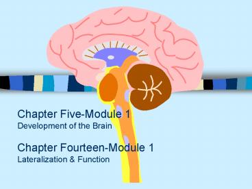
Title: Chapter Five-Module 1 Development of the Brain Chapter Fourteen-Module 1 Lateralization
1
Chapter Five-Module 1Development of the
BrainChapter Fourteen-Module 1Lateralization
Function
2
Development of the Brain-Growth and
Differentiation of the Vertebrate Brain
- Early Beginnings
- CNS begins to form at two weeks gestation
- Development of the neural tube (figure 5.2)
- At birth, brain weighs 350g, at one year 1,000g
(figure 5.3) - Growth and Development of Neurons
- Proliferation-production of new cells
- Migration-move toward final destination
- Differentiation-form axons and dendrites
- Myelination-addition of insulating sheath
3
Figure 5.2 Early development of the human
central nervous systemThe brain and spinal cord
begin as folding lips surrounding a fluid-filled
canal. The stages shown occur at approximately
age 2 to 3 weeks.
4
Figure 5.3 Human brain at five stages of
developmentThe brain already shows an adult
structure at birth, although it continues to grow
during the first year or so.
Video
5
Development of the Brain-Neuronal Survival
- Determinants of Neuron Survival
- Must make correct connections
- Must receive support from nerve growth factor
- neurotrophins act in several ways
- early in development cause cells to survive and
grow - increase the branching of incoming axons
- decrease pain and increase regrowth of damaged
axons - apoptosis-programmed cell death that occurs when
connections are not reinforced - Competition Among Axons as a General Principle
- We produce redundant synapses
- the most successful axons and combinations survive
6
Development of the BrainPathfinding Axons
- Pathfinding by Axons
- Chemical Pathfinding by Axons
- Example Weiss and the grafted salamander leg
- Specificity of Axon Connections
- Example Sperry and the rotated eye of newt
(figure) - Chemical Gradients
- cell surface molecule
- chemical attractants (e.g. TOPDV)
- Neurotrophins
7
Figure 5.7 Summary of Sperrys experiment on
nerve connections in newtsAfter he cut the optic
nerve and inverted the eye, the optic nerve axons
grew back to their original targets, not to the
targets corresponding to the eyes current
position.
8
Development of the Brain Fine-Tuning by
Experience
- Fine-Tuning by Experience
- Genetic Instruction are only approximate
- Effects of Experience on Dendritic Branching
- Enriched environments increase dendritic
branching (figure 5.10) dendritic spine growth
(5.11) thus a thicker cortex - What is an enriched human environment? Effects?
- Generation of New Neurons
- Can the adult brain generate new neurons?
- Olfactory cells must. Why?
- stem cells in the interior of the brain
- scientists have observed new cells in hippocampus
and cerebral cortex in monkeys of ages. - Possible meaning of new neural development?
9
Development of the Brain Effects of Experience
on Human Brain Structures
- Example music training on temporal lobe
development - identifying absolute pitch and temporal cortex
growth - Example somatosensory cortex (post-central
gyrus) in violin players - MEG D5 dipole strength, age of first playing,
and control groups (figure 5.13b) - Combinations of Chemical and Experiential Effects
- not always a clear 2-stage process of chemical
pathfinding and experiential strengthening - e.g., the identification by lateral geniculate
cells of activating retinal neurons (spontaneous
embryonic firing)
10
Development of the Brain The Vulnerable
Developing Brain
- Fetal Alcohol Syndrome
- decreased alertness, hyperactivity, varying
degrees of mental retardation, motor problems,
heart defects, and facial abnormalities - Fetal Nicotine Exposure
- low birthweight, SIDS, decreased intelligence,
hyperactivity - Fetal Cocaine Exposure
- decrease in IQ and language skills
- Module 1 Conclusions
11
Chapter Fourteen- Module 1Lateralization
12
Lateralization of Function
- Some Definitions
- Lateralization-Division of labor between the two
hemispheres - Commissures-Cross-over points of information in
the brain - Corpus Callosum
- Anterior Commissure
- Hippocampal Commissure
13
Figure 14.1 Two views of the corpus callosumThe
corpus callosum is a large set of axons conveying
information between the two hemispheres. (a) A
sagittal section through the human brain. (b) A
dissection (viewed from above) in which gray
matter has been removed to expose the corpus
callosum.
14
Figure 14.4 The anterior commissure and
hippocampal commissuresThese commissures allow
for the exchange of information between the two
hemispheres, as does the larger corpus callosum.
15
Visual Connections to the Hemispheres
- Visual Field-what is visible at any moment
- Right visual field--gtleft half of each
retina--gtleft hemisphere - Left visual field--gtright half of each
retina--gtright hemisphere - Cutting the Corpus Callosum
- Sometimes done to treat severe epilepsy
- Behavior is abnormal only when sensory stimuli
are limited to one side of the body
16
Figure 14.2 Connections from the eyes to the
human brain Route of visual input to the two
hemispheres of the brain. Note that the left
hemisphere is connected to the left half of each
retina and thus gets visual input from the right
half of the world the opposite is true of the
right hemisphere.
17
Split Hemispheres
- Competition
- Soon after surgery you may see competition
between activities on the two sides of the body - Hemispheric Specialization
- Left
- Speech
- Happiness
- Detail-oriented
- Right
- Emotional content of speech
- Recognizes emotions in others
- Expresses fear and anger
- Spatial Relationships
- Music perception
Animation
Recommended
