
Ultrasound Case: Muscle with a Hematoma PowerPoint PPT Presentation
1 / 3
Title: Ultrasound Case: Muscle with a Hematoma
1
Ultrasound Case Muscle with a Hematoma
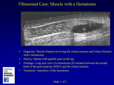
- Diagnosis Muscle Rupture involving the
Gastrocnemius and Soleus Muscles with a Hematoma. - History Patient with painful mass in the leg.
- Findings Long axis view of a hematoma (H)
situated between the medial head of the
gastocnemius (MHG) and the soleus muscles. - Treatment Aspiration of the hematoma.
Slide 1 of 3
2
Ultrasound Case Muscle with a Hematoma
- Diagnosis Muscle Rupture involving the
Gastrocnemius and Soleus Muscles with a Hematoma. - History Patient with painful mass in the leg.
- Findings Transverse view of a hematoma (H)
situated between the medial head of the
gastocnemius (MHG) and the soleus muscles. - Treatment Aspiration of the hematoma.
Slide 2 of 3
3
Ultrasound Case Muscle with a Hematoma
- Diagnosis Muscle Rupture involving the
Gastrocnemius and Soleus Muscles with a Hematoma. - History Patient with painful mass in the leg.
- Findings Needle within the hematoma during
ultrasound guided decompression. Ultrasound
allows continuous real-time monitoring. - Treatment Aspiration of the hematoma.
Slide 3 of 3
Recommended
