
Imaging and Multi-modality Navigation in Interventional Oncology PowerPoint PPT Presentation
1 / 76
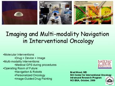
Title: Imaging and Multi-modality Navigation in Interventional Oncology
1
Imaging and Multi-modality Navigation in
Interventional Oncology
- Molecular Interventions
- Drug Device Image
- Multi-modality Interventions
- Medical GPS during procedures
- Operating Room of Future
- Navigation Robots
- Personalized Oncology
- Image-Guided Drug Painting
Brad Wood, MD NCI Center for Interventional
Oncology Intramural Research Program NCI BSA,
October, 2009
2
PET (Metabolic) Guided Procedures
3
Closing the Gap Between Diagnosis Therapy
4
Minimally Invasive Image GuidedConvergence of
Devices Imaging
Tumor Ablation Uterine Fibroid Embolization Stent
Grafts Brain Aneurysm Coiling Vertebroplasty Ballo
on Angioplasty Venous Ablation Carotid Stenting
Less Surgery
CO2Insufflation needle replaces laparoscope
Treatment needle
colon
Renal Cell Carcinoma
5
Center for Interventional Oncology Mission
- Close gap between Diagnosis Therapy
- Establish a collaborative environment to bring
together multidisciplinary partners to help
define minimally-invasive image-guided methods
for tx of locally-dominant cancer
6
(No Transcript)
7
Collaborative NetworkInterdisciplinaryInter-age
ncyTranslationalInternationalIndustry /
Extramural Academic / Government
http//www.cc.nih.gov/centerio/index.html
8
1955 NIH Open Heart Surgery w/ Extra-Corporal
Circuit
2009 NIH Percutaneous Liver Perfusion
300 PHPs in 120 pts 80 response rates for
neuroendocrine ocular melanoma
9
Imaging and Multi-modality Navigation in
Interventional OncologyOverview
- Molecular Interventions
- Drug Device Image
- Multi-modality Interventions
- Medical GPS during procedures
- Operating Room of Future
- Navigation Robots
- Personalized Local Regional Oncology
- Image-Guided Drug Painting
- RFA heat-deployed liposomal drug
- Image-able drug eluting bead RFA
- HIFU heat-deployed liposomal contrast drug
10
2009 NIH Medical GPS devices, Fusion-guided
procedures, Image-guided robotics
Early 20th Century Stereotactic Frame
11
Needle Ablation Complex Geometries Outcomes
Depend Upon Accuracy
12
Patient-Specific Treatment Plans
Risk to Adjacent Anatomy (Heart)
Risk of Heat Sink
13
- Automated RFA planning tool integrated with
navigation
Tracked Needle
Selected Target
US and CT view, with planned composite ablation
and tracked needle overlay
14
O.R. of the Future
- Navigation
- Visualization
- Automation
- Real-Time Fusion
15
(No Transcript)
16
GPS-Tumor AblationFrom Idea to Lab to Animal to
Patient toFDA approval to Market
Black Virtual Needle White Clandestine Cancer
Accuracy, Error benefit defined in gt200 patient
clinical trial
17
CT, US PET guided fusion biopsy in patient with
lymphoma
18
Molecular InterventionsDevice Image Drug
19
Prostate InterventionsIdea to Design to Lab to
Phantom to Animal to Patient
Sensor
20
Smart Needles use MRI Info outside of MRI
No need for MRI during procedure
21
GPS Fusion Makes the Dx
22
Automated Motion Correction
3.1 mm error
gt140 patient trial
83 pts w high suspicion MR had positive fusion
bx Aggressiveness correlated with imaging
23
Smart Surgical Equipment
24
Multi-Modality Surgery
25
Smart Surgery
- Tumor localization
- Faster resection
26
Steerable Bronchoscopy Catheter
27
Tracked Stent Grafts for Aortic Aneurysm Repair
28
Image to Tissue Correlation for Personalized
Oncology Drug Discovery
Image registration Sample collection
Biomarker Gene Protein
prognosis response sensitivity resistance metabol
ism
29
Image to Tissue Correlation for Personalized
Oncology Drug Discovery
- Biomarkers
- Identify target
- Verify delivery
- Predict response
- Toxicity
- Prognosis
- Individualize tx / Pt-specific cocktails
- Timing
- Sensitivity
- Resistance
- Drug Discovery
- Target
- Efficacy
30
PET Guided Interventions
31
Robots in IR
- Accuracy
- Less radiation
- Fast, Cost-effective
- Efficient
- Fewer needle attempts
- Tx planning
- Consistency
Better Outcomes
32
Bill Charboneau, Mayo
33
Integration of Robotics CT-guided Ablation
34
Drug Delivery Barriers
- IV vs IA
- Vessel wall
- Interstitium
- Cell membrane staying in cell (nucleus)
Blood vessels 3.3 kDa Dextran
3
2
4
4
4
2
1
4
2
4
3
35
Molecular Interventions targeted drug designed
for device
Tumor vasculature ideal size for nanomedicine
Drug Contrast
50-100 nm
36
Combination TargetingSmart IV Drug Thermal
Needle Device
Extravasation _at_ Edge of RFA
Vessel
Leaky Vessels
Residual Tumor
Ablation Needle
Dead Tumor Center
37
Physiologic, Thermal, Chemical Synergy
Leaky tumor vessels
Heat alters permeability
Cargo deployed _at_ 39-42 deg
Transition Temperature
38
Percent drug release in plasma over time at diff
temperatures
39
RFA and ThermoDoxin vitro feasibility
- Drug Release Independent of Heat Source
- Equivalent Cytotoxicity After Heat
30 min
JC Adenocarcinoma cells
Dox heated for 12 min, P gt 0.05
40
Paired heat transfer Pharmacokinetic model
Protein Binding/Transport into cells
- Transvascular Transport depends on
- Vessel Permeability (depends on drug molecule,
f(T)) - Vessel Surface Area
- Perfusion (f(T))
41
Modeling Perfusion vs Temp
41
42
RF ablationComparison Free DOX LTSL
- Increased drug delivery to thermal margin
Dieter Haemmerich
43
Imaging Drug EffectsThermoDox RFAIdea,
animal studies Phase I _at_ NIH Phase III 5
countries, 40 cancer centers
Pre-procedure
Intra-procedure
Day 71
12 month
- Enhancing rim corresponds to predicted drug
location
44
Drug Device (RFA)Effect on Treated Volumes
- Bland RFA -35.8 volume
- RFA LTSL 43.3 volume
RFA alone
RFA LTSL
45
RFA and ThermoDoxTime to progression
46
Drug eluting beads (DEB)
47
Image-able Drug Eluting Beads Pre-clinical,
bench, in-vivo
48
Imaging Drugs for Local Drug Dosingpersonalized
oncologyDistribution of bead correlates w/ true
bead location (image)
49
The spatial distribution of embolization beads is
directly related to bead size on micro-CT
- Small image-able beads (75-100 µm) found in
smaller peripheral arteries w/ many orders of
branching - Larger beads (100-300 µm) go central w/ gaps
between embolized arteries
50
Imaging Dynamic Drug Delivery Distribution of
drug correlates w/ bead location
2 hours post embolization Nuclei, Doxorubicin
51
30 Minutes Post Small Beads
52
24 Hours PostNecrosis colocalizes with drug
53
Doxorubicin Line Profile for Spatial Drug
Quantification
- Dox concentration is highest around beads
- Greatest concentration appears at 4 hrs
- Limited Dox at 24 hrs
54
Comparison of one many beads
- Greater concentration of Dox around more beads
55
2 Hr Confocal Microscopy subcellular
distribution
56
4 Weeks Post- DEB
Pre-Drug Eluting Beads (DEB)
57
Image Guided, Non-Invasive HIFU for Tissue
Destruction, Drug Delivery, or Hyperthermia
58
Pulsed HIFU enhanced delivery
MR contrast agent (Gd) muscle (rabbits)
FITC-dextran (500 kDa) SCC7 tumors (mice)
fluorescent Nanoparticles JC tumors (mice)
Genes - GFP (naked DNA) SCC7 tumors (mice)
ThermoDox ? growth inhibition mice
Velcade ? growth inhibition mice
TNFa ? growth inhibition SCC7 tumors (mice)
Radiolabled B3 Lewis Y Antibodies
Frenkel, NIH
59
Enhanced (systemic) delivery of Indium labeled
monoclonal antibody in a human Epidermoid tumor
model
systemic administration (tumors)
Khaibullina et al 2008 J Nuc Med
60
Enhanced inhibition of tumor growth HIFU drug
with narrow therapeutic window -Bortezomib
(Velcade)
systemic administration
Poff , Radiology
61
HIFU Thermal AblationMRI Thermometry to Sculpt
Treatment
62
HIFU Thermodox? Deposits more drug than HIFU
Doxil ?
Clin Cancer Res 2007
63
HIFU Thermodox? vs HIFU Doxil ?Regression
Study
Clin Cancer Res 2007
64
Drug Dose Paintingw/ MR-Image-able,
Heat-deployed Liposome
Water bath
Phantom
Phantom with LTSL
Heated zone
Un-heated
Heated
65
MR-Image-able, Heat-deployed Liposome
- 1/T1 linear function of Gd concentration
- Can differentiate lysed carrier from non-lysed on
MRI - Relaxivity of heated LTSL increased 66 (2.4 vs.
4.0 Mm-1s-1)
Maximum (and rapid) release of Dox was observed
at temperatures above 41ºC as measured by
spectrofluoroscopy
Un-heated
Heated
1/T1 vs. Gd concentration at 20ºC
66
HIFU causes release of contrast drug
Pre-hifu
Post-hifu to 41ºC
Post-hifu to 43ºC
- Noticeably higher signal
- Same Gd concentration
- Equal signal intensity baseline
- Much higher signal
67
MR-HIFU w/ image-able heat-deployed liposomal
carriers
- Real-time monitoring
- Precise spatiotemporal control of content release
- Noninvasive monitoring of contrast release,
temperature, potential for drug delivery
assesment - No cavitation
Locations of release in phantom
... overlayed with positions of prescribed cells
68
Feedback-controlled Liposomal Drug Delivery w/
MRI Guided HIFU
Drug CA Release Kinetics f(T) Pharmacokinetics
Treatment System
LTSL
- Uses perfusion, PS, temperature, drug release
kinetics, PK. - Adjusts treatment location and heating intensity
in real time to achieve a uniform, high drug
concentration in the tumor
Perfusion PermeabilityVasc. SA Temperature Contra
st Agent Release
HEAT
HIFU
69
Paired heat transfer pk model HIFU Drug
Tissue Drugconcentration
Temperature
10 mm
DRUG
TEMP
70
Modify HIFU for hyperthermia, drug delivery,
thermal ablation
- Poorly perfused regions ? poor delivery of drug
- Solutions
- Adjust T to perfusion for homogeneous delivery
- Ablate residual viable tumor w/ MRI-guided HIFU
Tumor 40ºC
T?T
Tumor 37ºC
Tumor 40ºC
Tumor 40ºC
Gd-LTSL
Ablate!
Killed
Surviving
Ablated
HIFU
71
Tissue AlterationImmunotherapy
Pre-RFA
2 months Post RFA
72
Tumor Specific Response
73
Results Tumor regression
74
Re-challenge Adoptive transferconfer tumor
immunity
N8
75
RFA Induces APC infiltration amplification of
tumor-specific immune response
Control RFA
RFA plus DC
CD11C IF staining
DAPI (blue) nuclei CD11C (green)
APC
76
Team Science
Matt Dreher, Dieter Haemmerich, Ankur Kapoor, Ari
Partanen, Jochen Kruecker, Sheng Xu, Sham Sokka,
Karun Sharma, Elliot Levy, Aradhana Venkatesan,
Nadine Abi-Jaoudeh, Mark Dewhirst, Pavel
Yarmelenko, Julie Locklin, Neil Glossop, Peter
Pinto, Marston Linehan, Kevin Camphausen,
Aradhana Kaushal, James Pingpank, John Karanian,
Bill Pritchard, Alberto Chiesa, Itzhak Avital,
Udai Kammula
http//www.cc.nih.gov/centerio/index.html
Recommended
