
References PowerPoint PPT Presentation
1 / 1
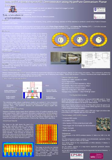
Title: References
1
SmartPET A Small Animal PET Demonstrator using
HyperPure Germanium Planar Detectors R.J.
Cooper(1), A.J. Boston(1), H.C Boston(1), J.R.
Cresswell(1), A.N, Grint(1), A.R. Mather(1), P.J.
Nolan(1),D.P. Scraggs(1), G. Turk(1), C.J.
Hall(2), I. Lazarus(2), A. Berry(3) T.
Beveridge(3), J. Gillam(3), R.A. Lewis(3) (1)
Department of Physics, University of Liverpool,
UK (2) CCLRC Daresbury, Warrington, Cheshire,
UK (3) School of Physics and Materials
Engineering, Monash University, Melbourne,
Australia
Abstract The SmartPET project aims to exploit
advances in the sensitivity, timing, position and
energy resolution of HPGe detectors to construct
a small animal Positron Emission Tomography (PET)
system. The development of sophisticated
digital acquisition techniques and the use of
Pulse Shape Analysis (PSA) 1 and Gamma Ray
Tracking (GRT) 1,2 will allow accurate position
and energy information to be extracted, allowing
scattered interactions to be identified and used
for image reconstruction.
Motivation In conventional PET systems 85 of
photons incident on the detectors will undergo
Compton scattering and will be rejected on the
basis of energy. The rejection of these events
leads to highly inefficient data collection.
This project aims to tackle the deficiencies in
current PET systems by utilising the excellent
energy resolution and position sensitivity
offered by highly segmented germanium detectors
to use a greater proportion of events (70).
This will provide increased patient throughput
and/or reduced patient dose while achieving
improved spatial resolution. The SmartPET system
will provide highly efficient dual modality
PET/SPECT imaging with fine spatial resolution
and the potential for PET/MRI fusion.
The SmartPET Detectors The SmartPET system is
based on two 60mmx60mmx20mm HyperPure germanium
(HPGe) crystals electrically segmented with
orthogonal 5mm strips providing a raw position
resolution of 5mmx5mmx20mm. The use of Pulse
Shape Analysis (PSA) 1 will improve this
allowing interaction positions to be defined to
an accuracy of 1mm3 1. This precise position
and energy information will then form the input
of Gamma Ray Tracking (GRT) 2 algorithms to
accurately track the path of gamma rays through
the detector thus facilitating the inclusion of
scattered events in the reconstruction data set.
Detector Characterisation Detector
characterisation at Liverpool aims to calibrate
the experimental response of an entire germanium
detector for both energy and position. This is
achieved by performing precision scans of the
detector using a range of sources to generate a
set of reference pulse shapes. Electric field
simulation 3 allows a theoretical pulse shape
database to be produced for comparison with
experimental data and online PSA.
- Detector Specification
- Depletion at -1300V, Operation at -1800V
- 12 x12 Segmentation
- AC contacts 0.3 mm thick separated by 180mm
- DC contacts 50mm thick separated by 300mm
- 1mm thick Aluminium entrance window
- Warm FET configuration, 300mV/MeV pre-amps
- Average energy resolution 1.5keV FWHM _at_ 122keV
The Liverpool Scanning System
The Digital DAQ System
All 24 Pre-amp outputs are fed into six,
4-channel GRT4 VME cards 5. These signals are
digitised over a dynamic range of 1V using
14-bit, 80MHz Flash ADCs allowing pulse shapes to
be stored for offline analysis. Each GRT4 channel
contains an array of FPGAs which derive the gamma
ray energy using a Moving Window Deconvolution
(MWD) algorithm 5 and provide a 48 bit
timestamp for event correlation. The Linux PC
controlled DAQ system uses a NIM logic trigger
and allows data to be written to DLT magnetic
tape and RAID disk array.
The scanning system allows a germanium crystal to
be scanned in 1mm increments using a range of
collimated gamma ray sources resulting in the
generation of a detector response matrix. The
smartPET detectors are characterised using a
digital data acquisition system 4.
Pulse Shape Analysis (PSA) Analysis of digitised
charge pulses allows calibration of the 3-D
position sensitivity of the entire crystal
volume. Depth of interaction information is
obtained from rise-time analysis 2 while the
x-y interaction position is ascertained using
image charge analysis 2. The kinematics of
Compton Scattering can then be used to
reconstruct interaction sites on an event by
event basis by the implementation of Gamma Ray
Tracking (GRT) algorithms.
- The Daresbury GRT4 VME Module
- 4 channel VME module
- 14 bit 80MHz FADC
- Xilinx Spartan 2 FPGAs containing circular
buffer - Traces tagged with 16 bit header and 48 bit
timestamp - Trigger in/out gate
- MIDAS user interface
Hall03 A Gamma Ray Tracking Detector for
Nuclear Medicine, C.J. Hall et al.
References 1 Performance of the GRETA prototype
detector, K. Vetter et al, NIM A Vol. 452
(2000) 2 Three-dimensional position sensitivity
in two-dimensionally segmented HP-Ge detectors,
K. Vetter et al, NIM A Vol. 452 (2000) 3 A
simple method for the characterisation of HPGe
detectors, P. Madina, C. Santos, D.
Villaume 4 The position response of a
large-volume segmented germanium detector,
M. Descovich et al, NIM A 5 The GRT4 VME
Pulse Processing Card for Segmented Germanium
Detectors, I. Lazarus et al
Image Charge Analysis
Rise Time Analysis
References Hall03 A Gamma Ray Tracking Detector
for Nuclear Medicine, C.J.Hall et al.
Recommended
