
The Microscope PowerPoint PPT Presentation
1 / 16
Title: The Microscope
1
The Microscope
2
The First Light Microscope
- ANTON VAN LEEUWENHOEK was the first person to
have a major contribution in the development of
the microscope. - He ground and then polished a ball of glass into
a lens that had a magnification of about 270X!! - He attached the lens to a metal holder and
focused on specimens using small metal screws.
3
SIMPLE MICROSCOPE
- Because his microscope had only one lens, it is
referred to as a SIMPLE MICROSCOPE. - With his simple microscope, Leeuwenhoek made many
discoveries about things we cannot see including
bacteria, human blood cells, and sperm!!
4
COMPOUND MICROSCOPE
- The COMPOUND MICROSCOPE was eventually invented
in order to get higher and higher magnifications.
- It uses a combination of two lenses and the total
magnification is the product of the two lenses. - Example Ocular (10X) x Objective (40X) 400X
Total Magnification
5
COMPOUND MICROSCOPE
6
ROBERT HOOKE
- He was the first scientist to use the word
CELLS after viewing a thin slice of cork under
a compound microscope. He thought they looked
like tiny jail cells. - The cells he was looking at were really dead and
all that remained were the rigid cell walls that
were composed of CELLULOSE.
7
ROBERT HOOKES MICROSCOPE
8
MAGNIFICATION RESOLUTION
- MAGNIFICATION on a microscope is what makes the
specimens appear to be larger. - RESOLUTION on a microscope gives us the ability
to distinguish fine details in a specimen.
9
Images Produced by Light Microscopes
Amoeba
Streptococcus bacteria
Anthrax bacteria
Plant cells
Human cheek cells
Yeast cells
10
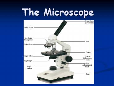
PARTS FUNCTIONS OF THE MICROSCOPE
- See Double-Sided Handout.
11
Beyond Light Microscopes
- Light microscopes are limited by their
resolution. - Light microscopes cannot produce clear images of
objects smaller than 0.2 micrometers - The electron microscope was invented in the
1930s by Max Knott and Ernst Ruska - Electron microscopes use beams of electrons,
rather than light, to produce images - Electron microscopes can view objects as small as
the diameter of an atom
12
(No Transcript)
13
Types of Electron Microscopes
- Transmission electron microscopes (TEMs) pass a
beam of electron through a thin specimen - Scanning electron microscopes (SEMs) scan a beam
of electrons over the surface of a specimen - Specimens from electron microscopy must be
preserved and dehydrated, so living cells cannot
be viewed
14
Images Produced by Electron Microscopes
Cyanobacteria (TEM)
Lactobacillus (SEM)
Campylobacter (SEM)
Deinococcus (SEM)
Avian influenza virus
House ant
Yeast
Human eyelash
15
Using Microscopes to Visualize the Three Shapes
of Bacteria
- Cocci (round)
- Bacilli (rod)
- Spirilla (spiral)
- Light microscope
Three shapes of bacteria taken with an SEM
Spirilla
Bacilli
Cocci
16
References
- http//education.denniskunkel.com/catalog/product_
info.php?products_id1123 - http//micro.magnet.fsu.edu/
- http//inventors.about.com/library/inventors/blrob
erthooke.htm
Recommended
