
Chapter 15 The Cardiovascular System: Blood Vessels PowerPoint PPT Presentation
Title: Chapter 15 The Cardiovascular System: Blood Vessels
1
Chapter 15The Cardiovascular System Blood
Vessels
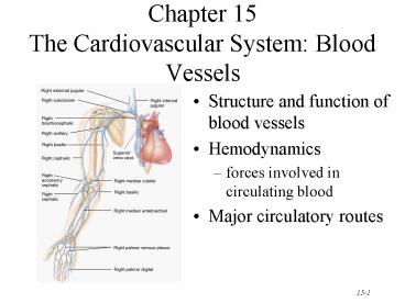
- Structure and function of blood vessels
- Hemodynamics
- forces involved in circulating blood
- Major circulatory routes
2
Anatomy of Blood Vessels
- Closed system of tubes that carries blood
- Arteries carry blood from heart to tissues
- elastic arteries
- muscular arteries
- arterioles
- Capillaries are thin enough to allow exchange
- Venules merge to form veins that bring blood back
to the heart - Veins carry blood back to the heart ( to the
right atrium)
3
Arteries
- Tunica interna (intima)
- simple squamous epithelium known as endothelium
- basement membrane
- internal elastic lamina
- Tunica media
- circular smooth muscle elastic fibers
- Tunica externa
- elastic collagen fibers
4
Sympathetic Innervation
- Vascular smooth muscle is innervated by
sympathetic nervous system - increase in stimulation causes muscle contraction
or vasoconstriction - decreases diameter of vessel
- injury to artery or arteriole causes muscle
contraction reducing blood loss (vasospasm) - decrease in stimulation or presence of certain
chemicals causes vasodilation - increases diameter of vessel
- nitric oxide, K, H and lactic acid cause
vasodilation
5
Elastic Arteries
- Largest-diameter arteries have lot of elastic
fibers in tunica media - Help propel blood onward despite ventricular
relaxation (stretch and recoil -- pressure
reservoir)
6
Muscular Arteries
- Medium-sized arteries with more muscle than
elastic fibers in tunica media - Capable of greater vasoconstriction and
vasodilation to adjust rate of flow - walls are relatively thick
- called distributing arteries because they direct
blood flow
7
Arterioles
- Small arteries delivering blood to capillaries
- tunica media containing few layers of muscle
- Metarterioles form branches into capillary bed
- to bypass capillary bed, precapillary sphincters
close blood flows out of bed in thoroughfare
channel - vasomotion is intermittent contraction
relaxation of sphincters that allow filling of
capillary bed 5-10 times/minute
8
Capillaries form Microcirculation
- Microscopic vessels that connect arterioles to
venules - Found near every cell in the body but more
extensive in highly active tissue (muscles,
liver, kidneys brain) - entire capillary bed fills with blood when tissue
is active - lacking in epithelia, cornea and lens of eye
cartilage - Function is exchange of nutrients wastes
between blood and tissue fluid - Structure is single layer of simple squamous
epithelium and its basement membrane
9
Types of Capillaries
- Continuous capillaries
- intercellular clefts are gaps between neighboring
cells - skeletal smooth, connective tissue and lungs
- Fenestrated capillaries
- plasma membranes have many holes
- kidneys, small intestine, choroid plexuses,
ciliary process endocrine glands - Sinusoids
- very large fenestrations
- incomplete basement membrane
- liver, bone marrow, spleen, anterior pituitary,
parathyroid gland
10
Venules
- Small veins collecting blood from capillaries
- Tunica media contains only a few smooth muscle
cells scattered fibroblasts - very porous endothelium allows for escape of many
phagocytic white blood cells - Venules that approach size of veins more closely
resemble structure of vein
11
Veins
- Proportionally thinner walls than same diameter
artery - tunica media less muscle
- lack external internalelastic lamina
- Still adaptable to variationsin volume
pressure - Valves are thin folds of tunica interna designed
to prevent backflow - Venous sinus has no muscle at all
- coronary sinus or dural venous sinuses
12
Varicose Veins
- Twisted, dilated superficial veins
- caused by leaky venous valves
- congenital or mechanically stressed from
prolonged standing or pregnancy - allow backflow and pooling of blood
- extra pressure forces fluids into surrounding
tissues - nearby tissue is inflamed and tender
- Deeper veins not susceptible because of support
of surrounding muscles
13
Anastomoses
- Union of 2 or more arteries supplying the same
body region - blockage of only one pathway has no effect
- circle of willis underneath brain
- coronary circulation of heart
- can occur in veins and venules as well
14
Blood Distribution
- 60 of blood volume at rest is in systemic veins
and venules - function as blood reservoir
- veins of skin abdominalorgans
- blood is diverted from it intimes of need
- increased muscular activityproduces
venoconstriction - hemorrhage causes venoconstriction to help
maintain blood pressure - 15 of blood volume in arteries arterioles
15
Systemic Circulation
- All systemic arteries branch from the aorta
- All systemic veins drain into the superior or
inferior vena cava or coronary sinus to return to
the right-side of heart
16
Arterial Branches of Systemic Circulation
- All are branches from aorta supplying arms, head,
lower limbs and all viscera with O2 from the
lungs - Aorta arises from left ventricle (thickest
chamber) - 4 major divisions of aorta
- ascending aorta
- arch of aorta
- thoracic aorta
- abdominal aorta
17
Aorta and Its Superior Branches
- Aorta is largest artery of the body
- ascending aorta
- 2 coronary arteries supply myocardium
- arch of aorta -- branches to the arms head
- brachiocephalic trunk branches into right common
carotid and right subclavian - left subclavian left carotid arise
independently - thoracic aorta supplies branches to pericardium,
esophagus, bronchi, diaphragm, intercostal
chest muscles, mammary gland, skin, vertebrae and
spinal cord
18
(No Transcript)
19
(No Transcript)
20
Coronary Circulation
- Right left coronary arteries branch to supply
heart muscle - anterior posterior interventricular aa.
21
Subclavian Branches
- Subclavian aa. pass superior to the 1st rib
- gives rise to vertebral a. that supplies blood to
the Circle of Willis on the base of the brain - Become the axillary artery in the armpit
- Become the brachial in the arm
- Divide into radial and ulnar branches in the
forearm
22
Common Carotid Branches
Circle of Willis
- External carotid arteries
- supplies structures external to skull as branches
of maxillary and superficial temporal branches - Internal carotid arteries (contribute to Circle
of Willis) - supply eyeballs and parts of brain
23
(No Transcript)
24
(No Transcript)
25
Abdominal Aorta and Its Branches
- Supplies abdominal pelvic viscera lower
extremities - celiac aa. supplies liver, stomach, spleen
pancreas - superior inferior mesenteric aa. supply
intestines - renal aa supply kidneys
- gonadal aa. supply ovariesand testes
- Splits into common iliacaa at 4th lumbar
vertebrae - external iliac aa supplylower extremity
- internal iliac aa supplypelvic viscera
26
(No Transcript)
27
Visceral Branches off Abdominal Aorta
- Celiac artery is first branch inferior to
diaphragm - left gastric artery, splenic artery, common
hepatic artery - Superior mesenteric artery lies in mesentery
- pancreaticoduodenal, jejunal, ileocolic,
ascending middle colic aa. - Inferior mesenteric artery
- descending colon, sigmoid colon rectal aa
28
(No Transcript)
29
Arteries of the Lower Extremity
- External iliac artery become femoral artery when
it passes under the inguinal ligament into the
thigh - femoral artery becomes popliteal artery behind
the knee
30
(No Transcript)
31
Veins of the Systemic Circulation
- Drain blood from entire body return it to right
side of heart - Deep veins parallel the arteries in the region
- Superficial veins are found just beneath the skin
- All venous blood drains to either superior or
inferior vena cava or coronary sinus
32
(No Transcript)
33
Major Systemic Veins
- All empty into the right atrium of the heart
- superior vena cava drains the head and upper
extremities - inferior vena cava drains the abdomen, pelvis
lower limbs - coronary sinus is large vein draining the heart
muscle back into the heart
34
Veins of the Head and Neck
- External and Internal jugular veins drain the
head and neck into the superior vena cava - Dural venous sinuses empty into internal jugular
vein
35
(No Transcript)
36
(No Transcript)
37
Venipuncture
- Venipuncture is normally performed at cubital
fossa, dorsum of the hand or great saphenous vein
in infants
38
(No Transcript)
39
(No Transcript)
40
(No Transcript)
41
(No Transcript)
42
(No Transcript)
43
(No Transcript)
44
(No Transcript)
45
Hepatic Portal System
- Subdivision of systemic circulation
- Detours venous blood from GI tract to liver on
its way to the heart - liver stores or modifies nutrients
- Formed by union of splenic, superior mesenteric
hepatic veins
46
Arterial Supply and Venous Drainage of Liver
47
Pulmonary Circulation
- Carries deoxygenated blood from right ventricle
to air sacs in the lungs and returns it to the
left atria - Vessels include pulmonary trunk, arteries and
veins - Differences from systemic circulation
- pulmonary aa. are larger, thinner with less
elastic tissue - resistance to is low pulmonary blood pressure
is reduced
48
Fetal Circulation
- Oxygen from placenta reaches heart via fetal
veins in umbilical cord. - bypasses liver
- Heart pumps oxygenated blood to capillaries in
all fetal tissues including lungs. - Umbilical aa. Branch off iliac aa. to return
blood to placenta.
49
Lung Bypasses in Fetal Circulation
Recommended
