
48x48 poster template - PowerPoint PPT Presentation
1 / 1
Title:
48x48 poster template
Description:
Department of Radiation Oncology, Emory University, Atlanta, Georgia, USA. Our method involves using planning computed tomography (PCT) images acquired via ... – PowerPoint PPT presentation
Number of Views:68
Avg rating:3.0/5.0
Title: 48x48 poster template
1
Quantitative evaluation of a cone beam computed
tomography (CBCT)-CT deformable image
registration method for adaptive radiation
therapy Joshua D. Lawson MD, Eduard Schreibmann
PhD, Ashesh B. Jani MD, and Tim Fox
PhD Department of Radiation Oncology, Emory
University, Atlanta, Georgia, USA
INTRODUCTION
Deformable or non-rigid registration is an
essential tool in both Adaptive Radiation Therapy
(ART) and Image Guided Radiation Therapy (IGRT)
to account for soft tissue changes during the
course of radiation therapy. The most common
evaluation method used to assess the accuracy of
deformable image registration is qualitative
human evaluation. We propose a methodology to
systematically measure the accuracy of an
algorithm in recovering artificially introduced
deformations in cases of rigid geometry, and use
this method to quantify the ability of a modified
basis spline (B-Spline) registration algorithm to
recover artificially introduced deformations. The
evaluation method is entirely computer-driven and
eliminates biased interpretation associated with
human evaluation it can be applied to any method
of image registration chosen.
MATERIALS METHODS
Our method involves using planning computed
tomography (PCT) images acquired via conventional
CT simulator as well as cone-beam CT (CBCT)
acquired daily via LINAC-mounted kilovoltage
image system in the treatment delivery room. The
deformation occurring between the PCT and daily
CBCT images was obtained using a modified version
of the B-Spline deformable model designed to
overcome the low soft tissue contrast and
artifacts/distortions observed in the CBCT
images. Clinical CBCT images and contours of
phantom and central nervous system (CNS) cases
were deformed (or warped) with known random
deformation. In registering the deformed with the
non-deformed image sets, we tracked the
algorithms ability to recover the original,
non-deformed set. Registration error was
measured as the mean and maximum difference
between the original and the registered surface
contours from outlined structures. Using this
approach, two sets of tests can be devised. To
measure the residual error related to the
optimizers convergence performance, the warped
CT image is registered to the unwarped version of
itself, eliminating unknown factors such as noise
and positioning errors. To study additional
errors introduced by artifacts and noise in the
CBCT, the warped CBCT is registered to the
original CT.
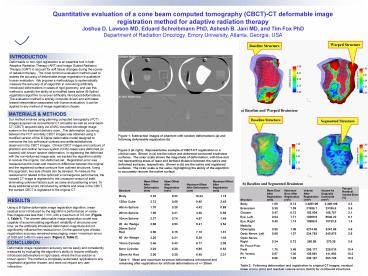
Figure 1 Subtraction images of phantom with
random deformations (a) and following deformable
registration (b) Figure 2 (at right)
Representative example of CBCT-CT registration in
a clinical case. Shown in (a) are the native and
deformed contoured brainstem surfaces. The color
scale shows the magnitude of deformation, with
blue and red representing areas of least and
farthest distance between the native and deformed
surfaces, respectively. Shown in (b) are the
native and registered surfaces. The color scale
is the same, highlighting the ability of the
algorithm to accurately recover the native
surface.
RESULTS
Using a B-Spline deformable image registration
algorithm, mean residual error introduced by the
algorithms performance on noise-free images was
less than 1 mm, with a maximum of 3.5 mm (Figure
1, Table 1). The chosen deformable image
registration model was capable of accommodating
significant variability of structures over time,
as the artificially introduced deformation
magnitude did not significantly influence the
residual error. On the second type of tests,
registration accuracy remained encouraging mean
/ maximum errors of 0.69 and 3.48 mm were seen
(Figure 2, Table 2).
CONCLUSION
Deformable image registration accuracy can be
easily and consistently measured by evaluating
the algorithms ability to recover artificially
introduced deformations in rigid cases, where the
true solution is known apriori. The method is
completely automated, applicable to any
registration algorithm chosen, and does not
require any user interaction.
Table 1 Mean and maximum structure
deformations introduced and remaining after
registration for artificial deformations of /-
20mm
Table 2 Following deformation and registration
to original CT images, residual linear errors
(mm) and residual volume errors (mm3) for
contoured structures
Recommended
CrystalGraphics Presentations


