MultiDimensional SolidState NMR of Proteins: Concepts - PowerPoint PPT Presentation
1 / 84
Title: MultiDimensional SolidState NMR of Proteins: Concepts
1
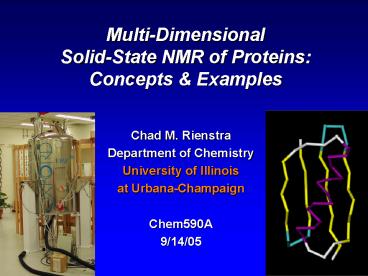
Multi-Dimensional Solid-State NMR of Proteins
Concepts Examples
- Chad M. Rienstra
- Department of Chemistry
- University of Illinois
- at Urbana-Champaign
- Chem590A
- 9/14/05
2
Outline
- Concepts
- Protein structure determination (Solution NMR)
- Chemical shift assignments
- Secondary structure determination
- Tertiary structure
- Solid-State NMR
- What is a solid for purposes of NMR?
- Resolution
- Sensitivity
- Applications Overview
3
Structure Determination by NMR
- Choose protein(s)
- Make protein
- synthesis
- bacterial expression
- Label 15N and 13C
- Optimize sample conditions
- Acquire and interpret spectra
- (1) chemical shift
- (2) torsion angles
- (3) distances
- Compute structure
4
Structures (Wüthrich)
5
The Multidimensional Paradigm
- Jeener, Ampere Summer School (1971).
- Ernst et al. (1974).(Nobel Prize 1991)
- Wüthrich (Nobel Prize 2002)
- Turn Hamiltonian termson and off as desired
- Resolve sites withchemical shifts
- Measure structural parameters in additional
dimension(s)
6
NMR for Chemistry
- H. Gutowsky, UIUC
- Applications of NMR to chemistry, biology, and
medicine derived from work - Based on two advances
- High resolution (site specificity)
- Chemical insight from observed frequencies
7
High Resolution 13C NMR
Silverstein, Bassler, and Morrill, Spectrometric
Identification of Organic Compounds, Fig 5.2
8
The Chemical Shift
- Not called NMR emission frequencies
- More usefulthan that!
- Electronicstructure
Silverstein, Bassler, and Morrill, Spectrometric
Identification of Organic Compounds, Appendix B
9
Chemical Shifts in Proteins
- FromWüthrichlecture
10
Unique Chemical Shifts in Proteins
- 1H and 13C shifts depend on
- 1. amino acid residue type
- 2. residue type of neighbors
- 3. conformation
- 4. hydrogen bonding
11
Chemical Shift Trends
- Color coded by carbon type
- Each residue hasdistinctive pattern
12
Residue Type ID
- Always clearG, A, S, T, P, I
- Usually clearL, V, D/N
- Aromaticsuse Cb-Cg todistinguish
- AmbiguousC, M, K, R, E/Q
13
Sample Preparation
- Unfolded protein (top)
- Folded protein (bottom)
- Chemical shift dispersion
14
High Resolution Solution NMR
- Basic ideas
- (1) site resolution
- (2) detailed chemical information at each site
- chemical shifts
- couplings
- relaxation rates
- dynamics
- function
- Gardner, Zhang, Gehring, and Kay J. Am. Chem.
Soc. 12011738 (1998) Maltose binding protein,
370 residues, 42 kDa complex with b-cyclodextrin
15
Assignments Backbone Walk
Solid N-CO-CX CO-N-CA/CB N-CA-CX
Solution H-N-CO H-N-(CO)-CA H-N-CA H-N-CA/CO
H-N-CA/CB
- Baldus, M., et al. Mol. Phys. 95, 1197- 1207
(1998). - Takegoshi, K. et al. Chem. Phys. Lett.
344, 631-637 (2001).
16
Chemical Shift Assignments
- 1. Identify amino acid type from chemical shifts
- 2. Connect to i-1 and i1 neighbors (also
identified by amino acid type) - 3. Iterate for entire protein until
self-consistent solution is found
17
Local NOEs Secondary Structure
- Helical region i to i3 contacts
18
Long Range Distances
- Constrain fold
- NOE nuclear Overhausereffect (1/r6)
19
Distance Geometry (Geography) Mapping
Molecular Structure
20
What is Solid-State NMR?
- What is a solid?
Easier to ask what isnt a solid?
21
SSNMR A Functional Definition
- For purposes of NMR, a solid is any material that
does not tumble faster than the NMR time scale
(ms to s)
ubiquitin tc 100 ps
lipid bilayer tc 1 s
micelle tc 10-100 ns
22
Dipolar Averaging
- Solution State
- rapid tumbling tc lt 10-20 ns
- molecular weight limit 50-100 kDa
- Solid State
- global motion frozen
- three approaches
- static powder
- macroscopically oriented
- magic-angle spinning
23
Types of Solids for NMR
- Chemical context
- Precipitated globular proteins
- Peptide aggregates
- Enzyme-substrate complexes
- Amyloid or synuclein fibrils
- Membrane proteins
- Trapped intermediates
- Physical appearance
- Powdered crystals (dry)
- Microcrystals (wet)
- Goopy precipitates
- Viscous solutions (slowly frozen)
- Membranes jelly or jello-like
?-amyloid fibrils, Tycko et al., Current Opinion
in Chemical Biology (2000).
24
Membrane Proteins gt30 of Sequenced Genomes
- PDB 26,811 structures (8/17/04)www.pdb.org
- 143 membrane protein structures (82
unique)blanco.biomol.uci.edu/Membrane_Proteins_x
tal.html
22,886 by X-ray 3,925 by NMR
- GPCRs
- Drug design
- Bioenergetics
- Cell recognition
- Fundamental biophysics
new folds
25
Next several slides are courtesy Stan Opella,
UCSD
26
1H - 1H spins in water in gypsum. 1H are dilute
by space.
27
1H - 15N spins in peptide plane. 15N is dilute
by space. 1H is abundant in large bath.
28
Structure determination of membrane proteins
Indirect mapping through "residual" effects.
Direct mapping through "static" effects.
15N Shift (ppm)
1H-15N Cplg (kHz)
15N Shift (ppm)
residue number
1H Shift (ppm)
29
Bicelle samples for structure determination of
membrane proteins.
- Native membrane environment.
- Phospholipids.
- Bilayers.
- Planar.
- Fully hydrated.
- Liquid crystalline phase.
- 37 oC
- Advantages over bilayers on glass plates.
- Sealed liquid samples.
- Solenoid RF coil
- No loss in filling factor.
30
15N NMR spectra of aligned MerFt (2TM, 60
residues).
Bilayers on plates
Flipped bicelles
Unflipped bicelles
Tyr (3 sites)
31
PISEMA spectra of membrane proteins in bicelles.
Pf1 coat Vpu TM domain
MerFt PISA Wheels 1TM, 46
residues 1TM, 36 residues 2TM,
60 residues
32
Hydrophobic mismatchLength of helix vs.
thickness of hydrophobic center of bilayers.
White and Wimley, Annu. Rev. Biophys. Biomol.
Struct. 28, 319 (1999)
33
Tilt angle compensates for hydrophobic
mismatch.No change in rotation angle.
T
34
Spectra of membrane proteins in bicelles have
little or no isotropic resonance intensity from
motionally-averaged sites.
35
Effect of flipping on MerFt spectra.
-NH3
36
Structures of membrane proteins determined by
solid-state NMR of aligned samples.
37
Assigned spectrum and preliminary
three-dimensional structure of MerFt in bicelles.
38
Magic-Angle Spinning Methods
- 80 of proteins cant be studied by X-ray or
solution NMR
membrane proteins (e.g., G-protein coupled
receptors)
microcrystalline globular proteins
peptide aggregates and fibrils
all of these proteins can be examined at high
resolution by employing 2D/3D magic-angle
spinning methods
39
Progress with Small Peptides
f-MLF-OH 2002
12 Structures RMSD 0.02 Å (backbone)
Rienstra, C. M. et al. Proc Natl Acad Sci USA 99,
10260-5 (2002).
Lansbury, P. T. et al. Nature Structural Biology
2, 990-998 (1995).
40
Amyloid Peptide Structures
- b-Amyloid
- Alzheimers disease
- Tycko (NIH)
- Griffin (MIT)
- Botto (Argonne)
- Transthyretin (TTR)
- senile dementia
- Jaroniec Griffin (MIT)
Petkova, A. T. et al. J. Mol. Biol. 335, 247-260
(2004). Jaroniec, C. P. et al. Proc. Natl. Acad.
Sci. USA 101, 711-716 (2004).
41
GB1
- 56 residues
- known structure
- rehearsal molecule
a-Synuclein
- 140 residues
- no known structure
- Parkinsons disease
42
Typical Experiment Late 1990s
- 13C-Asp retinal labeled in bacteriorhodopsin
- Slow, labor-intensive, myopic
43
Distance Geometry (Geography) Mapping
Molecular Structure
44
Ultra-High Resolution SSNMR
- 2,000 resolved peaks in one 2D spectrum
45
Outline
- Concepts
- Protein Structure Determination
- Solid-State NMR
- Application 1
- GB1
- Known protein structure
- Develop and demonstrate methods
- Application 2
- Synuclein
- Parkinsons disease
- Impossible to study by solution NMR or X-ray
46
Model System GB1
- Expresses well (100 mg/L)
- Easy purification (heat, one column)
- High quality crystal (1pga) and NMR (1gb1)
structures available - Thermostable (Tm 85 oC)
1 MET GLN TYR LYS LEU ILE LEU ASN GLY LYS 11
THR LEU LYS GLY GLU THR THR THR GLU ALA 21 VAL
ASP ALA ALA THR ALA GLU LYS VAL PHE 31 LYS GLN
TYR ALA ASN ASP ASN GLY VAL ASP 41 GLY GLU TRP
THR TYR ASP ASP ALA THR LYS 51 THR PHE THR VAL
THR GLU
47
Crystal Growth Rate v. Quality
Weeks to months
Seconds to minutes
Martin, R. W. Zilm, K. W. J. Magn. Reson. 165,
162-174 (2003).
Hours to days
48
GB1 Sample Preparation
- Start with (concentrated) solution conditions
- 5 mM (30 mg/mL)
- pH 5.5 phosphate buffer, 50 mM
- Add precipitant (2-methylpentane-2,4-diol
isopropanol, 21) - Nanocrystals grow in seconds to minutes
- Pellet into NMR rotor
49
Kinetics of Crystal Growth
- Single crystal
- (0.1 mm)3 cube -gt 1 x 10⁻⁴ m on each side
- 1 month -gt 2.6 x 10⁶ s
- 4 x 10-11 m / s (presuming linear, constant)
- In reality, crystal growth slows as
concentration of solution decreases - Nanocrystal (in this example)
- Growth time ltlt15 minutes 103 s
- Linear growth gives major dimension 4 x 10⁻⁸ m
- 400 Å on each side (10 times protein size)
- Lower bound (kinetics are in fact not zeroth
order)
50
Assay with 1D 15N Spectra
- 30/g 15NH4Cl
- 1 mmol 15 min 200 nmol 3-4 hrs.
51
GB1 13C 1D Spectrum (5 min.)
- 13C,15N
- 12 mg
- 128 scans
- No LB
- 11 kHzMAS
- 70 kHzTPPM
52
The NMR Hamiltonian Magnitude of Interactions
- Technical challenges
- Peak rf fields corona discharge (high voltage
breakdown) of air gap in coil - Peak MAS rates speed of sound atsurface of
rotor, material hardness
53
Magic-Angle Spinning
maximum structural information
maximum resolution and sensitivity
54
Resolution Technical Challenges
- Static CP spectrum of 10 mg protein (GB1)
- 128 scans (4 minutes)
- Scale adjusted (10x) relative to subsequent
55
Magic-Angle Spinning (1960s)
- 11 kHz MAS
- No 1H decoupling
- 128 scans
- Andrew, E. R. Bradbury, A. Eades, R. G.,
"Nuclear magnetic resonance spectra from a
crystal rotated at high speed", Nature (London)
1958, 182, 1659. - Lowe, I. J., "Free induction decays of rotating
solids", Phys. Rev. Lett. 1959, 2, 285-287.
56
CW Decoupling (1975)
- 75 kHz 1H decoupling field (CW)
- 11 kHz MAS
- Same 4 minute acquisition time
- Schaefer, J. Stejskal, E. O., "13C-NMR of
Polymers Spinning at the Magic Angle", J. Am.
Chem. Soc. 1976, 98, 1031.
57
TPPM Decoupling (1995)
- 75 kHz 1H field with phase modulation
- 11 kHz MAS
- 4 minutes
- Bennett, A. E. Rienstra, C. M. Auger, M.
Lakshmi, K. V. Griffin, R. G., "Heteronuclear
decoupling in rotating solids", J. Chem. Phys.
1995, 103, 6951-6958.
58
Why We Bother The Two Limits
single methyl signal
all aliphatic intensity
59
Dipolar Recoupling
- MAS averages couplings to zero
- Multiple pulse sequence restores dipolar
couplings
60
Recoupling Example 15N-13C Cross Polarization
- Non-zero time-averaged coupling
61
Assignments Backbone Walk
Solid N-CO-CX CO-N-CA/CB N-CA-CX
Solution H-N-CO H-N-(CO)-CA H-N-CA H-N-CA/CO
H-N-CA/CB
- Baldus, M., et al. Mol. Phys. 95, 1197- 1207
(1998). - Takegoshi, K. et al. Chem. Phys. Lett.
344, 631-637 (2001).
62
Residue Type ID
- Always clearG, A, S, T, P, I
- Usually clearL, V, D/N
- Aromaticsuse Cb-Cg todistinguish
- AmbiguousC, M, K, R, E/Q
63
An NMR Crossword Puzzle
15N
Franks et al., J.. Am. Chem. Soc. (submitted)
64
Comparison with Solution NMR
- Excellent agreement with solution
- General trends consistent with secondary
structure - How precise is this approach?
65
Chemical Shifts Solution v. Solid
0.58 0.52 ppm
0.22 1.11 ppm
0.09 0.56 ppm
0.87 2.00 ppm
66
Comparison with X-Ray Diffraction
- TALOS semi-empirical chemical shift analyssis
- 39 f / y pairs converged to good tolerance in
TALOS - 71 / 78 agree within error with crystal
structure
X-Ray Solid-State NMR
f
y
- Cornilescu, G. Delaglio, F. Bax, A., "Protein
backbone angle restraints from searching a
database for chemical shift and sequence
homology", J. Biomol. NMR 1999, 13, 289-302.
67
SPECIFIC CP for NC Distances
- - Baldus, M., et al. Mol. Phys. 95, 1197-1207
(1998).
68
Band Selective 15N to 13C TransferMethyl Only
Recoupling
filtered
- Strong couplingsalong backbone avoided
- Polarization transfer primarily to 13CH3signals
unfiltered
69
Three Comparable Couplings NAV
70
(No Transcript)
71
All Distances Within 6.5 Å
Leu5 N (6.2 Å)
Leu7 N (5.9 Å)
1pga reference structure (X-ray)
Ile6 N (4.3 Å)
Ile6 Cd1
Phe52 N (6.4 Å)
Thr53 N (6.4 Å)
72
GB1 gt5 Å Distances
- Distanceto Ile6 Cd1
60 ms
20 ms
40 ms
60 ms (w/ LB)
- Thr53 N(6.4 Å)
- Ile6 N(4.3 Å)
- Leu5/7 N(6.2/5.9 Å)
- Phe52 N(6.4 Å)
73
REDOR
- 19F-REDOR Shift NMR Spectroscopy
- 1H-19F-13C-15N 3.2 mm MAS probe
- 11.1 kHz, 0 or 512 p pulses on 19F
74
FRESH 15N-13C 2D DS DataDepends on 19F-15N
Distance
- K31
- T53
- V54
- I6
- E27, K28, V29
- L5, Q2, T17
- A23, A24
75
19F-15N Distances in 1pga
53
54
6
5
F
31
27
76
Tying Together Domains
- Connects b3 with a-helix b1 b4
- Potential for 13C 1. Higher g 2. Located
on side chains - (esp. CH3)
F
77
(No Transcript)
78
a-Synuclein
- Associated with neural plasticity
- Abundant and highly conserved
- Relatively small (140 residues)
- Natively unfolded
- Known to form fibrils
- Implicated in neurodegenerative disease
(Parkinsons, Alzheimers, etc.) - Collaboration with Julia George,Dept. of
Molecular and Integrative Physiology
79
a-Synuclein and Neuropathology
- Synuclein aggregates (Lewy bodies) are the
pathological hallmark of Parkinsons disease - Mutations in a-synuclein can cause familial,
early onset Parkinsons disease - A53T
- A30P
- extra copies of gene
- a-synuclein inclusions are also observed in
- Alzheimers
- Dementia with Lewy bodies
- Downs syndrome
- Multiple system atrophy
Spillantini et al (1997) Nature 388839
80
Neurodegenerative Diseases with Intracellular
Filamentous Lesions
81
a-Synuclein Hydrophobicity Plot
- Alternating domains of hydrophobicity and
hydrophilicity - Periodicity of 11
- Detects an underlying 11-mer repeat
- Suggests amphipathic helix
repeats
acidic tail
NH3
COO-
82
a-Synuclein Fibrils
- Empirical diagnostic
- Dependent on dye affinity
- No known relationships to atomic-resolution
structure
Conway, K. A., Harper, J. D. Lansbury, P. T.
Fibrils formed in vitro from alpha-synuclein and
two mutant forms linked to Parkinson's disease
are typical amyloid. Biochem. 39, 2552-2563
(2000).
83
SSNMR Spectra of aS Fibrils
- Incubate at 37 oC,50 mM phosphate,1 month
- Sub-ppm line widths
- CP dynamicsMotion near termini
- Kathy Kloepper
- Donghua Zhou
84
aS Fibrils 13C-13C 2D Spectrum


