
Bacteriology PowerPoint PPT Presentation
1 / 89
Title: Bacteriology
1
Bacteriology
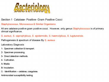
Section 1 Catalase- Positive- Gram Positive
Cocci Staphylococcus, Micrococcus Similar
Organisms All are catalase positive gram positive
cocci. However, only genus Staphylococcus is of
primary clinical significance. S. aureus, S.
saprophyticus, S. epidermidis, S. haemolyticus,
S. lugdunensis. Pathogenesis spectrum of
disease By S. aureus Laboratory Diagnosis 1-
Specimen collection transport. 2- Specimen
processing. 3- Direct detection methods. 4-
Cultivation. A- Media B- Incubation. C-
Identification catalase, coagulase. Antimicrobia
l susceptibility testing.
2
Negative tube slide coagulase
Positive tube slide coagulase
3
(No Transcript)
4
Section 2 Catalase Negative, Gram Positive
Cocci
Streptococcus, Enterococcus, Similar Organisms
Of the organisms considered in this chapter,
those that are most commonly encountered in
infections in humans include Streptococcus
pyogenes, S. agalactiae, S. pneumoniae, Viridans
streptococci, enterococci, usually Enterococcus
faecalis or E. faecum. The others are rarely
encountered or considered as contaminants
mistaken for viridans streptococci or
enterococci.
5
Morphology and Identification Individual cocci
are spherical or ovoid arranged in chains and
diploes, non-motile, some are capsulated,
anaerobes, facultative anaerobes, non-spore
formers. Some require 5-10 CO2 for growth.
Streptoccci are alpha hemolytic, Bata hemolytic
or nonhemolytic. Antigenic structure 1- Group
specific cell wall antigen Carbohydrate present
in the cell-wall of many streptococci and forms
the basis of serological groupings( Lancefield
groups A-H, K-U ). 2- M protein Major
virulence factor of group A Streptococcus
pyogenes. 3- T substance Has no relation to
virulence. 4- Nucleoprotein Called P
substances which probably make up most of the
streptococcal cell body. Toxins and Enzymes A-
Streptokinase(Fibrinolysin). B-
Streptodornase. C- Hyaluronidase. D-
Erythrogenic Toxins. E - Diphosphopyridine
nucleotidase. F- Hemolysins S. pyogenes
elaborates two hemolysins(streptolysins)
Streptolysin O (ASOT) and Streptolysin S(B
hemolysis)
6
Cassisication of Streptococci of Medical
Importance
A- Streptococcus pyogenes( Group A, B Hemolytic
Streptococci). B- Streptococcus agalactiae
These are group B streptococci. C- Groups C and
G. D- Enterococcus fecalis(E. faecum, E durans)
Group D. E- Streptococcus bovis
Nonenterococcal group D streptococci. F-
Streptococcus anginosis. G- Group N
Streptococci. H- Groups E,F,G, H and K-U
Streptococci. I- Streptococcus pneumoniae. J-
Viridans Strepococci (S. mitis, S. mutans, S.
salivaris, S. sanguis). K- Nutritionally Variant
Streptococci. L- Peptostreptococcus (Many
Species). Pathogenesis and Clinical
Findings Infections can be divided into several
categories A- Diseases caused by S.
pyogenes 1- Erysipelas 2- Cellulitis 3-
Necrotizing fasciitis(streptococcal gangrene). 4-
Puerperal fever. 5- Sepsis. 6- Streptococcal
sore throat
7
Beta hemolytic
Alpha haemolytic streptococci
8
7- Streptococcal pyoderma8- Infective
endocarditis acute and subacute.9-
Streptococcal toxic shock syndrome10- Scarlet
fever.Poststreptococcal Diseases1- Rheumatic
Fever2- Acute Glomerulonephritis Laboratory
Diagnosis 1- Specimen collection transport. 2-
Specimen Processing. 3- Direct Detection
Methods Antigen detection, gram stain. 4-
Cultivation A- Media of choice - blood agar. B-
Incubation conditions duration. C- Colonial
appearance. D- Identification. Serodiagnosis
ASOT Antimicrobial Susceptibility Testing
Therapy Similar Organisms Leuconostoc,
Lactococcus, Globiacatella, Pediococcus,
Aerococcus, Gamella,Helcoccus, Alloiococcus.
9
(No Transcript)
10
Streptococci
Impetigo Tonsillitis
11
(No Transcript)
12
(No Transcript)
13
Section 3 Non B ranching, Catalase
Positive, Gram Positive Bacilli
Bacillus Similar Organisms
Bacillus spp. related genera Brevibacillus
Paenibacillus all are aerobic, gram positive,
spore forming rods. Genus Bacillus Spore-
forming gram positive strict aerobic capsulated
bacilli, 1x3-4 u size, arranged in long chains
spores may central, subterminal or terminal,
depending on the species. Most members are
saprophytic prevelent in soil, water, air and
vegetation such B. cereus, and B. subtilius.
Some are insect pathogens. Cause the disease
Anthrax in animals in which the organism is
transmitted through eating vegetations containing
the spores. Human is infected through contact
with animals or their products. Disease in
Human A- Cutaneous Anthrax(malignant pustule)
Generally occurs on exposed surfaces of the arms,
face and neck through wound contamination by the
spores of the organism. About 95 of the cases
with amortality rate 20 . B- Inhalation
Anthrax(wool sorter disease) About 5 of the
cases with 85-90 mortality.
14
C- Gastrointestinal Anthrax Is very
rare. Laboratory Diagnoses Specimen
processingDirect detection methods gram
stain.Cultivation. A- Media blood agar. B-
Incubation conditions duration C- Colonial
appearance. D- Identification.Antimicrobial
susceptibility testing therapy
The anthrax bacillus, Bacillus anthracis, was the
first bacterium shown to be the cause of a
disease. In 1877, Robert Koch grew the organism
in pure culture, demonstrated its ability to form
endospores, and produced experimental anthrax by
injecting it into animals.
15
Bacillus cereus
Bacillus anthracis. Gram stain. The cells have
characteristic squared ends. The endospores are
ellipsoidal shaped and located centrally in the
sporangium. The spores are highly refractile to
light and resistant to staining.
16
Listeria, Corunebacterium, Similar Organisms
All are catalase positive, gram positive rods,
not acid fast, do not branch, do not form
spores.
Corynebacterium diphtheriae
Morphology and Identification Corynebacteria are
0.5-1u in diameter and several micrometers long..
Metachromatic granules(metaphosphate) are
irregularly distributed within the rods giving
them beaded appearance. The rods tend to be
parallel or at acute angles to one another.
Colonies on blood agar are small, granular and
gray and may have small zone of haemolysis. Four
biotypes Gravis, mitis,intermedius and
belfanti. Pathogenesis The disease Diphtheria is
caused by lysogenic C. diphtheriae(toxin
producer). It is a droplet infection in which
the organism pass through the nasopharynx .
PathologyPseudomembrane over the tonsils,
pharynx, larynxDamage by toxins to heart muscle,
liver, kidneys, and adrenals. Also nerve damage
resulting in paralysis of the soft palate, eye
muscles or extermities.Clinical FindingsFever,
sore throat, dyspnea because of the obstruction
caused by the membrane. Later ondifficulties
with vision, soeech, swallowing, or movement of
the arms or legs. Var gravis is more severe.
Symptoms ten to subside spontaneously.
17
Stained Corynebacterium cells. The "barred"
appearance is due to the presence of
polyphosphate inclusions called metachromatic
granules. Note also the characteristic
"Chinese-letter" arrangement of cells.
18
Laboratory diagnosis 1- Specimen collection
transport. 2- Specimen processing. Direct
detection methods. 3- Cultivation. A- Media of
choice potassium tellurite, tinsdale medium. B-
Incubation conditions duration. C- Colonial
appearance. D- Identification.
Listeria monocytogenes
Listeria monocytogenes are facultative anaerobic
non-sporing Gram-positive motile rods that are
catalase positive. They are widespread throughout
nature, having been isolated from the environment
and from many animals. L. monocytogenes grows
under refrigeration temperatures from 1C up to
44C. It grows at pH values of between 4.6 and
9.6 and their minimum water activity value for
growth is 0.90.
L. monocytogenes is easily destroyed by heat and
food poisoning outbreaks are relatively rare.
There are several species in the genus listeria,
L. monocytogenes is important as a cause of a
wide spectrum of disease in animals and
humans. Laboratory diagnosis
19
(No Transcript)
20
Other organisms Kurthia spp. Brevibacterium
spp. Dermabacter hominis. Turicella
otitidis. Arthrobacter spp. Microbacterium
spp. Cellulomonas spp. Exiguobacterium spp.
Section 4 Non Branching, Catalase Negative,
Gram Positive Bacilli Erysiplothrix
rhusiopathiae, Lactobacillus spp., Gardnella
vaginalis, Arcanobacterium spp. All are
catalase negative, non-spore-forming,
gram-positive rods. Some may exhibit rudimentary
branching. Laboratory diagnosis
21
Section 5 Branching or Partially Acid-Fast,
Gram-Positive Bacteria
Nocardia, Streptomyces, Rhodococcus, Orskovia,
Similar Organisms
The actinomycetes are a large diverse group of
gram positive bacilli. Their cells elongate to
form branching, filamentous forms. Some
organisms form filaments, or hyphae, on the agar
surface or into the agar, whereas others extend
into the air. These organisms either aerobic,
facultative anaerobic, or obligate anaerobic.
Aerobic actinomycetes belong to the order
actinomycetales. There are more than 40 genera,
not all of them are pathogens. Actinomycetes
whose cell walls contain mycolic acid are
therefore partially acid-fast those whose cell
walls do not contain mycolic acid are therefore
non-acid-fast. In general, the aerobic
actinomycetes are not frequently isolated in the
clinical laboratory, but they are causes of
serious human disease. Partially Acid-Fast
Aerobic Actinomycetes Nocardia gram positive
often with a beaded appearance, variably acid
fast, catalase positive, strictly aerobic
form branched filaments as they grow as they
age they gragment into pleomorphic rods or
coccoid elements. Currently 11 validly described
species are included in the genus. N.
asteroides, N. nova, n. brasiliensis. Rhodococcus,
Gordona, Tsukamurella
22
(No Transcript)
23
Non-Acid-Fast Aerobic Actinomycetes
Streptomyces, Actinomadura, Dermatophilus,
Nocardiopsis, Orskovia, Rothia, the
Thermophilic Actinomycetes Gram positive
branching filaments that do not have mycolic acid
present in their cell envelopes are therefore
non-acid- fast. This group of actinomycetes are
heterogeneous are encountered infrequently in
the clinical laboratory. Laboratory
Diagnosis Specimen collection, Transport, and
Processing Direct Detection Methods Cultivation Id
entification Antimicrobial Susceptibility Testing
Therapy
24
Section 6 Gram-Negative Bacilli and Coccobacilli
(MacConkey-Positive, Oxidase- Negative)
Enterobacteriaceae
25
Enteric Gram Negative Rods
Enteric Gram Negative Rods
Family Enterobacteriaceae
Large heterogeneous group of gram negative rods
whose natural habitat is the intestinal tract of
humans and animals. Facultative anaerobes,
oxidase negative, ferment glucose with the
production of acid or acid and gas, reduce
nitrate to nitrite. Most are motile with
peritrichous flagella, some have a polysaccharide
capsule. According to lactose fermentation,
enterobacteriaceae are classified into 1- Rapid
lactose fermenters ferment lactose in 24
hours. Escherichia coli Klebsiella
pneumoniae Enterobacter aerogenes 2- Late
lactose fermenters ferment lactose in 48 hours
or do not ferment lactose. Edwardsella, Serratia,
Citrobacter, Arizona, Providencia, Erwinia. 3-
Non-lactose fermenters Shigella, Salmonella ,
Proteus
26
(No Transcript)
27
Diseases Caused by Enterobacteriaceae Other Than
Salmonella and Shigella A- E. coli 1- Urinary
tract infection. 2- E. coli associated diarrheal
diseases a- EPEC b- ETEC C- EHEC d- EIEC e-
EAEC 3- Sepsis. 4- Meningitis. 5- Otitis
media 6- Wound infection B- Klebsiella-Enterobac
ter-Serratia Proteus-Morganella-Providencia
and Citrobacter Respoiratory tract, urinary
tract, sepsis, otitis media
28
The Shigellae
S. dysenteriae, S. sonnei, S. flaxonari, S.
boydii. Bacillary dysentery Morphology and
Growth Characteristics Pathogenesis and
Pathology Toxins Clinical Findings Diagnostic
Laboratory Tests Immunity Treatment Epidemiology,
prevention and control.
The Salmonella
S. typhi, S. paratyphi A and B , S. cholerasuis,
S. typhimurium, S. entertidis. Morphology and
Identification. Classification.
29
Pathogenesis and Clinical Findings S. typhi, S.
paratyphi A and B , S. cholerasuis, S.
typhimurium, S. entertidis are of human origin.
The vast majority are animal pathogens that
constitute the reservoir for human
infection. Three main types of disease in
human A- The Enteric Fevers B- Bacteremia with
Focal Lesions C- Enterocolitis Diagnostic
Laboratory Tests 1- Specimens. 2- Culture. 3-
Identification by biochemical tests and
serology. 4- Agglutination test( Widal
test). Immunity Treatment Epidemiology
30
Some enteric organisms are present as normal
flora in the intestinal tract of human and
animals causing infections in other tissues and
organs. Others are pathogens 1- E. coli
associated with diarrheal disease. 2-
Shigella. 3- Salmonella. Antigenic
Structure Enterobacteriaceae have a complex
antigenic structure. gt 150 different heat stable
somatic O Ag. gt 100 heat labile K (capsular )
Ag. gt 50 H (flagellar ) Ag. Bacteriocines Protein
antibiotics like substance produced by many gram
negative bacteria in which their production is
controlled by a plasmid. They are active against
strains of the same species and closely related
species but not the producer strain. Colicins by
E. coli. Marcescins by Serratia
marcescins. Pyocins by Pseudomonas aeruginosa.
31
(No Transcript)
32
(No Transcript)
33
(No Transcript)
34
(No Transcript)
35
(No Transcript)
36
Acintobacter, Chryseomonas, Flavomonas,
Stenotrophomonas
All are oxidase negative, grow on MacConkey, but
unlike Enterobacteriaceae, which ferment glucose,
these organisms either oxidize glucose or do not
utilize glucose. Acintobacter spp. Widely
distributed in nature including the hospital
environment. May become normal flora of skin
respiratory tract of hospitalized
patient. Stenotrophomonas maltophilia Widely
distributed in nature including the hospital
environment. May become normal flora of skin
respiratory tract of hospitalized patient. CDC
group No-1 Oropharynx of animals. Not part of
human flora. Laboratory Diagnosis Specimen
collection transport Specimen processing Direct
detection methods Cultivation identification
37
Section 7 Gram Negative Bacilli
Coccobacilli(MacConkey-Positive, Oxidase
Positive)
Pseudomonas, Burkholderia, Similar Organisms
Aerobic, straight slender gram negative rods
whose cells range from 1-5 um in width. All
species except B. mallei are motile. Burkholdria
cepacia Present in the environment (soil, water,
plant), survive well in hospital environment, not
part of human flora. B. Pseudomallei Present in
the environment , not part of human flora. B.
Mallei Cause glanders disease in horses, donkeys,
mules. Transmitted to human. Not part of human
flora. Ralstonia pickettii Present in the
environment , not part of human flora.
38
Pseudomonas aeruginosa Present in the environment
, rarely part of human flora. P alcaligenes, P.
pseudoalcaligenes, P. dinitrificans Present in
the environment , not part of human flora.
Rarely encountered in clinical specimen. P.
fluorescens, P. putida, P. stutzeri Present in
the environment , not part of human
flora. Brevundimonas vesicularis Present in the
environment , not part of human flora.
39
Non-Fermenting Gram Negative Bacilli
The Pseudomonas Group Gram negative, motile,
aerobic rods some of which produce water-soluble
pigments. Occur widely in soil, water, plants
and animals. According to rRNA/DNA homology and
common cultural characteristics pseudomonads are
classified into the following groups 1-
Fluorescent group
Pseudomonas aeruginosa
P. fluorescens
P. putida 2- Nonfluorescent group
P.
stutzeri
P. mendocina 3- Burkholderia 4- Comamonas
species Acidovorax species 5-
Brevundimonas species 6- Streptotrophomonas
maltophilia
40
Pseudomonas aeruginosa
A- Morphology Identification B- Culture C-
Growth characteristics Antigenic Structure
Toxins Pathogenesis Clinical Findings Diagnostic
Laboratory Tests A- Specimens B- Smears C-
Culture Treatment Epidemiology
41
(No Transcript)
42
(No Transcript)
43
(No Transcript)
44
Achromobacter, Rhizobium, Ochrobacterium,
Similar Organisms Are environmental inhabitants,
oxidase positive, grow on macConkey agar,
oxidize glucose, rarely found in human
infection. Laboratory Diagnosis Specimen
Collection Transport No special considerations
are required. Specimen Processing No special
considerations are required. Direct
Detection Gram Stain. Cultivation Media Blood
agar, chocolate agar,MacConkey. Incubation at
35c in 5-10 C02 or ambient air for 24
hours. Identification Biochemical reactions,
commercial kits.
45
Chrysobacterium, Sphingobacterium, Similar
Organisms Are environmental inhabitants, oxidase
positive, most grow on MacConkey agar, oxidize
glucose, rarely found in human infection. Most
are yellow pigmented. Laboratory
Diagnosis Specimen Collection Transport No
special considerations are required. Specimen
Processing No special considerations are
required. Direct Detection Gram
Stain. Cultivation Cultivation Media Blood
agar, chocolate agar,MacConkey. Incubation at
35c in 5-10 C02 or ambient air for 24
hours. Identification Biochemical reactions,
commercial kits.
46
Alcaligenes, Bordetella (nonpertussis),
Comomonas, Similar Organisms MacConkey
positive, oxidase positive, nonglucose utilizers.
All not part of human flora. Present in the
environment. Achromobacter xylosoxidans Alcaligen
es faecalis Bordetella bronchiseptica Delftia
acidovorans Oligelia uretheralis Psychrobacter
spp. Roseomonas spp. Shewanella
putrefaciens Specimen Collection Transport No
special considerations are required. Specimen
Processing No special considerations are
required.
47
Direct Detection Gram Stain. Cultivation Cultivati
on Media Blood agar, chocolate agar,MacConkey.
Incubation at 35c in 5-10 C02 or ambient air for
24 hours. Identification Biochemical reactions,
commercial kits.
48
Vibrio, Aeromonas, Plesiomonas shigelloides,
Chromobacterium violaceum All are oxidase
positive, glucose fermenting, gram negative
bacilli that grow on MacConkey agar.
49
The Vibrios The vibrios are among the most common
bacteria in surface waters worldwide. Curved
aerobic rods, motile with one polar
flagellum. Medically Important Vibrios Vibrio
cholerae Morphology and Identification Comma-shape
d, curved rods 2-4um long, actively motile with
one polar flagellum. Culture and growth
characteristics Media TCBS, pH
alkaline(8.5-9.5), oxidase negative. Most
vibrios are halophilic(6 NaCl). Antigenic
Structure Biologic Classification O139 and O1
V. cholerae cause classic cholera, occasionally,
non-O1/non-O139 V. cholerae causes cholera-like
disease. Serogroup O1 have the serotypes Ogawa
and Inaba. Two biotypes, classic and El
Tor. Enterotoxins Pathogenesis and
Pathology Clinical Findings
50
Diagnostic Laboratory Tests A- Specimens B-
Smears C- Culture D- Specific
Tests Immunity Treatment Epidemiology,
Prevention, Control V. Parahaemolyticus Halophil
ic bacterium that causes gastroenteritis
following ingestion of contaminated seafood such
as raw fish or shellfish. Other Vibrios
Aeromonas Aeromonads are 1-4 um long and are
motile. Gastroenteritis. Four species, A.
hydrophilia. Plesiomonas Motile with one polar
flagellum. Gastroenteritis.
51
(No Transcript)
52
Section 8 Gram Negative Bacilli Coccobacilli
(MacConkey Negative, oxidase
Positive Sphingomonas paucimobilis Similar
Organisms Fail to grow on MacConkey, oxidase
positive, oxidize glucose. Exists in the
environment, not part of human flora. Infections
include bacteremia, wound urinary tract
infections. Laboratory Diagnosis Specimen
Collection Transport No special considerations
are required. Specimen Processing No special
considerations are required. Direct
Detection Gram Stain. Cultivation Cultivation Medi
a Blood agar, chocolate agar. Incubation at
35c in 5-10 C02 or ambient air for 24
hours. Identification Biochemical reactions,
commercial kits
53
Moraxella Elongated Neisseria Eikenella
corrodans Similar Organisms Pasteurella
Similar Organisms Actinobacillus, Kingella,
Cardiobacterium, Capnocytophaga, Similar
Organisms
54
Section 9 Gram Negative Bacilli
Coccobacilli ( MacConkey Negative, Oxidase
variable ) Haemophilus Group of small gram
negative rods that require enriched media,
usually containing blood or its derivatives, for
isolation. 1- Haemophilus influenzae Morphology
and Identification Short 1.5um capsulated
coccobacilli and short rods. Culture Growth
Characteristics X V factors. Chocolate
agar. Antigenic Structure Pathogenesis Clinical
Findings Diagnostic Laboratory Tests A-
Specimens B- Direct Identification C-
Culture Immunity Treatment / Epidemiology,
Prevention, Control. Haemophilus aegyptius H.
Aphrophilus H. Ducreyi Causes chancroid(soft
chancer
55
(No Transcript)
56
Campylobacter, Arcobacter, Helicobacter All
these organisms are small curved, motile, gram
negative bacilli. With few exceptions, the
majority of these bacteria also have a
requirement for a microaerophilic
atmosphere. Campylobacter Currently, 18 species
various subspecies are recognized in the genus
Campylobacter . Campylobacter Arcobacter spp.
Are relatively slow growing, fastidious, in
general, asaccharolytic. Campylobacter jejuni
C. coli Morphology Identification Gram negative
comma, S shape motile with one polar
flagellum. Culture and Cultural
Characteristics Microaerophilic, grow at 37c or
42c, Skirrows medium. Clinical Findings Diagnostic
Laboratory Tests Specimen stool within 2 hr or
use Cary-Blair transport medium. Blood for
culture. Direct Detection gram
stain. Cultivation Identification C. Fetus Other
Campylobacters
57
(No Transcript)
58
(No Transcript)
59
Helicobacter In 1983, spiral-shaped organisms
resembling Campylobacteria were isolated from the
stomach, were named Campylobacter pylori.
Based on many studies, the genus Helicobacter was
established in 1989 was named Helicobacter
pylori. At least 20 species are included in this
genus, the majority of which colonize mammalian
stomachs or intestines. The genus Helicobacter
consists of curved, microaerophilic,
gram-negative rods, with most species having
strong urease activity. Species isolated from
human are Helicobacter pylori Gram negative
rods motile with multiple polar flagella,
microaerophilic, oxidase positive. Clinical
Findings Diagnostic Laboratory Tests Specimen
Stool, Stomach biopsy. Direct detection Smear
prepared from stomach biopsy stained by Giemsa,
gram, rapid urea test for stomach biopsy , urea
breathing test, PCR for stool. Cultivation Media
Skirrows, chocolate agar. Incubation
Microaerophilic at 37c for one week.
60
Identification Colonial morphology, gram stain,
urea test, oxidase, catalase. Serodiagnosis ELISA
for detection IgG IgA antibodies for H. pylori
in patient serum. Treatment Triple drug therapy
which includes metronidazole, a bismuth salt,
either amoxicillin or tetracycline.
61
Legionella Pneumophilia Morphology
Identification Gram negative, motile, fastidious
rods 0.5-1 um wide 2-50 um long, poorly stained
by gram stain, require a medium supplemented with
L-cysteine buffered at pH 6.9 for optimal
growth. Nearly 42 species of the genus
Legionella. Clinical Findings Laboratory
Diagnosis Specimen Collection Transport Sputum,
pleural fluid, blood, lung, transbronchial, or
other biopsy material. Specimen Processing
biologic safety, decontamination of specimens
containing normal flora by dilute HCl,
homogenization of tissues. Direct
detection Direct smear preparation stained by
Giemsa. DFA, ELISA, PCR. Cultivation Media
Buffered charcoal yeast extract
agar(BCYE). Incubation 37c for 2 weeks before
dicarding. Colonies may appear in 3 or 4
days. Identification Serodiagnosis
IFA Treatment / Epidemiology Control
62
(No Transcript)
63
The Brucellae Obligate parasites of animals
and humans and characteristically located
intracellularly.. Morphologically and
Identification Short,nonmotile, aerobic gram
negative rods and coccobacilli 1.2 um in length
that stain poorly by conventional gram stain.
Many isolates require 5-10 CO2 for growth.
There are 6 known species, but only the following
cause human disease 1- B. abortus cause
abortion in cattle. 2- B. melitensis cause
abortion in sheep goat. 3- B. suis cause
abortion in swine. 4- B. canis cause abortion
in dogs 7 cats. Clinical Findings Diagnostic
Laboratory Tests A- Specimens blood early in
infection, bone marrow other tissues B-
Culture blood culture bottles (Castaneda),
commercial blood culture systems such as Bactec
lysis centrifugation system. Identification C-
Serology serum agglutination test (titer
gt1160), ELISA (needs further evaluation). Immunit
y Treatment Prevention , Control
64
The Bordetellae Several species Bordetella
pertussis Morphology and Identification Nonmotile
gram negative short rods Pathogenesis and
Pathology Clinical Findings Diagnostic Laboratory
Tests Specimen Nasopharyngeal aspirate or
swab Direct detection DFA stain Cultivation Medi
a Bordet-Gengou with incubation at 37c for 12
days Identification Immunity Treatment /
Prevention, Epidemiology Control. B.
Parapertussis B. bronchoseptica
65
(No Transcript)
66
Section 11 Gram Negative Cocci
Gram Negative Bacteria
The Neisseriae
- Gram negative cocci usually in pairs, nonmotile,
nonspore forming o.8u in diameter, facultative
anaerobes, or aerobes. All neisseriae are
oxidase positive. - Nonpathogens
- N. flavescens, N. flava, N. subflave, N. sicca,
N. lactamica. - Two important pathogens
- N. gonorrhoeae(gonococcia)
- N. meningitidis(meningococcia)
- N. Gonorrhoeae
- Antigenic structure
- Pathogenesis and clinical findings
- Gonococci attack mucous membranes of the
genitourinary tract, eye, rectum, and throat,
producing acute suppuration that may lead to
tissue invasion, this is followed by chronic
inflammation and fibrosis.
67
Disease in Males
Disease in Females
Disease of the Newborn
Ophthalmia neonatorum Diagnostic Laboratory
test 1- Specimen 2- Smear 3-
Culture Immunity Treatment Epidemiology
68
N. meningitidis
Antigenic Structure Pathogenesis and Clinical
Findings Diagnostic Laboratory Tests 1-
Specimens 2- Smears 3- Culture 4-
Serology Immunity Treatment Epidemiology
69
(No Transcript)
70
(No Transcript)
71
(No Transcript)
72
Infections Caused by Anaerobic Bacteria The
infections are usually polymicrobial caused by
anaerobic, aerobes and facultative anaerobes.
Anaerobic bacteria are present as normal flora on
the skin and mucosal surfaces and in high
concentration in the mouth and gastrointestinal
tract. Infection occurs when contamination of
normal sterile body sites with aerobic and
anaerobic bacteria occurs Physiology Growth
Conditions of Anaerobic Bacteria. Anaerobic
Bacteria in Human Infections. Gram Negative
anaerobes A- Gram Negative Bacilli 1-
Bacteroides 2- Prevotella 3- Porphyromonas 4-
Fusobacteria B- Gram-Negative Cocci Veilonella
73
Anaerobic Bacteria A- Gram-Positive Bacilli 1-
Actinomyces 2- Lactobacillus 2-
Propionibacterium 4- Eubacterium,
Bifidobacterium, and Arachenia 5- Clostridium B-
Gram-Positive Cocci Peptostreptococcus Pathogenes
is of Anaerobic Infections Polymicrobial Nature
of Anaerobic Infections Diagnosis of Anaerobic
Infections Treatment of Anaerobic Infections
74
(No Transcript)
75
(No Transcript)
76
(No Transcript)
77
Mycobacteria Rod-shaped aerobic bacteria, not
stained by gram stain Three species cause
tuberculosis in human Mycobacterium
tuberculosis M. Bovis M. Africanum Mycobacterium
tuberculosis Morphology and Identification Thin
straight rods o.4x3 um Culture Growth
Characteristics Pathogenesis Pathology Primary
Infection Reaction Types of Tuberculosis. Immunit
y Hypersensitivity. Clinical Findings Tuberculin
Test Diagnostic Laboratory Tests. Treatment. Epid
emiology/ Prevention Control
78
Mycobacterium avium Complex (MAC or MAI) Other
Mycobacteria Mycobacterium laprae Discovered by
Hansen in 1873. Clinical Findings Lepromatous
type. Tuberculoid type. Diagnosis. Treatment. Epid
emiology. Prevention Control.
79
(No Transcript)
80
(No Transcript)
81
Spirochetes Other Spiral Microorganisms Large
heterogeneous group of spiral motile bacteria.
Two families 1- Spirochaetaceae Three free
living genera. 2- Treponemataceae Three human
pathogens Treponema, Borrelia,
Leptospira. Treponema pallidum Morphology
Identification Slender spirals o.2um wide
5-15um length. Motile. Culture Growth
Characteristics Pathogenesis, Pathology,
Clinical Findings A- Acquired Syphilis
Primary, secondary, tertiary. B- Congenital
Syphilis Diagnostic Laboratory Tests A-
Specimens B- Darkfield examination C-
Immunofluorescence D- Serology
82
Congenital syphilisI. Early
Runny nose (rhinitis) known as snuffles, a
macular rash and
mucus patches
83
Hutchinson's teeth - peg-shaped upper incisors
Frontal bosses and saddle nose
Late congenital syphilis Gumma - thin, atrophic
scar from a previous gumma
84
T. pallidum
1- Nontreponemal antigen tests A- VDRL, RPR B-
Complement fixation test(Wassermann, Kolmer). 2-
Treponemal antibody tests. A- Fluorescent
treponemal antibody. B- Hemagglutination
test. Immunity Treatment Epidemiology /
Prevention, Control. Diseases Related to
Syphilis Bejel Yaws Pinta Rabbit Syphilis
85
Borrelia recurrentis - Relapsing fever
Morphology Identification Irregular spirals
10-30 um long o.3 um wide. Culture Pathology,
Pathogenesis, Clinical Findings Diagnostic
Laboratory Tests Treatment/ prevention,
epidemiology Control. Leptospira Morphology
Identification Culture Pathogenesis, Clinical
Findings Diagnostic Laboratory Tests. Treatment/
prevention, epidemiology Control.
86
(No Transcript)
87
Mycoplasmas Cell-Wall-Defective Bacteria
- Mycoplamas
- gt150 species. At least 15 are of human origin.
Four are oif primary importance. - Morphology Identification
- Culture Growth Characteristics
- Mycoplasmal Infection
- Diagnostic Laboratory Tests.
- Treatment
- Treatment/ prevention, epidemiology Control.
- Mycoplasma pneumoniae Atypical pneumonia
- Pathogenesis Clinical Findings
- Laboratory tests
- Treatment/ prevention, epidemiology Control.
- M. Hominis
- Ureaplasma urealyticum
- M. Genitalium
- L forms
88
Rickettsial Diseases
Rickettsiae are pleomorphic obligate
intracellular coccobacilli, short rods(o.3x1-2
um)or cocci(o.3 um in diameter). Are not stained
by gram stain but are visible under the light
microscope using Giemsa stain or other stains.
Rickettsiae can be cultured in living tissue
only. Yolk sac of embryonated chick eggs and
tissue culture. Mode of Transmission of
Rickettsial Diseases All rickettsiae need an
arthropod vector for their transmission except Q
fever. Clinical Findings Rickettsial infections
are characterized by fever, headache, malaise,
prostration, skin rash, and enlargement of spleen
and liver. However, no skin lesions in Q
fever. A- Typhus Group 1- Epidemic typhus 2-
Endemic typhus B - Spotted fever Group C- Scrub
typhus D- Q Fever Treatment Epidemiology
89
Chlamydiae
Small cells o.3 um in diameter, obligate
intrcellular parasites as they lack mechanisms
for the production of metabolic energy which is
provided by the host. Their cell wall resemble
that of gram negative bacteria. Classification Thr
ee species 1- Chlamydia trachomatis A-
Trachoma B- Genital Infection Inclusion
Conjunctivitis. C- Respiratory Tract Infection D-
lymphogranuloma venereum 2- C. pneumoniae
Respiratory Infections. 3- C. psittaci
psittacosis(Ornithosis).
Recommended
