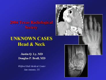
2004 Texas Radiological Society UNKNOWN CASES Head - PowerPoint PPT Presentation
1 / 15
Title:
2004 Texas Radiological Society UNKNOWN CASES Head
Description:
noncontrast head CT performed at another institution was reportedly ... Craniofacial bones commonly involved-frontal, sphenoid, ethmoid, maxillary, mandible ... – PowerPoint PPT presentation
Number of Views:63
Avg rating:3.0/5.0
Title: 2004 Texas Radiological Society UNKNOWN CASES Head
1
2004 Texas Radiological SocietyUNKNOWN
CASESHead Neck
- Justin Q. Ly, MD
- Douglas P. Beall, MD
- Wilford Hall Medical Center
- San Antonio, TX
2
HEAD NECK
- 42-year-old female
- chronic migraines
- noncontrast head CT performed at another
institution was reportedly unremarkable - family h/o intracranial aneurysms MRI/MR
angiography of COW was performed to exclude
aneurysm
3
(No Transcript)
4
(No Transcript)
5
(No Transcript)
6
2004 Texas Radiological SocietyDiagnosis
forHead Neck Case
- Justin Q. Ly, MD
- Douglas P. Beall, MD
- Wilford Hall Medical Center
- San Antonio, TX
7
Imaging Findings
- Heterogenous, mildly expansile clivus mass on
T1WI - Moderately-intense uptake on bone scan
- Ground-glass attenuation on CT
8
Fibrous Dysplasia of Clivus
- DDX
- Chordoma
- Chondrosarcoma
- Plasmacystoma
- Lymphoma
- GCT
- Cavernous hemangioma
- Carcinomas (adenocystic
- or nasopharyngeal)
- Mets
- Pagets
- low T1, high T2 signal
- In this case, signal not as
- ? as you would expect to see
- w/aggressive neoplastic process
9
(No Transcript)
10
Fibrous Dysplasia of Clivus
- 1st described by Lichtenstein 1938
- Benign, presents in 1st 2 decades
- Etiol unknown
- Marrow replaced by fibro-osseous connective
tissue replacing mature bone with structurally
weak, immature woven bone - Classification
- Monostotic (long bones of extremities, ribs,
vertebrae, craniofacial bone) 70 - Polyostotic 30
- Albrights-polyostotic, skin hyperpigmentation,
endocrine dysfxn (hyperthryoid, preocicous
menstruation females)
11
Fibrous Dysplasia of Clivus
- Craniofacial bones commonly involved-frontal,
sphenoid, ethmoid, maxillary, mandible - Clivus involvement in monostic form rare
- Presentation asymptomatic, headache
12
Fibrous Dysplasia of Clivus
- XR-cystic, sclerotic, mixed forms (Leeds et al)
- CT-amorphous ground glass or smudgy
appearance, thinning of cortex - MR
- Expansile
- Heterogeneous-homogenous, low to intermediate T1
and low to high T2 signal - Contrast enhancement with variable signal
- Signal reflects overall cellularity, varying
collagen content, extent of bone trabec, cyst
formation - May have cystic components
- Scleroticlow T1, T2 signal
- Low signal may also be seen with fibrous tissue
- Intermediate T2 signal high metabolic activity
(Utz et al.)
Leeds at al. Fibrous dysplasia of the skull and
its differential diagnosis. Radiology
196278570-582. Utz et al. MR appearance of
fibrous dysplasia. J Comput Assist Tomogr
198913845-851
13
Fibrous Dysplasia of Clivus
- Path
- Fibrous connective tissue with trabeculae of
immature bone w/o surrounding osteoblasts - Termination of active phaseincreasingly ossified
- Histologic degree of activity closely corresponds
to radiologic findings (Ameli et al) - Disease ceases to progress or evolves slowly
after bone maturation
Ameli NO, et al. Monostotic fibrous dysplasia of
the cranial bones. Report of fourteen cases.
Neurosurg Rev 1981471-77.
14
Fibrous Dysplasia of Clivus
- Tx
- Dependent on activity level, symptoms/cranial
nerve compromise, location in skull - If active phasetotal excision recommended (Ameli
et al) - Generally good prognosis, small risk of malignant
change - Monostotic craniofacial lesions-0.05 malign
transformation - Osteosarc, fibrosarc, chondrosarc
15
2004 Texas Radiological SocietyUNKNOWN CASES
- Justin Q. Ly, MD
- Douglas P. Beall, MD
- Wilford Hall Medical Center
- San Antonio, TX
Click here to return to case list


