
Urine Analysis, Urine MCS PowerPoint PPT Presentation
Title: Urine Analysis, Urine MCS
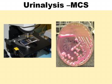
1
Urinalysis MCS
2
- Objective
- What is Urinary Track Infection (UTI)?
- Why Urinalysis MCS is Requested?
- Bacterial Causes of UTI
- Risk Factors For Developing a UTI
- Collection, Transportation and Reception
- Processing Urine analysis and Culture
- Culture media, additional tests and
identification of common Gram-ve org ve in
Urine - Urine parasites
- Recommendation
3
- What is Urinary Track Infection (UTI)?
- Any infection of the part of the urinary system
including kidney, bladder, ureters and the
urethra.
4
- Why it's done
- A urinalysis is a common test that's done for
several reasons - To check your overall health. Your doctor may
recommend a urinalysis as part of a routine
medical exam, pregnancy checkup, pre-surgery
preparation, or on hospital admission to screen
for a variety of disorders, such as diabetes,
kidney disease and liver disease. - To diagnose a medical condition. Your doctor may
suggest a urinalysis if you're experiencing
abdominal pain, back pain, frequent or painful
urination, blood in your urine, or other urinary
problems. A urinalysis may help diagnose the
cause of these symptoms. - To monitor a medical condition. If you've been
diagnosed with a medical condition, such as
kidney disease or a urinary tract disease, your
doctor may recommend a urinalysis on a regular
basis to monitor your condition and treatment. (4)
5
Bacterial Causes of UTI Bacteria are by far the
most frequent group of organisms causing urinary
tract infections. Organisms that are known to
commonly cause UTI are E. Coli Staphylococcus
aureus Staph. Saprophyticus Proteus
species Streptococcus faecalis (group D
Strep) Klebsiella species Pseudomonas Spp MTB,
Chlamydia trachomatis, Mycoplasma, Candida spp
and Leptospira interrogens
6
Risk Factors For Developing a UTI 1) UTI is more
common in women than in men because a womens
urethra is located closer to their rectum than
mans. Female urethra is shorter so it is easier
for bacteria to reach the bladder. 2) Pregnant 3)
Post menopausal women with bladder or uterine
prolapse 4) Tumors and kidney or bladder stone
5) Diabetes mellitus 6) Using diaphragm as
contraception 7) Patients with urinary
catheter 8) Enlarge prostate in man 9)
Immunocompromised patients 10) Any medical
condition involving bladder or kidney
7
Collection, Transportation and Reception
The urinary catheter Urine specimens for
laboratory investigations can be collected from
catheterized patients as shown (left). The second
port is for putting fluids into the bladder
(right). Urine from the drainage bag should not
be tested because it may have been standing for
several hours.
Type of Specimens
- Midstream urine (MSU)
- Clean catch
- Adhesive bag
- Suprapubic Aspiration
- Catheter sample
8
Processing Urine analysis and Culture
Urine analysis 1.Microscopically,
Macro Bacteria, parasite, fungi etc 2.Dip stick
9
- Urine analysis
- 1- Microscopy
- 2- Dip stick
- 3- Culture
10
Culture media
blood agar
MacConkey agar
CLED agar
a differential medium
an enriched medium
Selective medium
11
Laboratory examination of urine
Quantitative (Colony counts)
a urine sample is streaked on surface of Blood
Agar plate and CLED agar / Mc Conkey agar with a
special loop calibrated to deliver a known
volume.
Over night incubation
Isolation of colonies, Biochemical tests, Drug
susceptibility test,
Over night incubation
RESULT
12
GRAM NEGATIVE GRAM POSITIVE
Escherichia coli Enterococcus
Klebsiella Staphylococcus saprophyticus
Proteus Streptococcus agalactiae (group B)
Other Enterobacteriaceae (Enterobacter,Citrobacter.) Staphylococcus aureus1 (Associated with staphylococcemia(
Pseudomonas aeruginosa
- Other organisms
- Candida albicans
- Schistosoma haematobium
- Tricomonas vaginalis
13
MacConkey's agar showing both lactose and
non-lactose fermenting colonies. Lactose
fermenting colonies are pink whereas non-lactose
fermenting ones are colourless or appear same as
the medium.
14
CLED agar
Selective culture medium for detection and
isolation Of Escherichia coli and coliform
bacteria in urine
15
MICROSCOPIC APEARANCEGram negative bacilli
16
gram negative bacilli
17
MacConkeys agar plate showing growth of
Lactose fermenter pink coloniese.g. E. coli
18
E coli
Indole Reactions Negative
Positive
19
Indol Test
20
MacConkeys agar plate showing growth of
Lactose fermenter pink coloniesKlebsiella
21
CLED agar plate showing growth of mucoid
coloneis Klebsiella
22
MacConkeys plate showing growth of Non -
Lactose fermenter pale coloniese.g. Proteus
23
Blood culture palate showing Swarming growth
of Proteus
24
Urease Test
proteus is Urease positive Urease splits urea
into ammonia and alkalinizes the urine with
production of crystals
25
Proteus growth Swarming
CLED (Cystine-Lactose-Electrolyte-Deficient) -
inhibits the proteus swarm
26
Pseudomonas aeruginosa
27
(No Transcript)
28
(No Transcript)
29
Biochemical Identification
Enterococcus species
- Bile Esculin hydrolysis
Both Group D streptococci and enterococci produce
a positive (left) bile Esculin hydrolysis test.
30
MICROSCOPIC APEARANCEGram positive cocci in
clusters most likely staphylococci
FROM CULTURE
SMEAR FROM SPESIMEN Pus cells Gram
positive cocci in clusters
31
- To differentiate between Staphylococcus aureus
Staphylococcus epidermidis use the following
test - 1.COAGULASE TEST
- Tube coagulase test
- Slide coagulase test
- 2. DNAase TEST
32
1-COAGULASE TEST Slide coagulase
test
33
2-DNAase TEST
34
Blood agar plate showing growth of
Staphylococcus aureus Colonies are
golden yellow in color
35
Staphylococcus spp
Staphylococcus aureus Staphylococcus
epidermidis Golden colonies (yellowish)
white colonies
36
CATALASE TEST
Procedure Mix the colony in a drop of hydrogen
peroxide (H2O2)
37
COAGULASE TEST Tube coagulase
test
38
Differential Characteristics
39
NOVOBIOCIN TEST
Staphylococcus saprophyticus (resistant-Novobiocin
)
Staphylococcus epidermidis (sensitive-Novobiocin )
40
Antibiotic sensitivity test Agar diffusion
method
41
Candida albicans
- Growth on Sabouraud's Dextrose Media
- Candida albicans on blood agar
42
1.CHLAMYDOSPORE TEST.
43
2.GERM TUBE TEST
44
Schistosoma haematobium
Schistosoma haematobium(urine eggs 115-170 x
45-65 micrometers)(primates)
45
- Recommendation
- CLED Agar
- Hydrogen Perioxide
- Opticin
- Bacitercin
- Nutrient Agar
46
Reference 1) Dr S. R. Dutta, PMGH Microbiology
SOP (2011), Port Moresby, NCD. 2) Monica
Cheesbrough, District Laboratory Practice in
Tropical Countries, 2nd Ed, 2006, Cambridge
University Press, Cape Town- South Africa. 3)
David P Kateete, Cyrus N Kimani, Fred A Katabazi
Identification of Staphylococcus aureus DNase
and Mannitol salt agar improve the efficiency of
the tube coagulase test. Am J Clinical
Microbiology and Antimicrobials 2010, 923 4)
http//www.mayoclinic.org/tests-procedures/urinaly
sis
47
Presentation By
RABBIE NIGEL BERO Western Highland Provincial
Health Authority, MT HAGEN, Western Highland
Province, Papua New Guinea
THANK YOU
Recommended
