
Electrophoresis PowerPoint PPT Presentation
Title: Electrophoresis
1
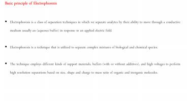
- Basic principle of Electrophoresis
- Electrophoresis is a class of separation
techniques in which we separate analytes by their
ability to move through a conductive medium
usually an (aqueous buffer) in response to an
applied electric field. - Electrophoresis is a technique that is utilized
to separate complex mixtures of biological and
chemical species. - The technique employs different kinds of support
materials, buffers (with or without additives),
and high voltages to perform high resolution
separations based on size, shape and charge to
mass ratio of organic and inorganic molecules.
2
- In the course of electrophoresis, two electrodes
are immersed in two separate buffer chambers.
The two chambers are connected such that charged
particles can migrate from one chamber to the
other. - By using a power supply, electric potential
difference is generated between the two
electrodes. - As a result, electrons flow from one of the
electrodes, the anode, towards the other
electrode, the cathode.
3
When charged molecules in the two chambers are
placed in an electric field, they migrate toward
either the positive (anode) or negative (cathode)
pole according to their charge and Neutral
species do not experience the electrical field
remain stationary.
4
Different ions migrate at different speeds
dictated by their sizes and by the number of
charges they carry. As a result, different ions
can be separated from each other by
electrophoresis. It is very important to
understand the basic physics describing the
dependence of the speed of the ion as a function
of the number of charges on the ion, the size of
the ion, the magnitude of the applied electric
field and the nature of the medium in which the
ions migrate. By understanding these basic
relationships, the principles of the many
different specific electrophoresis methods become
comprehensible.
5
The fundamental principle of electrophoresis is
illustrated in above Figure. The mathematical
description of the force during electrophoresis
is simple. An electric force Fe is exerted on the
charged particle. The magnitude of the electric
force equals the product of the charge q of the
particle and the electric field E generated
between the two electrodes It can be easily
calculated using the value of the voltage (volt)
set by the electric power supply and the distance
of the two electrodes (cm). As soon as the
electric field is applied and the charged
particles are accelerated by the electric force,
a drag force (Fd) called friction will also be
immediately exerted on the particles by the
medium.
?? ?? ????
6
At the typically very low speed of particle
migration during electrophoresis, the force Fd is
a linear function of the velocity (v) of the
particle, as described by Equation below The
ratio of the force and the velocity is defined as
the frictional coefficient (f). The value of f is
a function of the size and shape of the particle
and the viscosity of the medium. The larger the
particle and the more obstructing the medium, the
higher the value of f.
?? ?? ????
7
When electrophoresis is started, particles
accelerate instantaneously to a velocity (v) at
which the magnitude of the drag force equals the
magnitude of the (opposite) accelerating electric
force
????????
Once the magnitude of the two opposing forces
becomes equal, the resultant force becomes zero.
Therefore, each particle will move at a
constant velocity characteristic of the given
particle at the given accelerating potential and
medium.
8
There are several forms of electrophoresis
S.N. Technique Buffer held by
1 Paper paper
2 Gel a porous gel of agarose or polyacrylamide
3 Capillary capillary tube
We generally focus on the capillary
electrophoresis
In capillary electrophoresis we inject the
sample into a buffered solution retained within a
capillary tube but the principle remains the same
for all kinds of electrophoresis. When an
electric field is applied across the capillary
tube, the samples components migrate as the
result of two types of action Electrophoretic
mobility and Electroosmotic mobility.
9
Electrophoretic mobility is the solutes response
to the applied electrical field. The other
contribution to a solutes migration is
electroosmotic flow, which occurs when the buffer
moves towards the cathode Electrophoretic
Mobility The velocity with which a solute moves
in response to the applied electric field is
called its electrophoretic velocity, ? it is
defined as where µep is the solutes
electrophoretic mobility, and E is the magnitude
of the applied electrical field.
?? ???? ?? ???? E
10
A solutes electrophoretic mobility is defined
as where q is the
solutes charge, ? is the buffer
viscosity, and r is the solutes
radius. Electrophoretic mobility and, therefore,
electrophoretic velocity, increases for more
highly charged solutes and for solutes of smaller
size. Particles having different
electrophoretic mobility, i.e. those that migrate
at different speeds in the same medium and
electric field, can be separated by
electrophoresis.
?? ???? ?? 6??????
11
When an electrical field is applied to a
capillary filled with an aqueous buffer we
expect the buffers ions to migrate in
response to their electrophoretic mobility.
Because the solvent, H2O, is neutral we
might reasonably expect it to remain
stationary. What we observe under normal
conditions, however, is that the buffer solution
moves towards the cathode. This phenomenon is
called the electroosmotic flow. Electroosmotic
flow occurs because the walls of the capillary
tubing are electrically charged. The surface of a
silica capillary contains large numbers of
silanol groups (SiOH). At pH levels greater than
approximately 2 or 3, the silanol groups ionize
to form negatively charged silanate ions (SiO)
12
Electric double layer
Formed due to partial neutralization of silanol
in fixed layer
Due to cations from buffer bind strongly with
silanol
Cations in the diffuse layer migrate toward the
cathode. Because these cations are solvated, the
solution is also pulled along, producing the
electroosmotic flow.
13
The anions in the diffuse layer, which
also are solvated, try to move toward the
anode. Because there are more cations than
anions, however, the cations win out and the
electroosmotic flow moves in the direction of the
cathode. The rate at which the buffer moves
through the capillary, what we call its
electroosmotic flow velocity, ?eof, is a function
of the applied electric field, E, and the buffer
s electroosmotic mobility, µeof Electroosmotic
mobility is defined as where ?? is the buffer
dielectric constant, ?? is the zeta potential,
and ? is the buffer viscosity.
?? ?????? ?? ?????? ?? ?? ?????? ????
4????
14
Total Mobility A solutes total velocity, , as it
moves through the capillary is the sum of its
electrophoretic velocity and the electroosmotic
flow velocity.
?? ?????? ?? ???? ?? ??????
?? ?????? ?????????????? gt ?? ?????? ??
?????? ???????????????? ?? ?????? ??
?????? ???????????? lt ?? ??????
Each species has the same electroosmotic flow,
?eof. Cations elute first because they have a
positive electrophoretic velocity, ?ep Anions
elute last because their negative electrophoretic
velocity partially offsets the electroosmotic
flow velocity. Neutrals elute with a velocity
equal to the the electroosmotic flow
15
Migration Time Another way to express a solutes
velocity is to divide the distance it travels by
the elapsed time where l is the distance
between the point of injection and the detector,
and tm is the solutes migration time.
Combining the above two equations and
solving for tm leaves us with
?? ?????? ?? ?? ??
?? ?????? ?? ?????? ??( ?? ???? ?? ??????
)??
?? ?? ?? ( ?? ???? ?? ?????? )??
16
Instrumentation
17
Instrumentation 1) Capillary 2575?m ID fused
silica capillary with a thin outer coating of
polyimide 2) Injection volume 1 ?l for a 50 cm
long, 50 ?m ID 3) Detector a. Optical
absorbance detector b. Laser based absorbance
detector c. Refractive index detection d.
Thermooptical absorbance e. Fluorescence
detector f. Chemiluminescence detector g.
Electrochemical detector Conductivity,
Amperometric h. Radioactivity detector I.
Hyphenated detection CE-MS 4) Power supply 5)
Buffer and additives
18
(No Transcript)
19
- Application
- Electrophoresis is used in forensics, molecular
biology, genetics, microbiology and biochemistry.
- The results can be analyzed quantitatively by
visualizing the gel with UV light and a gel
imaging device. - The image is recorded with a computer operated
camera, and the intensity of the band or spot of
interest is measured and compared against
standard or markers loaded on the same gel. - Depending on the type of analysis being
performed, other techniques are often implemented
in conjunction with the results of gel
electrophoresis, providing a wide range of
field-specific applications.
20
Qualitative analysis can be conducted by
comparing the patterns produced to standards.
This example is a molecular weight determination
of proteins but other materials can be evaluated.
21
Mediums in electrophoresis and their applications
Recommended
