
III' Biological Membranes PowerPoint PPT Presentation
1 / 71
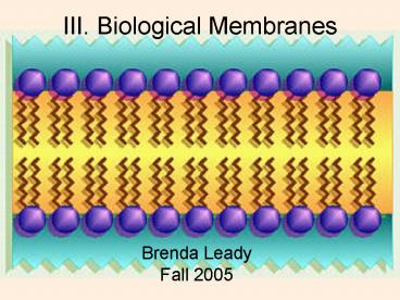
Title: III' Biological Membranes
1
III. Biological Membranes
- Brenda LeadyFall 2005
2
- A cell can be divided into
- Plasma membrane
- A.k.a. cytoplasmic membrane/ cell membrane
- Cytoplasm
- nucleus
3
Plasma membrane
- Separates inside from outside
- Intracellular fluid
- ICF
- Inside cells
- Extracellular fluid
- ECF
- Outside cells
ECF
ICF
4
Fig 3.2
5
Fluid Mosaic Model
- Thin lipid bilayer with proteins
- Constantly changing mosaic
- Phospholipids (most), cholesterol and glycolipids
6
Fig 3.3
7
Phosopholipid
- Polar head is hydrophilic
- Nonpolar tail is hydrophobic
- Self orienting and self assembling
- Assemble with heads out and tails in
8
Surfaces differ
- Differ in kinds and amounts of lipids
- Integral membrane proteins
- Embedded in membrane
- Most transmembrane
- Transport (channels, carriers), receptors
- Peripheral Membrane Protein
- Attached only loosely
- Easily dislodged
- May function in cell structure, enzyme or movement
9
Fig 3.4
10
Membrane Transport
- Interstitial fluid
- ECF derived from blood
- Bathes cells
- Constant traffic between cell and interstitial
fluid - Membrane is selectively/ differentially permeable
- Lets some things in or out but not others
11
Active/ Passive
- Passive processes
- Substances cross membrane without energy input
from the cell - Active processes
- Require cell to use ATP to move substances
12
Passive Processes
- Diffusion
- Filtration
13
Diffusion
- Important in every cell
- Tendency for molecules or ions to scatter evenly
throughout the environment - All molecules are in constant motion bumping into
each other - Move from areas of higher concentration to areas
of lower concentration - Along or down a concentration gradient
14
Fig 3.6
15
(No Transcript)
16
Why do substances move?
- They are being hit by solvent molecules
- Like balls on a pool table
17
Speed influenced by
- Concentration difference
- Greater difference faster diffusion
- Size of molecule
- Smaller molecules diffuse faster
- Temperature
- Warmer temperatures make diffusion go faster
18
Eventually
- Concentrations equal out and NET diffusion stops
- Molecules are in constant motion and so movement
NEVER stops
19
Plasma membranes allow molecules to pass if
- Lipid soluble
- Small enough to pass through membrane channels
- Assisted by carrier molecules
20
Types of diffusion
- Simple
- Facilitated
- Osmosis
21
Fig 3.7
22
Simple Diffusion
- Unassisted diffusion of very small or lipid
soluble substances - Oxygen, carbon dioxide, fat soluble vitamins,
alcohol
23
Fig 3.7
24
Facilitated Diffusion
- Passive transport process using carriers or
water-filled channels - Glucose, other sugars, amino acids, and ions
25
Carrier
- Transmembrane intergral protein with specificity
for certain large substances (sugars and amino
acids for example) - Envelopes and releases substances to other side
- Limited by number of carriers available
26
Fig 3.7
27
Channel
- Transmembrane protein that transports water or
ions - Some always open, other not
- Also limited by number available
28
Fig 3.7
29
Osmosis
- Diffusion of water through a semipermeable
membrane - Crosses membrane on its own or through channels
- Aquaporins
- Occurs when water concentration differs on 2
sides of a membrane
30
- If water concentration is equal, no net movement
membrane
31
- If solute concentration different, water
concentration is different - If one side has more water, it moves to the other
side with less - Solute follows its concentration gradient
Direction of salt movement
membrane
80 salt 20 water
10 salt 90 water
Direction of water movement
32
Fig 3.8
33
However
- If membrane is not permeable to solute,
- Water moves but NOT the solute
- Volume changes on one side
34
(No Transcript)
35
osmosis
36
Tonicity
- Ability of a solution to change the shape or tone
of cells by altering internal water volume
37
- Isotonic- same tonicity
- Cell does not shrink or swell
- 0.9 saline
- Hypertonic
- Solution more concentrated
- Cell shrinks due to water loss
- Crenate
- Hypotonic
- Solution less concentrated than cell
- Cells swell due to water gain
- Swell and pop (lyse)
- Hyper- and hypo- can also refer to the
relationship of the solution to the cell - You have to be told if you are referring to the
cell or the solution
38
Fig 3.9
39
(No Transcript)
40
(No Transcript)
41
Filtration
- Generally only across capillary walls
- Forces water and solutes through a capillary wall
using pressure - Passive process based on a PRESSURE gradient
- Higher pressure to lower pressure
- Well see this again later
42
Active Processes
- Active transport
- Vesicular transport
43
Active processes
- Must use ATP to move the substances
- Unable to pass using passive means
- Too large, not lipid soluble, against gradient
44
Active transport
- Requires carrier proteins to move against/ up a
concentration gradient - From where there is less to where there is more
- Transports only specific substances
45
Fig 3.10
46
Vesicular Transport
- Large particles, macromolecules and fluids
- Exocytosis- moving stuff out of cell
- Endocytosis- moving stuff into cell
- Phagocytosis- cell eats debris or invaders
- Forms phagosome, fuses with lysososme for
digestion - Transcytosis- in one side and out the other
47
Fig 3.12
Exocytosis
48
(No Transcript)
49
Exocytosis
Endocytosis
50
Phagocytosis
51
Absorption of Digested Food
52
Figure 23.21
53
(No Transcript)
54
Absorption of Digested Food
- Carbohydrates
- Broken down into monosaccharides
- Facilitated diffusion or active transport
- Proteins
- Broken down into amino acids
- Facilitated diffusion or active transport
55
(No Transcript)
56
- Lipids
- Triglycerides broken down into monoglycerides and
fatty acids - Associate with bile slats to form micelles
- Move to epithelial surface and use simple
diffusion into cell - Without micelles, float on watery chyme and never
contact cells - Inside cells, remade into triglycerides and
coated with proteins to form chylomicron - Processed by Golgi for export outside cell
- A few free fatty acids enter blood but
chylomicrons enter lacteal (lymph capillary) - Eventually lymph dumps into blood
- Chylomicrons broken down to free fatty acids and
glycerol that can enter body cells
57
(No Transcript)
58
- Nucleic acids
- Broken down into pentose sugars, nitrogenous
bases and phosphate ions - Active transport into cells and into blood
59
MembraneReviewQuestions
60
What type of transport does require the cell to
spend ATP?
- Diffusion
- Osmosis
- active transport
- facilitated diffusion
61
I add sodium chloride to a beaker of water
SALT
- What is the solute?
- The solvent?
Water
62
Diffusion is
- Substances travel down a concentration gradient
- Solvent travels down a concentration gradient
- Substances travel up a concentration gradient
- Solvent travels up a concentration gradient
63
Osmosis is movement of the solvent across a
membrane(2 are correct)
- From an area with more solvent to an area with
less solvent - From an area with less solvent to an area with
more solvent - From an area with more solute to an area with
less solute - From an area with less solute to an area with
more solute
64
A
C
E
B
D
F
Figure 1
Figure 2
Figure 3
- In figures 1, 2 and 3, is the cell hypertonic,
hypotonic or isotonic to the solution? - Figure 1
- Figure 2
- Figure 3
hypertonic
hypotonic
isotonic
65
A
C
E
B
D
F
Figure 1
Figure 2
Figure 3
- In figures 1, 2 and 3, is the solution
hypertonic, hypotonic or isotonic to the cell? - Figure 1
- Figure 2
- Figure 3
hypotonic
hypertonic
isotonic
66
A
C
E
B
D
F
Figure 1
Figure 2
Figure 3
- In each figure, which arrow depicts how the
solute will move (the dots)? - Figure 1
- Figure 2
- Figure 3
A
D
both
67
A
C
E
B
D
F
Figure 1
Figure 2
Figure 3
- In each figure, which arrow depicts how the
solvent will move? - Figure 1
- Figure 2
- Figure 3
B
C
both
68
A
C
E
B
D
F
Figure 1
Figure 2
Figure 3
- In each figure, which arrow depicts the direction
of osmosis? - Figure 1
- Figure 2
- Figure 3
B
C
both
69
A
C
E
B
D
F
Figure 1
Figure 2
Figure 3
- In each figure, which arrow depicts the direction
of diffusion? - Figure 1
- Figure 2
- Figure 3
A
D
both
70
A
C
E
B
D
F
10 salt
7 glucose
6 albumin
15 glucose
5 salt
6 albumin
Figure 1
Figure 2
Figure 3
- In each figure, which arrow depicts the direction
of diffusion? - Figure 1
- Figure 2
- Figure 3
A
D
both
71
A
B
10 salt
5 salt
Figure 1
- What would make diffusion go FASTER?
- Temperature- warmer or colder?
- Concentration- 8(lower) or 20(higher)
- Molecule size- larger or smaller
Recommended
