
Diagnoses Case1 PowerPoint PPT Presentation
1 / 12
Title: Diagnoses Case1
1
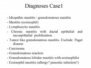
Diagnoses Case1
- - Idiopathic mastitis / granulomatous mastitis
- - Mastitis (eosinophil)
- - Lymphocytic mastitis
- - Chronic mastitis with ductal epithelial and
myoepithelial proliferation - - Tumor like granulomatous mastitis. Exclude
Paget disease - - Carcinoma
- - Granulomatous reaction
- - Granulomatous lobular mastitis with
eosinophilia - - Eosinophil mastitis (allergy? parasitic
infection?)
2
Diagnoses Case2
- Solid intraductal papilloma, with myoepithelial
ductal proliferation - - Ductal adenoma/complex sclerosing papillary
lesion - - Intraductal sclerosing papilloma
- - Solid intraductal papilloma
- - Papillary intraductal carcinoma arising in a
papilloma - - In situ papillary carcinoma
- -Carcinoma papillare intracysticum et invasion
with DCIS - - Intracystic papilloma with malignant change
- - Encysted papillary carcinoma
3
Diagnoses Case3
- Hamartoma mammae
- - Columnar cell change with mucin filled cysts
and ADH - - Proliferative mastopathy
- - Ductal papillomatosis
- - Fibrocystic change with columnar cell
hyperplasia - - Fibrocystic change with benign epithelial cell
proliferation, with microcalcification - -Complex sclerosing laesion. No malignancy. Mild
dysplasia in the intraductal papillomatosis
component - - Hypersecretory hyperplasia
- - FCC DIN1a, c
- - DIN1a, mucin filled ducts, calcification
4
Diagnoses Case4
- Lobular carcinoma, solid variant, with apocrin
differenciation - - Invasive cancer (ductal, NST)
- - Collision lobular and ductal carcinoma, with
pagetoid epidermal invasion - - Invasive ductal carcinoma with skin
infiltration - - Invasive ductal carcinoma (diff. melanoma
malignum?) - - Carcinoma ductale exulceratum valde infiltrans
with skin propagation, with lymphangio-invasion,
and partial neuroendocrin differentiation - - Melanoma or ducta lcancer, or mixed
- - Paget carcinoma
5
Diagnoses Case5
- Pleomorph lobular carcinoma
- - Lobular invasive carcinoma lymphocytic
inflammation or lymphoid tumor - - Invasive lobular carcinoma, pleomorphic
- - Invasiv lobular carcinoma sclerosing
lymphocytic lobulitis - - Mastitis granulomatosa, with infiltrative
ductal-lobular carcinoma in the background - - Inflammatory lobular-ductal cc.
- - Mixed lobular and ductal cancer
- - IDC
- - Pleomorph carcinoma high grade DCIS
6
Diagnoses Case 6
- Secretory carcinoma
- - Adenoid cystic carcinoma
- - Breast hamartoma
- - Cystic-secretory hyperplasia
- - Sclerosing adenosis (pleomorph adenoma-like)
- - Tubular carcinoma. Excision incomplete.
- - Sclerosing adenosis well differentiated
tubular carcinoma - - Apocrin carcinoma
- - Mucoepidermoid carcinoma
7
Diagnoses Case 7
- Invasive ductal carcinoma, with partial DCIS-like
appearance, with extensive angioinvasion - - Invasive carcinoma (ductal, NST with clear cell
areas (glycogen rich?) - - Acinus sejtes carcinoma
- - Invasive glycogen-rich carcinoma
- - Ductal carcinoma- in situ and invasive
- - Comedo carcinoma and invasive ductal carcinoma
- - Carcinoma lobulare in situ (comedo-like foci)
- - Metastasis (clear cell carcinoma)
- - Clear cell carcinoma
8
Diagnoses Case 8
- - Malignant phylloid tumor
- - Malignant spindle cell tumour of the breast
- - Malignant phyllodes tumour
- - Sarcoma NOS
- - Spindle cell carcinoma
- - Myofibroblastoma
- - Well differentiated liposarcoma, spindle cell
type - - Phylloid tumor (borderline)
- - Phyllodes tumor - malignant low grade
- - Schwannoma
- - Pericytoma
- - Myoepithelioma
- - Spindle cell myoepithelioma
9
Diagnoses - Case 9
- - Intraductal papillary carcinoma
- - Papillary DCIS with minor cribriform
component - - Papillary intraductal carcinoma
- - Cystic papillary in situ carcinoma
- - Intracystic papillary carcinoma
- - In situ papillary carcinoma
10
Diagnoses Case - 10
- - Complex sclerotising laesion
- B2 (sclerosing adenosis, cyst, usual type
hyperplasia as parts of the fibrocystic changes
spectrum, a diagnosis probably lacking
correlation with the clinical presentation) - Fibrocystic change with hyperplastic lesions
- - Sclerosing adenosis
- - Benign (sclerotising adenosis)
- - Intraductal dysplasia
- - Mastopathia fibrocystica
- - Epitheliosis
- - UDH
- - Adenosis
- - Radial scar. B3
11
Diagnoses Case 11
- -Invasive well differentiated ductal carcinoma
- - B5 Invasive carcinoma
- - Cribriform or tubular carcinoma
- - Invasive ductal carcinoma, NOS
- - Invasive ductal carcinoma
- - Carcinoma cribriforme
- - Carcinoma lobulare-ductale
- - Carcinoma
- - IDC
- - Invasive carcinoma with DCIS component. B5
12
Diagnoses Case 12
- -not suitable for diagnosis
- - B5 Invasive carcinoma
- - Carcinoma invasivum mammae
- - Tissue unsatisfactory for interpretation
- - not assessable, may be benign
- -Inflammatory carcinoma (poorly differentaiated
ductal carcinoma) in a background of
granulomatous mastitis. - - Malignant soft tissue tumor
- - Small fragment malignant epithelial tumor,
Immunhistochemistry necessary - - Malignant tumor. B5
Recommended
