
Archetypal Retrograde: Endocytosis PowerPoint PPT Presentation
1 / 41
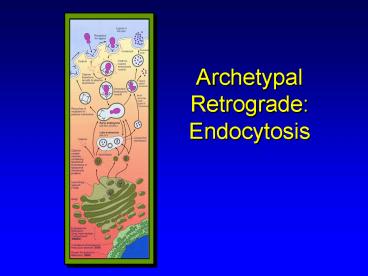
Title: Archetypal Retrograde: Endocytosis
1
Archetypal Retrograde Endocytosis
2
Endocytosis
- Obtain raw materials for cell functions
- Defend organism from noxious chemicals and
harmful biologicals
3
Endocytosis
- Phagocytosis
- Macropinocytosis
- Pinocytosis
- Can involve specific receptors on surface or can
be passive - Receptors concentrate uptake
- In all cases some extracellular material
internalized passively
4
Endocytosis
- Plasma membrane invaginates to form pit
- Opening of pit constricts to form narrow neck
- Apposing membranes fuse to seal pit into vesicle
5
Phagocytosis
6
Pinocytosis
Material can be randomly captured or
concentrated by specific receptors Receptor
mediated endocytosis Membrane receptors
concentrate extracellular material for
specific uptake and trafficking
7
Endosomes Lysosomes
- Membrane vesicles endosomes/phagosomes --
deliver ingested microorganisms and other
material destined for destruction to lysosomes
(1). - Fusion of lysosome with these vesicles creates a
2 lysosome. - Fusion exposes these endocytosed substrates to
hydrolases which - degrades the ingested material.
Secondary Lysosome
Endosome
8
Lysosomal Storage Disease
Golgi
Primary Lysosome
Hydrolase
Digestion
Endo- cytosis
Secondary Lysosome
9
Lysosomal Storage Disease
Golgi
Hydrolase
Mutant Enzyme
Primary Lysosome
Endo- cytosis
Secondary Lysosome
Cholesteryl Ester
Wolmans Fibroblast
10
Lysosomal Storage Disease
Golgi
Hydrolase
Primary Lysosome
X
Abnormal Trafficking
Endo- cytosis
Secondary Lysosome
Cholesteryl Ester
Wolmans Fibroblast
11
Smooth Endoplasmic Reticulum
- Short anastomosing tubules that are NOT
associated with ribosomes - Well developed in cells that synthesis and
secrete steroids - Referred to as sarcoplasmic reticulum in
skeletal and cardiac muscle - segregates Ca essential for contraction
12
Smooth Endoplasmic Reticulum
Steroid synthesis Neutralization of poison
Lipid metabolism Phospholipid synthesis
Glycogen utilization
13
Peroxisomes (Microbodies)
- Small, membrane-limited spherical bodies
- 0.5 to 1.0 um in diameter
- contain oxidative enzymes -- catalase
- Involved in the formation and breakdown of
intracellular hydrogen peroxide - used for killing phagocytosed bacteria
- Involved in beta oxidation of fatty acids
14
Peroxisomes (Microbodies)
In many animals, but not as prominate in humans,
peroxisomes contain a central dark nucleoid body
of urate oxidase.
15
Mitochondria
- Involved in aerobic energy production and storage
- Regulate ion content of cytoplasm.
- Store factors important in regulated cell death
(apoptosis) - Defects lead to cancer,
- neurodegenerative diseases and auto-immununity
16
Mitochondria
- Vary in size, shape and number
- Move freely within cytosol and tend to aggregate
in areas with high energy demands
17
Mitochondria
- Outer membrane
- permeable
- contains porin, a pore-forming protein which
allows free passage of small molecules - contains enzymes that convert lipids into forms
that can be metabolized by mitochondria
18
Mitochondria
- Inner membrane
- separated from outer by intermembranous space
- thinner and thrown into folds -- cristae
- aerobic respiration and electron transport takes
place here (contains cytochromes and enzymes
involved in ATP production)
19
Mitochondria
- Inner Matrix
- rich in proteins
- contains dense granules which are the storage
site for divalent cations - contains one or more strands of double stranded
circular DNA - enzymes for Krebs cycle and fatty acid beta
oxidation abundant in matrix
20
Mitochondria
- Inner Matrix DNA
- Maternally derived
- Produces only a small portion of the protein in
mitochondria, rest come via nuclear DNA
transcription of RNA and protein synthesis in
cytosolic polyribosomes. - Imported proteins have a mitochondrial targeting
sequence.
21
Mitochondria
- Cells with higher energy demands have more
mitochondria and more cristae per mitochondria
22
Non-membrane Limited Organelles
Cytoskeleton and Inclusions
23
Cytoplasmic Matrix
- Proteins organized (microtrabecular lattice)
- Very little unbound water, cytoplasm more like
runny jello than glass of water - Some areas more solid than others
24
Cytoskeleton
- Supporting framework which maintains shape and
polarity of cell
Fluorescent staining of Actin (green) and lipid
inclusions (blue) in smooth muscle cells
25
Cytoskeleton
- Important for cell movement
- Can be very dynamic structures under constant
remodeling to accommodate shape changes necessary
to confront environment - Critical component of cilia and flagella (motile
structures)
26
Cytoskeleton
- Intracellular Movement
- Contraction can move cytoplasm
- Attach to other organelles and with molecular
motors coordinate their movement - Outside-In signaling
- Bind to integral membrane proteins and transduce
signals from outside environment
27
Components of the Cytoskeleton
28
Cytoskeleton
- Microtubules
- Filaments
- Microfilaments
- Intermediate Filaments
- Microtrabecular components
29
Microtrabecular Lattice
- Hypothetical
- Very little unbound water in cell
- Proteins and bound water organized in lattice
structure - Provides framework for organizing biochemical
reactions
30
Microfilaments
- Two recognized Types
- Thick (13-16 nm diameter) Filaments- Myosin
- Thin (6-8 nm diameter) Filaments- Actin
- Major proteins of muscle cells where they are
critical components of contraction apparatus - Actin (and probably myosin) found in almost all
cell types
31
Actin Microfilaments
- 2 strings of bead-like subunits twisted together
in a rope - globular subunits are stabilized by Ca and ATP
- form stable subunits with myosin
32
Actin is a major component of microvilli
- Cylindrical, membrane-bound cytoplasmic
projections - Core of 25-30 actin microfilaments crosslinked by
villin anchored into terminal web - complex of actin and spectrin molecules
33
Intermediate Filaments
- Intermediate in size (10-15 nm)
- Reasonably stable elements serving primarily
structural function - Bind other cytoskeletal and intracellular
structures to one another
34
Microtubules
- Elongate macromolecules made up of globular
protein subunits (hollow cylinder) - Composed of heterodimer of alpha tubulin and beta
tubulin subunits in helix
35
Microtubules
- Polymerization directed by microtubule organizing
centers - Cilia
- Basal bodies
- Centrosome
36
Microtubules
- Polymerization can be highly dynamic (mitotic
spindle) or can be relatively stable (cilia) - Change in length accomplished by fast growth at
one end () while other end grows slowly (-) or
disassembly at negative end. - Change in length controlled by environment and
Microtubular Associated Proteins.
37
Microtubules
- Functions
- Cell shape and movement (microtubules are stiff)
- Maintenance of cell polarity
- Intracellular transport of secretory granules
- Chromosome movement during division
- Beating of cilia and flagella
38
Centrioles
- Small cylindrical paired structures located in
centrosome - 9 triplets of microtubules arranged around a
central axis - Each triplet consists of 1 complete and 2
incomplete microtubules fused
39
Centrioles
40
Cilia
- Short, fine, hair-like, beating structures
- Associated with basal bodies
- thin, dark-staining band at base of cilia
- Similar to centriole
- result of centriole replication
41
Cilia
- Contain an organized core of microtubules
- 9 2 arrangement
- Contains a pair of dynein arms which make a
temporary bridge with the B microtubule of
adjacent doublet
Recommended
