
Characterizing Erythrocyte Membrane Proteins by SDSPAGE Part 1 PowerPoint PPT Presentation
1 / 19
Title: Characterizing Erythrocyte Membrane Proteins by SDSPAGE Part 1
1
Characterizing Erythrocyte Membrane Proteins by
SDS-PAGE Part 1
2
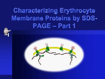
Structure of Blood
Neutrophil (white blood cell) Platelets Red
blood cells Plasma Lymphocyte (white blood cell)
3
Origin of the Formed Elements
4
Erythrocyte Cytoskeleton
Carbohydrate residues
Anion exchanger
Band 4.1
Lipid bilayer
Glycophorin C
Ankyrin
Palidin
a-Spectrin
b-Spectrin
Band 4.9
Adducin
Actin
Tropomodulin
Tropomyosin
5
Etiology of Spherocytosis
HEREDITARY CONDITION OR UNKNOWN MUTAGEN
Normal pluripotent stem cells
Pluripotent stem cells with genetic defect
Phenotypically indistinguishable
Commitment, differentiation
Commitment, differentiation
Spherocyte
Normal erythrocyte
Premature destruction
Hemolysis
spleen
Normal three to four month life span
Anemia, blindness, kidney damage, stroke,
heart damage, premature death
hypothetical
6
Plan of Attack
sick
Cultured stem cells w/defect
Defective bone marrow stem cells
Compare erythrocyte cytoskeletal proteins
Develop erythrocytes in vitro
Obtain whole blood
Replace defective genes
Replace stem cells
normal
Develop erythrocytes in vitro
Altered stem cells
Perfect match?
7
Applied, Basic, and Pure Science
- Applied
- targets a specific problem
- limited applicability to other areas/problems
- valueless unless completely successful
- Basic
- seeks fundamental knowledge in a relevant area
- provides foundation in many areas
- Pure
- can go in any direction
- any possibility can be realized
8
Project Outline
- Blood fractionation
- Isolate erythrocytes from other elements
- Rupture and wash erythrocytes
- Estimate protein concentrations
- SDS-PAGE
- Prepare (denature) samples
- Conduct electrophoresis
- Stain and analyze gels
9
Blood Fractionation
Diluted whole blood
Remove diluted plasma
Resuspend pellet in water, causing cells to
rupture (lyse)
Centrifuge low speed
Wash cells
Red cell pellet
Centrifuge high speed
Several wash steps
Lysed material in suspension
Final membrane pellet
Remove diluted cytoplasm
Membrane pellet
Experimental Biosciences
10
Buffers and Osmosis
direction of water movement
HYPO-osmotic (swollen erythrocyte)
ISO-osmotic (normal biconcave erythrocyte)
HYPER-osmotic (shrunken, or crenated erythrocyte)
11
Centrifugation
Balance caps with tubes
Rotor failure
or else...
Load opposite sides
12
Prepare Washed Erythrocytes
Whole blood sample
Diluted whole blood
Initial separation
Second wash
Second separation
Washed pellet
mark tube
Suspended in buffered NaCl
Centrifuged 500 x g
Removed SN and resuspended
Centrifuged 500 x g
Removed SN
Experimental Biosciences
13
Aliquots and Fraction Yields
First low speed supernatant
Lysate
Record total volume
Measure and record total volume (includes aliquot
volume)
Lyse washed erythrocytes
500 x g
First high speed supernatant
Membrane pellet
Measure and record total volume
Measure and record total volume
After final wash
10,000 x g
14
Lyse the Red Cell Pellet
- Suspend pellet in dilute buffer
- Cells take up water and quickly rupture
- The process is called lysis
- Membranes remain suspended in diluted cytoplasm
15
Isolate the Membranes
Washed red cells
Lysed cell suspension
Initial separation
Wash the membranes free of hemoglobin
Suspended in buffered water
Centrifuged 10,000 x g
Removed SN and resuspended
Centrifuged 10,000 x g
Repeated washes
Experimental Biosciences
16
Recover the Membranes
Final supernatant
Leave stuck to tube
Keep pellet surface level while removing
supernatant
OR
Acceleration
Erythrocyte membranes
Remove using applicator stick
Fibrous pellet
17
Bradford vs. Biuret Assay
- Similarities
- Require protein standards
- Require spectrophotometer
- Require standard curve
- Differences
- Principle of color change
- Bradford is 100x more sensitive
- Bradford requires 3 min incubation
18
Preparing for the Project
- Outline fractionation procedure step by step
- dilutons and buffers
- centrifugation steps
- when to collect aliquots record volumes
- Outline the protein assay
- dilutions for aliquots and standards (pre-lab 2)
- assay procedure
19
Recordkeeping and Teamwork
- Take advantage of the partnership
- divide up the major tasks
- double check each others work
- Take timely notes
- work should be reproducible
- dont wait until the work is finished
- check on each other but dont copy
Recommended
