Mark Markowski, Bowen Cai, Edward Gelmann PowerPoint PPT Presentation
1 / 1
Title: Mark Markowski, Bowen Cai, Edward Gelmann
1
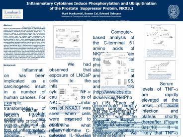
Inflammatory Cytokines Induce Phosphorylation and
Ubiquitination of the Prostate Suppressor
Protein, NKX3.1
Mark Markowski, Bowen Cai, Edward
Gelmann Department of Oncology and Medicine,
Lombardi Comprehensive Cancer Center, Georgetown
University School of Medicine, 3800 Reservoir
Road NW, Washington, D.C. 20007-2197
Computer-based analysis of the C-terminal 51
amino acids of NKX3.1 contain three potential
phosphorylation sites, all proximal to amino acid
200, at serines 185, 195, and 196
(http//www.cbs.dtu.dk/services/NetPhos/) (15).
Each of these serines was individually mutated to
an alanine residue to abrogate each putative
phosphorylation site. In addition we made
compound serine?alanine mutant constructs. The
serine mutants were tested both for protein
turnover and for sensitivity to TNF-a (Fig 3C
bottom). Mutation of serine 185 doubled the
half-life of NKX3.1 and also increased protein
half-life after TNF-a exposure from 25 to 40
minutes. However, NKX3.1(S185A) retained
sensitivity to TNF-a, suggesting that serine 185
had a major influence on protein degradation, but
was not targeted by TNF-a. Mutation of either
serine 195 or serine 196 prolonged protein
half-life. The serine 195 mutation attenuated and
the serine 196 mutation abrogated the effect of
TNF-a on protein degradation since there was no
change in the half-life of NKX3.1(S196A) after
exposure to TNF-a. Mutation of both serines 195
and 196 enhanced the protein half-life and
resistance to TNF-a more than the effect of the
serine 196 mutant alone. The compound mutant with
altered serine 185 and serine 195 showed a
half-life of 110 min, but retained an effect of
TNF-a. In contrast the serine 185, 196 compound
mutant had a prolonged protein half-life and
essentially no TNF-a sensitivity. Lastly,
simultaneous mutation of serines 185, 195, and
196 resulted in a protein with no sensitivity to
TNF-a and a half-life similar to the
NKX3.1(1-183) construct. Thus the effect of
C-terminal truncation on protein turnover and
TNF-a sensitivity was recapitulated by mutation
of three serines. To determine whether any of the
serine mutations affected nuclear localization of
NKX3.1 and thereby affected protein turnover we
expressed each MYC-tagged NKX3.1 construct in
LNCaP cells and determined subcellular
localization by immunohistochemistry. We observed
that all MYC-tagged serine?alanine constructs
localized to the nucleus of LNCaP cells (data not
shown).
Abstract Inflammation of the prostate is a
risk factor for the development of prostate
cancer. In the aging prostate regions of
inflammatory atrophy are foci for prostate
epithelial cell transformation. Expression of the
suppressor protein NKX3.1 is reduced in regions
of inflammatory atrophy and in preinvasive
prostate cancer. Inflammatory cytokines TNF-a and
IL-1ß accelerate NKX3.1 protein loss by inducing
rapid ubiquitination and proteasomal degradation.
The effect of TNF-a is mediated via the
C-terminal domain of NKX3.1 where phosphorylation
of serine 196 is critical for cytokine-induced
degradation. Mutation of serine 196 to alanine
abrogates phosphorylation at that site and the
effect of TNF-a on NKX3.1 ubiquitination and
protein loss. This is in contrast to control of
steady state NKX3.1 turnover which is mediated by
serine 185. Mutation of serine 185 to alanine
increases NKX3.1 stability by inhibiting
ubiquitination and doubling the protein
half-life. A third C-terminal serine at position
195 has a modulating effect on both steady state
protein turnover and on ubiquitination induced by
TNF-a. Thus, cellular levels of the NKX3.1 tumor
suppressor are affected by inflammatory cytokines
that target C-terminal serine residues to
activate ubiquitination and protein degradation.
Our data suggest that strategies to inhibit
inflammation or to inhibit effector kinases may
be useful approaches to prostate cancer
prevention.
Figure 1 C-terminus of NKX3.1 affects protein
stability. A. PC-3 cells transfected with
expression plasmids pcDNA-NKX3.1, or
pcDNA3.1-NKX3.1(1-183) plasmids were exposed to
50 µM cycloheximide for 2 hr. NKX3.1 levels were
assessed by western blotting with anit-NKX3.1
antibody 2 h post treatment. Transfection
efficiency was monitored by cotransfection with a
GFP expression plasmid. B. NKX3.1 and
NKX3.1(1-183) levels were quantitated in LNCaP
cells 2 h after treatment with cycloheximide. C.
LNCaP cells were transfected with pcDNA3 or
pcDNA3.1NKX3.1(1-183) and treated with 100 nM
bortezomib for 6 h. Both endogenous and exogenous
NKX3.1 levels were determined through western
blot analysis with anti-NKX3.1 antibody. D.
NKX3.1 and NKX3.1(1-183) expression in PC-3 cells
was analyzed 6 hr after exposure to bortezomib.
The top panel shows a short exposure and the
bottom panel a long exposure of the same blot.
The higher molecular weight bands, in this blot
detected by NKX3.1 antibody, are consistent with
polyubiquitinated NKX3.1.
.
Figure 4 TNF-a induces phosphorylation of
NKX3.1. A. LNCaP cells, transfected with
MYC-tagged NKX3.1, were pretreated with 100 mM
cycloheximide for 15 min and then exposed to 40
ng/ml TNF-a for 15 min. In the right panel
blotting was done after one aliquot was treated
with CIP. Cells were harvested for
immunoprecipitation with polyclonal anti-MYC
antibody followed by western blotting with either
monoclonal anti-MYC or anti-phosphoserine
antibody. In B. the effect of C-terminal
truncation and in C. S196 and S195 mutations
abolish NKX3.1 phosphorylation induced by TNF-a.
D. MYC-tagged NKX3.1 constructs as indicated were
expressed in LNCaP cells subjected to CHX
treatment for 30 min. Immunoprecipitation was
with anti-MYC antibody and immunoblotting was
done with either anti-phosphoserine antibody or
anti-MYC antibody.
We had observed that exposure of LNCaP cells to
the inflammatory cytokine TNF-a caused rapid loss
of NKX3.1. Similar loss of NKX3.1 was seen when
cells were exposed to another inflammatory
cytokine IL-1ß (Fig 2A). In contrast, no effect
on NKX3.1 levels was seen in response to the
proliferative cytokine IL-6. LNCaP cells are
known to express IL-6 receptors and respond to
IL-6 (13). A MYC-tagged NKX3.1 fusion protein had
a half-life similar to endogenous NKX3.1 in LNCaP
cells treated with cycloheximide indicating that
both endogenous and exogenous MYC-tagged NKX3.1
were subjected to similar mechanisms of protein
turnover. TNF-a accelerated degradation of
full-length MYC-tagged protein. In contrast,
truncation of the C-terminal domain prolonged
protein half-life and conferred resistance to the
effect of TNF-a on protein loss (Fig 2B). The
MYC-tagged NKX3.1 fusion protein was
ubiquitinated in response to TNF-a, but the
C-terminal truncated protein was resistant to
ubiquitination (Fig 2C). Polyubiquitination most
commonly occurs at lysine residues. The
C-terminal domain of NKX3.1 has lysines at
positions 193 and 201. Mutation of either or both
lysine residues to arginines had no detectable
effect either on steady-state turnover or on
TNF-a-induced degradation of NKX3.1 (data not
shown). TNF-a causes apoptosis of LNCaP cells,
but the effect is not seen until more than 48 hr
after exposure to the cytokine (14). To determine
whether caspase activation contributed to NKX3.1
turnover after exposure to TNF-a we treated cells
with the pancaspase inhibitor zVAD-FMK and saw no
effect on the degradation of NKX3.1 within 24 hrs
of exposure to 40 ng/ml TNF-a (data not shown).
Background
Inflammation has been implicated as a
carcinogenic insult in a number of human cancers.
For example, transformation of human prostate
epithelial cells occurs adjacent to foci of
inflammatory atrophy. Inflammation causes the
generation of reactive oxygen species that
increase the risk of oxidative damage of DNA and
generation of mutations (1). Inflammation of the
prostate is a risk factor for the development of
prostate cancer (23). One of the earliest events
in prostate cellular transformation is reduced
expression of the haploinsufficient prostate
specific suppressor protein, NKX3.1. The NKX3.1
gene is subject to loss at chromosome 8p21 and/or
methylation (4). Intracellular levels of the
NKX3.1 protein are reduced in prostate
intraepithelial neoplasia, a noninvasive
precursor to prostate cancer (4) and in regions
of inflammatory atrophy that are precursors to
malignant transformation in the prostate (5).
Control of NKX3.1 protein levels are under the
influence of many factors including those that
result in N-terminal threonine phosphorylation
that results in prolongation of protein half-life
(6). This paper addresses the mechanism by which
NKX3.1 is reduced in regions of prostatic
inflammation. The prostate-specific homeodomain
protein NKX3.1 is expressed in the adult almost
exclusively in the nuclei of luminal prostate
epithelial cells (7). Gene targeting studies in
mice have shown that haploinsufficiency of Nkx3.1
is semidominant since Nkx3.1/- mice develop
prostatic dysplasia with longer latency than
Nkx3.1-/-mice and loss of a single allele
cooperates with Pten loss to accelerate the
development and increase the severity of prostate
cancer (89). That prostate epithelial cells are
subjected to a dose-response of Nkx3.1 protein
levels is underscored by proportionately altered
expression of downstream Nkx3.1 transcription
targets in Nkx3.1/- mice (10). In human prostate
cancer NKX3.1 expression is reduced in primary
disease (4) and completely abrogated in most
metastatic foci, suggesting a continued selection
for loss of the protein during prostate cancer
progression (7). We show here that inflammatory
cytokines TNF-a and IL-1ß accelerate NKX3.1
protein loss by inducing rapid ubiquitination and
proteasomal degradation. The C-terminal domain
distal to the homeodomain of NKX3.1 is not the
site of ubiquitination, but is targeted at
specific serine residues, presumptive
phosphorylation sites, to mediate either
steady-state or cytokine-mediated protein
degradation.
Figure 5 Determinants of NKX3.1 ubiquitination
in steady state turnover and after exposure to
TNF-a. Ubiquitination of wild type and mutant
MYC-tagged NKX3.1 after cells were treated with
bortezomib with or without subsequent exposure to
TNF-a was analyzed in LNCaP cell extracts. An
expression vector for polyhistidine-tagged
ubiquitin was cotransfected into the LNCaP cells.
Ubiquitinated proteins were pulled downed by Ni2
beads and analyzed by western blotting using an
anti-MYC antibody.
Serum levels of TNF-a are rapidly elevated at
the onset of acute infection and plateau shortly
thereafter (Figure 6a).(16) It is likely that
TNF-a causes local tissue destruction at these
high concentrations. As the infection resolves,
the concentration of TNF-a gradually returns to
baseline.(16) In contrast to the drastic rise in
TNF-a serum concentration, the decrease in TNF-a
serum concentration occurs at a slower rate.(16)
Since decreased levels of TNF-a have been shown
facilitate cell growth, the delay in the return
of TNF-a concentrations to baseline may allow for
tissue remodeling.(17) The physiological
purpose of TNF-a-induced NKX3.1 loss may be to
temporarily allow for epithelial cell
proliferation and repair of damaged tissue. As
the inflammatory process resolves, TNF-a levels
decrease and NKX3.1 levels return to normal
(Figure 6b). The balance of TNF-a and NKX3.1
levels may be perturbed during chronic
inflammatory reactions. In the setting of
chronic inflammation with prolonged exposure to
inflammatory cytokines, NKX3.1 levels may be
persistently diminished in prostate epithelial
cells. Decreased NKX3.1 levels may predispose
the epithelium to malignant transformation.
Figure 3 Determinants of NKX3.1 steady
state and TNF-a-induced turnover. A. C-terminal
truncation mutants of NKX3.1 were expressed as
MYC-tagged constructs and cotransfected with a
GFP expression plasmid. Cells were treated with
cycloheximide for 1 hr and processed for western
blotting with anti-MYC antibody. FLfull-length.
B. The effect of bortezomib on levels of full
length and C-terminal truncated MYC-tagged NKX3.1
expression constructs was assayed by western
blot. FLfull-length. C. The MYC-tagged NKX3.1
deletion constructs and point-mutants were
analyzed for half-life after 0, 30, 60 and 120
min of exposure to cycloheximide or to
cycloheximide 40 ng/ml TNF-a. NKX3.1 proteins
were detected by western blotting. Maps of the
mutant constructs are shown at the left. Protein
half-lives during turnover or after exposure to
TNF-a are shown at the right. At least three
separate determinations were done for each value.
Mean /- standard deviations are shown for each
half-life.
To demonstrate directly that the C-terminus of
NKX3.1 was the target for TNF-a induced
phosphorylation, we performed immunodetection of
phosphoserine on MYC tagged NKX3.1. LNCaP cells
were transfected with a MYC-NKX3.1 expression
vector and pretreated with cycloheximide prior to
15 min exposure to TNF-a. As shown in the left
panel of Figure 4A, exposure to TNF-a induced the
presence of phosphoserine residues that were
sensitive to treatment with CIP (right panel
Figure 4A). TNF-a induced serine phosphorylation
only on the C-terminus as shown in Figure 4B
since C-terminal truncation abolished
immunodetection with anti-phosphoserine antibody.
In contrast, deletion of the N-terminal domain
upstream from the homeodomain did not affect
TNF-a-induced serine phosphorylation. Mutation of
serine 196 to alanine specifically abrogated
TNF-a-induced serine phosphorylation. The effect
was not changed by concurrent mutation of serine
195 to alanine (Figure 4C). We were also able to
show that in the presence of cycloheximide alone
mutation of serine 185 to alanine diminished
detection with the anti-phosphoserine antibody
whereas mutation of serines 195 and 196 or
C-terminal truncation to amino acid 192 had no
effect on NKX3.1 serine phosphorylation in the
presence of cycloheximide. Lastly, the
C-terminal serine mutations were also found to
decrease the polyubiquitination of NKX3.1 in the
presence of bortezomib and in response to TNF-a
(Fig 5). Thus the C-terminal serines determined
both ubiquitination and protein loss after
exposure of LNCaP cells to TNF-a.
Figure 6 TNF-a levels in normal and chronic
inflammation. A. TNF-a concentration plotted
over the course of both normal and chronic
inflammatory processes. TNF-a concentration is
divided into three sections and the effect of
each concentration range on cellular outcome is
indicated. B. Chronological change in NKX3.1
expression paralleling TNF-a concentration during
both a normal (top) and chronic (bottom)
inflammatory reaction.
Results
Figure 2 TNF-a and IL-1ß induce NKX3.1
degradation. A. Endogenous NKX3.1 levels in
LNCaP cells after exposure to 40 ng/ml TNF-a,
IL-1ß or IL-6. Relative levels of NKX3.1 were
normalized to ß-actin levels and quantitated in
the graph. B. TNF-a is shown to accelerate NKX3.1
protein turnover, but have no effect on
NKX3.1(1-183). MYC-tagged NKX3.1 expression
plasmids were transfected into LNCaP cells. Cells
were treated with 100 µM cycloheximide for 1 hour
and then exposed to 40 ng/ml TNF-a. C. The effect
of TNF-a on ubiquitination of MYC-tagged NKX3.1
and NKX3.1(1-183) in LNCaP is shown. Cells were
transfected with MYC-tagged NKX3.1 expression
plasmids and with a polyhistidine-tagged
ubiquitination expression plasmid. Cells were
treated for 1 hr with bortezomib and then for 6
hr with TNF-a. Cells lysates were exposed to
Ni2-charged agarose beads and then subjected to
western blotting with anti-MYC antibody. Input
levels of NKX3.1 were determined by western blot
analysis of each total cellular lysate prior to
the addition of Ni2 beads.
Cellular NKX3.1 protein levels are critical for
maintenance of the prostate epithelial phenotype.
We therefore characterized intracellular protein
turnover to determine the half-life of NKX3.1. In
PC-3 prostate cancer cells exogenously expressed
NKX3.1 has a half-life of approximately one hour
(Fig 1A). Members of the NK family of homeodomain
proteins such as Nkx2.5 and Nkx3.1 have been
shown to have increased levels of expression and
of protein activity after removal of the peptide
domain that lies C-terminal to the homeodomain
(1112). Therefore we measured the half-life of a
C-terminal truncated NKX3.1 lacking the 51 amino
acids downstream from the homeodomain and
observed a prolonged half life (Fig 1A). LNCaP
prostate cancer cells are one of the few cell
lines that express endogenous NKX3.1. In LNCaP
cells exogenous MYC-tagged NKX3.1 has a half life
of approximately 60 min whereas the C-terminal
truncated protein had a half life of nearly 4
hours (Fig 1B). We also examined turnover of
endogenous NKX3.1 in LNCaP cells by treating
cells with bortezomib, a reversible proteasome
inhibitor that prolonged half-life of endogenous
NKX3.1, but had no effect on the level of
exogenous C-terminal truncated protein (Figure
1C). Bortezomib also blocked turnover of
exogenous NKX3.1 expressed in PC-3 prostate
cancer cells. Moreover, in the presence of
bortezomib higher molecular weight moieties of
NKX3.1 were seen that represented
polyubiquitinated NKX3.1 accumulating in PC-3
cells. Under the same conditions no
ubiquitination of NKX3.1(1-183) was seen (Figure
1D).
To determine what region of the C-terminal
domain influenced NKX3.1 stability we engineered
a series of MYC-tagged deletion constructs whose
stability were tested in LNCaP cells. Deletion at
amino acids 216, 208, or 200 had no effect on
steady-state turnover of NKX3.1. Deletion at
amino acid 192 prolonged half-life to a lesser
degree than seen with deletion to amino acid 183
(Figure 3A). The NKX3.1 constructs truncated at
amino acids 192 and 183 were also less sensitive
to the effect of bortezomib on protein turnover
(Fig 3B). Truncation at amino acid 192 resulted
in an increased protein half-life and abrogation
of the effect of TNF-a (Fig 3C top). MYC-NKX3.1
truncation extended to amino acid 183 caused
further increase in protein half-life.
- Conclusions
- NKX3.1 levels are regulated by phosphorylation
dependent ubiquitination. - Serine 185 mediated steady-state cellular
turnover and serine 196 regulated TNF-a induced
protein degradation with serine 195 having a
modulating effect on serines 185 and 196. - Inflammation may accentuate the degradation of
the tumor suppressor protein, NKX3.1,
predisposing the epithelium to malignant change. - This data suggests that anti-inflammatory agents
may have a role in prostate cancer prevention. - Identification of the kinase that targets the
NKX3.1 C-terminal domain may be a future target
for a for a prostate cancer prevention or
treatment strategy.
References 1. De Marzo AM et al Nat Rev Cancer
2007 Apr7(4)256-69. 8. Bhatia-Gaur R
et al Genes and Development 199913966-77.
15. Blom N et al J Mol Biol 1999 Dec
17294(5)1351-62 2. Blumenfeld
W et al Am J Surg Pathol 1992 Oct16(10)975-81.
9. Kim MJ et al Proc Natl Acad Sci U S
A 2002 Mar 599(5)2884-9. 16.
Boontham P et al Surgeon 2003 1(4)187-206.
3. De Marzo AM et al Urology 2003 Nov62(5
Suppl1)55-62. 10. Magee JA et al Cancer
Cell 2003 Mar3(3)273-83.
17. Balkwill F et al Lancet 2001
357(9255)539-545. 4. Asatiani E et al Cancer
Res 2005 Feb 1565(4)1164-73. 11. Chen
CY et al J Biol Chem 1995270(26)15628-33. 5.
Bethel CR et al Cancer Res 2006 Nov
1566(22)10683-90. 12. Carson
JA et al J Biol Chem 2000 Dec 15275(50)39061-72.
6. Li X et al Mol Cell Biol 2006
Apr26(8)3008-17.
13. Hobisch A et al Cancer Res 1998 Oct
1558(20)4640-5. 7 Bowen C et al, Cancer Res
2000 Nov 160(21)6111-5.
14. Kimura K et al Cancer Res 199959(7)1606-14.

