
Ultrasound Case: Chronic Stenosing Tenosynovitus PowerPoint PPT Presentation
1 / 2
Title: Ultrasound Case: Chronic Stenosing Tenosynovitus
1
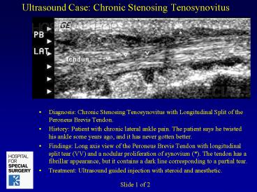
Ultrasound Case Chronic Stenosing Tenosynovitus
- Diagnosis Chronic Stenosing Tenosynovitus with
Longitudinal Split of the Peroneus Brevis Tendon. - History Patient with chronic lateral ankle pain.
The patient says he twisted his ankle some years
ago, and it has never gotten better. - Findings Long axis view of the Peroneus Brevis
Tendon with longitudinal split tear (VV) and a
nodular proliferation of synovium (). The tendon
has a fibrillar appearance, but it contains a
dark line corresponding to a partial tear. - Treatment Ultrasound guided injection with
steroid and anesthetic.
Slide 1 of 2
2
Ultrasound Case Chronic Stenosing Tenosynovitus
- Diagnosis Chronic Stenosing Tenosynovitus with
Longitudinal Split of the Peroneus Brevis Tendon. - History Patient with chronic lateral ankle pain.
The patient says he twisted his ankle some years
ago, and it has never gotten better. - Findings Power doppler image shows increased
vascularity of the inflamed synovium surrounding
the abnormal Peroneus Brevis tendon. - Treatment Ultrasound guided injection with
steroid and anesthetic
Slide 2 of 2
Recommended
