
Abstract PowerPoint PPT Presentation
1 / 1
Title: Abstract
1
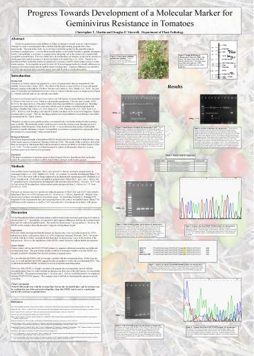
Progress Towards Development of a Molecular
Marker for Geminivirus Resistance in
Tomatoes Christopher T. Martin and Douglas P.
Maxwell, Department of Plant Pathology.
Abstract Each year geminiviruses cause
millions of dollars in damage to tomato crops in
Central America. Attempts to create a resistant
plant with a suitable fruit through breeding
programs have been unsuccessful. The goal of
this study was to develop a molecular marker for
the gene that controls resistance to
geminiviruses. A successful molecular marker
would enable breeders to quickly determine a
tomatos susceptibility or resistance to
geminiviruses and greatly aid in the creation of
a commercially acceptable resistant hybrid.
Previous studies have shown that there are
hotspots on the tomato genome in which genes that
control resistance to disease are likely to be
found (Pan et al., 1999). Therefore we
hypothesized that a molecular marker for
geminivirus resistance could be found within a
hotspot on the tomato genome. To accomplish our
goal we used a PCR-based tagging method to
identify differences in sequences between
resistant and susceptible tomato breeding lines.
Sequence differences are indicative of a DNA
introgression from a resistant species and could
be used as a molecular marker.
Figure 2 Sample RFLP map of Chromosome 1 (Pan et
al., 1999). This figure is the upper portion of
the RFLP map for chromosome 1. Resistance genes
are in bold on the right side. RFLP markers are
in smaller type, also on the right side.
Figure 1 Tomato plant exhibiting geminivirus
symptoms. Common symptoms include leaf curling
and crumpling, yellow color, and a reduced size
of the plant and its fruit.
- Introduction
- Background
- Tomatoes in Central America are plagued by a
series of geminiviruses that are transmitted by
the whitefly, Bemisia tabaci (Jones, 2003). The
effect of the disease is near total loss of crops
and annual damages ranging in the millions of
dollars (Morales and Anderson, 2001 Nakhla et
al., 2004). In some areas of Nicaragua and
Guatemala losses have been so extensive that the
crop is no longer grown (Figure 1). Suitable
resistant cultivars are currently unavailable. - Lycopersicon hirsutum and Lycopersicon chilense
are wild species of tomato that have shown
resistance to Tomato yellow leaf curl virus,
which is a monopartite geminivirus (Vidavsky and
Czosnek, 1998). However, the shape and size of
the plants fruits make them unsuitable for
commercial use. Breeding programs have been
underway for some time with the goal of creating
a resistant hybrid plant that produces a healthy
fruit (Chen et al., 2003 Mejia et al., 2004
Narasegowda et al., 2003 Scott et al., 1995).
However, each breeding cycle takes five months
and there can be an incorrect diagnosis of plant
resistance due to escapes. Thus far, breeding
programs have been ineffective in producing a
successful resistant hybrid for Central America. - Therefore, in order to more quickly produce a
resistant hybrid, a molecular marker for the
resistance gene is needed. The molecular marker
could be used to track the resistance gene
through successive generations with Polymerase
Chain Reaction (PCR). A successful molecular
marker would enable breeders to quickly determine
a tomatos susceptibility or resistance to
geminiviruses and greatly aid in the creation of
a commercially viable resistant hybrid. - Biological Rationale
- Restriction fragment length polymorphism
(RFLP)-based probes have been used to help
develop a map of the tomato genome (Solanaceae
Genomics Network, 2004). The results of this
work have shown that there are hotspots in which
genes that control resistance to disease are
likely to be found (Figure 2) (Pan et al., 1999).
For this research, we defined hotspots as a
place on the genome where two or more resistance
genes are located in close proximity. - Hypothesis
- The high concentration of resistance genes in
these hotspots led us to hypothesize that a
molecular marker for geminivirus resistance could
be found within a hotspot on the tomato genome.
Results
600bp
300bp
Figure 3 Initial Primers developed for
chromosomes 1 and 7. Eleven different primer
combinations were run with Heinz 1706 DNA under
standard reaction conditions developed in the
Maxwell lab (Czosnek et al., 2004). The
PCR-amplified DNA was run on an electrophoresis
gel of 1.5 agarose in 0.5X TBE buffer, stained
with ethidium bromide, and visualized with a
Kodak Gel Logic 200 Imaging System. These primers
were designed around RFLP markers TG301 and
TG149, which are located on chromosomes 1 and 7,
respectively. TG301 Primers are in lanes 2-8 and
TG149 primers are in lanes 9-12. Lane 13 is a
positive control and lane 14 is a negative
control. Several of the primer pairs developed
for chromosome 1 gave bands suitable for use in
sequencing reactions, but primer pair
P301F3/P301R2 gave the best band and was used for
a sequencing reaction. None of the primers
developed for chromosome 7 gave bands. As a
result, a second set of primers were designed
around chromosome 7.
Figure 4 Second set of primers for chromosome 7.
Eight different primer combinations were run
with Gh13B DNA under standard reaction conditions
developed in the Maxwell Lab (Czosnek et al.,
2004). The PCR-amplified DNA was run on an
electrophoresis gel of 1.5 agarose in 0.5X TBE
buffer, stained with ethidium bromide, and
visualized with a Kodak Gel Logic 200 Imaging
System. These primers were designed from RFLP
markers TG662 and TG143, which are both located
on the long arm of chromosome 7. The TG143
primers are in lanes 2-5 and the TG662 primers
are in lanes 6-9. Lane 10 is a positive control
and lane 11 is a negative control. Two of the
primer pairs designed from TG143 gave bands
suitable for use in sequencing reactions, but
none of the TG662 primers gave bands suitable for
use in sequencing reactions. P143F2/P143R1 gave
the best band, and was used for a sequencing
reaction.
Table 1 Primers used for sequencing
- Methods
- We used the tomato breeding lines, Gh13, Gc9, and
Gc173, that are resistant to geminiviruses in
Guatemala (Mejía et al., 2004 Nakhla et al.,
2004). As a control, we used the breeding line
Heinz 1706. Heinz 1706 is the tomato cultivar
being sequenced in an international sequencing
project (Budiman et al., 2000 Ozminkowski,
2004), and is susceptible to geminiviruses
(Maxwell, D., pers. com.). Gh13 is the F7
generation and is a homogeneous breeding line
with resistance derived from L. hirsutum. Gc173
and Gc9 are at least F8 breeding lines with
resistance genes introgressed from L. chilense by
J. W. Scott (Scott et al., 1995). - Hotspots on chromosomes two and eleven within the
genome of Gh13, Gc9, and Gc173 were tested to
determine if there was a DNA introgression of L.
hirsutum or L. chilense, respectively. Hotspots
were chosen based on their concentration of
resistance genes. We tested these hotspots by
obtaining PCR fragments for the experimental
lines and comparing them to the control,
susceptible tomato, Heinz 1706. Differences in
the sequences as small as 3-4 were indicative of
an introgression from a wild species.
600bp
- Discussion
- We had hypothesized that a molecular marker could
be found in the resistance gene hotspots located
on chromosomes1 or 7. Specifically, we expected
to find sequence differences between the
resistant tomato lines and our control
susceptible tomato. The results for chromosome 7
are inconclusive. However, the INDEL in the
sequence from chromosome 1 supports our
hypothesis in part. - Implications
- Previous studies had indicated that the hotspot
on chromosome 1 was a possible location for a DNA
introgression from a wild species (Pan et al.,
1999 Solanaceae Genomics Network, 2004). Our
results correlate with these studies, and
indicate that the hotpot on chromosome 1 may be
the location of the introgression. However, the
significance of the INDEL cannot be known without
further investigation. - Future Studies
- Future studies will use the P301F3/P301R2 primers
to sequence additional susceptible and additional
resistant plants lines. The goal of these
studies would be to determine whether or not the
INDEL was strongly correlated with plants that
showed resistance to geminiviruses. - It is possible that the INDEL will not strongly
correlate with the resistant plant lines. If
this were the case, we would find that the INDEL
appeared in the susceptible as well as the
resistant plant DNA. This would indicate that
the INDEL we found was not an acceptable
molecular marker. - However, if the INDEL is strongly correlated with
geminivirus resistant plants and not with the
susceptible plants, then we could conclude an
introgression from one of the wild species was
responsible for the INDEL. The parent resistant
plants L. hirsutum and L. chilense would then
need to be sequenced with the P301F3/P301R2
primers. This sequence data would tell us which
plant the introgression had come from. - Final Conclusion
- If the INDEL holds true with the resistant lines
but not the susceptible lines, and its existence
can be confirmed in one of the parent breeding
lines, then this INDEL can be used as a molecular
marker for resistance to geminiviruses.
Figure 5 P301F3/P301R2 primers for the hotspot
on chromosome 1. The primers were based on RFLP
marker TG301, which is located on the short arm
of chromosome 1. PCR reactions were run under
conditions developed in the Maxwell lab (Czosnek
et al., 2004). The PCR-amplified DNA was run on
an electrophoresis gel of 1.5 agarose in 0.5X
TBE buffer, stained with ethidium bromide, and
visualized with a Kodak Gel Logic 200 Imaging
System. Lane 2 is a negative control, and lanes
3-6 contain the PCR reaction mixture. The DNA
produced strong bands at 550 bp. As a result, it
was directly sequenced.
Figure 6 Sequence data from P301F3/P301R2
primers for chromosome 1. Analysis of sequence
data for the individual plants was accomplished
using the CHROMAS software. The above picture is
representative of the type of sequence that the
P301F3/P301R2 primers produced. The peaks were
strong, and the overall sequence was very clear.
As a result, the sequences of the different plant
lines were compared against one another.
Figure 7 Analysis of sequence from P301F3/P301R2
primers for chromosome 1. Comparison of the
sample sequences from the four different plant
lines was accomplished with the DNAMAN software
(Lynnon Corp., Quebec, Canada). This analysis
showed a 99 identity among the sequences. The
only discrepancy appeared in the form of an INDEL
at roughly the 415th bp.
References 2004. Tomato-Arabidopsis Synteny
Map. Solanaceae Genomics Network. Cornell
University. o_arabidopsis/synteny_map.html (November 5,
2004). Budiman, MA., Mao, L., Wood, TC., and
Wing, RA. 2000. A deep-coverage tomato BAC
library and prospects toward development of an
STC framework for genome sequencing. Genome Res.
10129-136. Chen, J.T., Hanson, P.M., Kuo, G.,
and Opena, R.T. 2003. Genetic improvement of
summer fresh market tomatoes. J. Agri. Assoc.
China 483-102. Jones, D.R. 2003. Plant
viruses transmitted by whiteflies. Eur. J. Plant
Path. 109195-219. Mejía, L., Teni, R.E.,
Vidavski, F., Czosnek, H., Lapidot, M., Nakhla,
M.K., and Maxwell D.P. 2004. Evaluation of
tomato germplasm and selection of breeding lines
for resistance to begomoviruses in Guatemala.
Acta Hort. (in press). Morales, F.J. and
Anderson, P.K. 2001. The emergence and
dissemination of whitefly-transmitted
geminiviruses in Latin America. Arch. Virol.
146415-441. Nakhla, M., Sorenson, A., Mejía,
L., Ramírez, P., Karkashian, J.P., and Maxwell,
D. 2004. Molecular Characterization of
Tomato-Infecting Begomoviruses in Central America
and Development of DNA-Based Detection Methods.
International Plant Virology Laboratory.
A-Final.htm (October 5, 2004). Narasegowda,
M.M., Czosnek, H., Vidavski, F., Tarba, S., Milo,
J., Leviatov, S., Mallithimmaiah, V.H.,
Seetharam, P.A., Subbappa, K.R., and Muniyappa,
V. 2003. Comparison of resistance to tomato
leaf curl virus (India) and tomato yellow leaf
curl virus (Israel) among lycopersicon wild
species, breeding lines and hybrids. Eur. J.
Plant Path. 1091-11. Omnikowski, R. 2004.
Pedigree of variety Heinz 1706. Report of the
Tomato Genetics Cooperative 54 27. Pan, Q.,
Liu, Y., Budai-Hadrian, O., Sela, M.,
Carmel-Goren, L., Zamir, D., and Fluhr, R. 1999.
Comparative genetics of nucleotide binding
site-leucine rich repeat resistance gene
homologues in the genomes of two dicotyledons
tomato and arabidopsis. Genetics Society of
America 88309-322. Scott, J.W., Stevens, M.R.,
Barten, J.H.M., Thome, C.R., Polston, J.E.,
Schuster, D.J. and Serra, C.A. 1995.
Introgression of resistance to whitefly-transmitte
d geminiviruses from Lycopersicon chilense to
tomato. Taxonomy, Biology, Damage Control and
Management, Ed. by D. Gerling and R.T. Mayer,
Intercept Ltd., Andover, UK. p.
357-367. Vidavsky, F. and Czosnek, H. 1998.
Tomato breeding lines resistant and tolerant to
tomato yellow leaf curl virus issued from
Lycopersicon hirsutum. Phytopathology 88910-914.
300bp
Figure 8 P143 F2/P143R1 primers for the hotspot
on chromosome 7. The primers were based on RFLP
marker TG143, which is located on the long arm of
chromosome 7. PCR reactions were run under
conditions developed in the Maxwell lab (Czosnek
et al., 2004). The PCR-amplified DNA was run on
an electrophoresis gel of 1.5 agarose in 0.5X
TBE buffer, stained with ethidium bromide, and
visualized with a Kodak Gel Logic 200 Imaging
System. The DNA produced medium strength bands at
roughly 300 bp. As a result these bands were
directly sequenced.
Figure 9 Sequence data from P143F2/P143R1
primers for chromosome 7. The quality of the
sequence data was evaluated using the CHROMAS
software. The above picture is representative of
the overall sequence. The P143F2/P143R2 primers
produced strong peaks, but the net sequence was
unclear. It appears that these primers produced
two different DNA fragments of roughly equal
size. As such, this sequence was not usable.
Recommended
