
2.2 Pericoronal radiolucencies PowerPoint PPT Presentation
Title: 2.2 Pericoronal radiolucencies
1
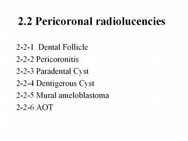
2.2 Pericoronal radiolucencies
- 2-2-1 Dental Follicle
- 2-2-2 Pericoronitis
- 2-2-3 Paradental Cyst
- 2-2-4 Dentigerous Cyst
- 2-2-5 Mural ameloblastoma
- 2-2-6 AOT
2
2-2-1 Dental follicle
- Well-defined, unilocular,
- round-shaped radiolucence
- A well-corticated margin
- Surround the crown of
- an impact tooth
- Size
3
2-2-1 Dental follicle
- dental folliclea supporting tissue
- in the period of tooth developing
- cap stage?bell stage
- impaction of upper canine
4
2-2-2 Pericoronitis
- locate at distal aspect of lower 3rd molar
- extend from distal aspect of the lower 3rd
molars crown to the ramus of mandibular bone - radiolucence
- poor-defined
- surrouned by
- soft tissue (gingiva)
- the crown of tooth 38.
5
2-2-2 Pericoronitis
- Sitedistal aspect of lower 3rd molar
- The space for developing lower 3rd molar is not
enough. - Failure degeneration of enamel epithelium
- (??ppt)
Recommended
