
Apoptza v imunitnm systmu PowerPoint PPT Presentation
1 / 53
Title: Apoptza v imunitnm systmu
1
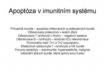
Apoptóza v imunitním systému
- Prirozená imunita apoptóza infikovaných a
pokozených bunek - Diferenciace prekurzoru v kostní dreni
- Diferenciace T lymfocytu v thymu negativní
selekce - Diferenciace B lymfocytu kontrola VDJ
rekombinace - Rovnováha mezi Th1 a Th2 populacemi
- Efektorová cytotoxicyta T lymfocytu a NK bunek
- Poruchy apoptózy autoimunita, hyperreaktivita,
malignity - odtranení starých bunek, které splnily svou
funkci
2
Insights into Pathogen Immune Evasion Mechanisms
Anaplasma phagocytophilum Fails to Induce an
Apoptosis Differentiation Program in Human
Neutrophils.Borjesson DL, Kobayashi SD, Whitney
AR, Voyich JM, Argue CM, Deleo FR.Department of
Veterinary Population Medicine, College of
Veterinary Medicine, University of Minnesota, St.
Paul, MN 55108.Polymorphonuclear leukocytes
(PMNs or neutrophils) are essential to human
innate host defense. However, some bacterial
pathogens circumvent destruction by PMNs and
thereby cause disease. Anaplasma phagocytophilum,
the agent of human granulocytic anaplasmosis,
survives within PMNs in part by altering normal
host cell processes, such as production of
reactive oxygen species (ROS) and apoptosis. To
investigate the molecular basis of A.
phagocytophilum survival within neutrophils, we
used Affymetrix microarrays to measure global
changes in human PMN gene expression following
infection with A. phagocytophilum. Notably, A.
phagocytophilum uptake induced fewer
perturbations in host cell gene regulation
compared with phagocytosis of Staphylococcus
aureus. Although ingestion of A. phagocytophilum
did not elicit significant PMN ROS,
proinflammatory genes were gradually
up-regulated, indicating delayed PMN activation
rather than loss of proinflammatory capacity
normally observed during phagocytosis-induced
apoptosis. Importantly, ingestion of A.
phagocytophilum failed to trigger the neutrophil
apoptosis differentiation program that typically
follows phagocytosis and ROS production.
Heat-killed A. phagocytophilum caused some
similar initial alterations in neutrophil gene
expression and function, which included delaying
normal PMN apoptosis and blocking Fas-induced
programmed cell death. However, at 24 h,
down-regulation of PMN gene transcription may be
more reliant on active infection. Taken together,
these findings suggest two separate antiapoptotic
processes may work concomitantly to promote
bacterial survival 1) uptake of A.
phagocytophilum fails to trigger the apoptosis
differentiation program usually induced by
bacteria, and 2) a protein or molecule on the
pathogen surface can mediate an early delay in
spontaneous neutrophil apoptosis.
3
Figure 1. Morphological features of autophagic,
apoptotic and necrotic cells. (a) Normal, (b)
autophagic, (c) apoptotic (d) and necrotic cells.
Whereas the morphologic features of apoptosis are
well defined, the distinction between necrotic
and autophagic death is less clear. The
bioenergetic catastrophe that culminates in
cellular necrosis also stimulates autophagy as
the cell tries to correct the decline in ATP
levels by catabolizing its constituent molecules.
Thus, vacuolation of the cytoplasm is observed in
both autophagic cells (b) and in cells stimulated
to undergo programmed necrosis (d). By contrast,
ATP levels are maintained in normal (a) and
apoptotic cells (c) consistent with the limited
number of autophagic vacuoles in their cytoplasm.
The scale bar represents 1 µm.
4
Figure 2. Viral infection can induce programmed
necrosis. Binding of the inflammatory cytokine,
TNF, to its receptor results in either apoptosis
or in programmed necrosis in cells in which the
apoptotic pathway is disabled. Many viruses carry
genes that inhibit apoptosis thereby prolonging
the life of their host cell and foiling the
cell's attempt to limit its use as a
virus-producing factory by initiating apoptosis.
A recent study 15 indicates that viruses also
carry genes to suppress programmed necrosis, the
back-up mechanism that infected cells use for
suicide. These observations establish a
physiologic role for programmed necrosis in
mammalian cells.
5
Figure 3. DNA damage and PARP activation runs a
metabolic test on cells resulting in programmed
necrosis selectively in proliferating cells. (a)
DNA damage results in the depletion of
cytoplasmic NAD owing to the PARP-dependent
modification of nucleosomal proteins with
ADP-ribose chains enzymatically derived from NAD.
These modifications expose the damaged DNA and
assist in the targeting of DNA repair complexes
to the site of the damage. The depletion of
cytoplasmic NAD results in the inhibition of
glycolysis as NAD is required in the glycolytic
pathway for the conversion of glyceraldehyde
3-phosphate to 1,3-bisphosphoglycerate. Thus,
following PARP activation and NAD depletion
glucose can no longer be converted to the
pyruvate needed to fuel oxidative phosphorylation
in the mitochondrion. (b) The loss of the ability
to oxidize glucose for energy creates a situation
in which cells must oxidize alternative
substrates such as amino acids and fatty acids.
Proliferating cells are committed to using amino
acids and lipids to building new proteins and
membranes respectively, and do not have the
metabolic programs in place to switch to using
these substrates to fuel ATP production. Thus,
ATP levels decline below the level compatible
with the operation of plasma membrane ion
transporters and the cell dies by necrosis.
Vegetative cells, in contrast, are not committed
to a high rate of macromolecular synthesis and
are able to divert amino acids and fatty acids
into pathways leading to their oxidation in the
mitochondrion. This maintains intracellular ATP
levels and allows DNA repair.
6
Protivirová odpoved bunek
- Apoptóza (aktivace checkpoint proteinu, Viry
produkují IAPs inhibitors of apoptosis napr.
Bcl-2 homology, inaktivátory kaspáz,inakt. P53,
neutralizace TNF beta, sekrece homologu TNF
receptoru, internalizace FAS, virové FLIPs
FLICE (kasp. 8) inhibitory proteins
7
(No Transcript)
8
(No Transcript)
9
(No Transcript)
10
(No Transcript)
11
(No Transcript)
12
(No Transcript)
13
(No Transcript)
14
(No Transcript)
15
(No Transcript)
16
(No Transcript)
17
Figure 23-50. Current models of the intracellular
pathways leading to cell death by apoptosis or to
trophic factor mediated cell survival in
mammalian cells. The details of these pathways in
any given cell type are not yet known. (a) In the
absence of a trophic factor. Bad, a soluble
pro-apoptotic protein, binds to the
anti-apoptotic proteins Bcl-2 and Bcl-xl, which
are inserted into the mitochondrial membrane. Bad
binding prevents the anti-apoptotic proteins from
interacting with Bax, a membrane-bound
pro-apoptotic protein. As a consequence, Bax
forms homo-oligomeric channels in the membrane
that mediate ion flux. Through an as-yet unknown
mechanism, this leads to the release of
cytochrome c from the space between the inner and
outer mitochondrial membrane. Cytochrome c then
binds to the adapter protein Apaf-1, which in
turn promotes a caspase cascade leading to cell
death. (b) In the presence of a trophic factor
such as NGF. In some cells, binding of trophic
factors stimulates PI-3 kinase activity, leading
to activation of the downstream kinase Akt, which
phosphorylates Bad. Phosphorylated Bad then forms
a complex with the 14 - 3 - 3 protein. With Bad
sequestered in the cytosol, the antiapoptotic
Bcl-2/Bcl-xl proteins can inhibit the activity of
Bax, thereby preventing the release of cytochrome
c and activation of the caspase cascade. Adapted
from B. Pettman and C. E. Henderson, 1998, Neuron
20633.
18
T Helper Cell Differentiation and Function
Antigen
APC
IL-12
19
A Model Depicting the Role of Apoptosis in Th1
and Th2 Balance
DC1
DC2
TCR
?
MHCAg
MHCAg
IL-12
Thp
IL-4
Stat 4 ERM T-bet
Stat 6 GATA-3 c-Maf IRS
Th2
Th1
FLIP
Suicide Fratricide
TRAIL
Fratricide
CD95L
20
(No Transcript)
21
(No Transcript)
22
(No Transcript)
23
(No Transcript)
24
2 pathways by which a lymphocyte can kill a
target cell
nucleus
nucleus
taken from Raff Nature 1998396119 Fig 2
25
(No Transcript)
26
A natural killer (NK) cell attacking a cancer
cell. The NK cell is the smaller cell on the
left. This scanning electron micrograph was taken
shortly after the NK cell attached, but before it
induced the cancer cell to kill itself. (Courtesy
of J.C. Hiserodt, in Mechanisms of Cytotoxicity
by Natural Killer Cells R.B. Herberman and D.
Callewaert, eds.. New York Academic Press,
1995.)
27
Mice with deficiencies or mutations in CTL death
pathways
28
Cytotoxic Lymphocyte Granule Components
- Perforin/Cytolysin
- Granzymes
- Granzyme A, Granzyme B, Granzymes C-H
- Serglycin/Chondroitin sulfate proteoglycan
- Granulysin
- Lysosomal enzymes
- Proteases, Cathepsins BHLC(W)
- Cathepsins DE
- Glycosidases, Other degradative
enzymes - Chemokines
- MIP1-a, MIP1-b, RANTES
- Calreticulin
- Granule membrane proteins
- CTLA-4
- LAMPs
- Man-6-PR
29
Granzymes A subfamily of serine proteases found
in the secretory granules of hematopoietic cells
Hallmark properties 1) 25-30kd glycoproteins
with a PHSRPYM motif near the NH2 terminus which
interacts with granule proteoglycans 2)
Endopeptidase activity with neutral pH optima,
suggesting they operate after exocytosis
Cytotoxic T lymphocyte granules contain 1)
Granzyme A, with tryptase specificity cleaving
after arg or lys residues. Measured by the
sensitive BLT esterase assay. 2) Granzyme B, with
an unusual aspase specificity cleaving after
asp residues. Can process and activate caspases
3,7,8,910. 3) Granzymes C-G, expressed at lower
levels, with poorly defined chymase
specificity. Not detectable in CTL in vivo.
30
Figure 10.19. T cells of the mucosal immune
system bearing gd T-cell receptors and an
activating NK receptor recognize and kill injured
enterocytes. Infection or other injury to
enterocytes, the epithelial cells lining the
lumen of the gut, stimulates a stress response,
which causes expression on the cell surface of
two atypical MHC class IB molecules, known as
MIC-A and MIC-B. Intraepithelial T cells carrying
the NK receptor NKG2D bind MIC-A and MIC-B and
induce apoptosis in the injured enterocytes. The
dying enterocyte is removed from the epithelium
and the local tissue injury is repaired.
31
(No Transcript)
32
(No Transcript)
33
(No Transcript)
34
(No Transcript)
35
(No Transcript)
36
(No Transcript)
37
Dissecting the Death Receptor Pathway A
deficiency of caspase-8 is embryonic lethal in
mice possibly due to a cardiac defect. Embryonic
fibroblasts obtained from caspase-8-deficient
mice are completely resistant to death receptor
induced apoptosis. 56 Caspase-8, therefore, is an
essential element of the death receptor pathway
despite reports that caspase-2 might be able to
substitute for it in receptor-associated
apoptosis. 126 Studies using transgenic mice that
express CrmA, an inhibitor of caspase-8, in
lymphocytes show that these lymphocytes are
resistant to death receptor-induced apoptosis. 54
Mice that have defects in FasL or Fas show a
similar resistance to death receptor-induced
apoptosis, but in addition these mice develop
T-cell hyperplasia and high levels of
autoantibodies. 33 Mice deficient in FADD die
during embryogenesis with a phenotype similar to
that of caspase-8-deficient mice. 50, 53
Interestingly, mature T cells that express a
dominant interfering mutant of FADD (FADD-DN) or
lack FADD show a reduced proliferative potential
in response to mitogens or antigens. 50, 52 In
contrast, lymphocytes from transgenic animals
expressing CrmA proliferate normally in response
to mitogenic stimulation, as do lymphocytes from
mice with defective FasL or Fas. 51, 54 Although
the phenotype of the caspase-8-deficient mouse
lends support to the theory that caspase-8 may be
involved in the control of cell proliferation,
the CrmA transgenic studies indicate that
caspase-8 does not have a critical role in cell
proliferation. Thymocyte development and
selection is dysregulated when the function of
FADD is blocked. At an early stage of development
CD3 4 8 pro-T cells differentiate to
become CD48 thymocytes after assembly of a
functional T cell receptor b chain. Those cells
that are unable to assemble a functional TCR b
chain are culled at the pre-TCR checkpoint, but
this is not the case in thymocytes expressing
FADD-DN or in FADD-deficient pro-T cells from
chimeric mice. 132, 133 Interestingly, this
phenomenon is not seen in mice lacking Fas, which
indicates that other (death) receptors must be
involved in this culling process. The normal
proliferation of thymocytes as they progress from
the CD3 4 8 pro-T to the CD348 thymocyte
stage is severely impaired by FADD-DN expression.
133 Thus, FADD plays a critical role in cell
death and cell proliferation at the pre-TCR
checkpoint. It has been suggested that there is
an element of cross talk between death
receptor-induced apoptotic signalling and the
intrinsic apoptotic program. Evidence suggests
that activated caspase-8 can cleave Bid (a
pro-apoptotic BH3-only Bcl-2 family member) to a
truncated form, which is then able to activate
the intrinsic pathway and thus amplify the
apoptotic program. 105, 134, 135 Bid-deficient
mice show some resistance to Fas-induced
hepatocyte apoptosis but their lymphocytes are
normal and remain sensitive to Fas-induced
killing. 136 Thus, Bid may play a role in
amplifying the death receptor signal through the
intrinsic Bcl-2 apoptotic pathway in some but not
all cells. Indeed, since Bid can also be cleaved
by caspases other than caspase-8, 105, 134, 136
it may play a more general role as an amplifier
in apoptosis signalling.
38
(Box 3 from Hengartner MO, Nature 2000407770)
39
Trafficking of cytolytic granules to the plasma
membrane in CTL
- Natural mutations (Human Mice)
- Chediak-Higashi syndrome (lyst-function
unknown)-beige mice - Griscelli syndrome (Rab27a- vesicle
docking)-ashen mice - Familial haemophagocytic lymphohistiocytosis
(FHL) - (pfp Munc13-4 mutations
- 4. Hermansky-Pudlak syndrome (Adaptor protein-3
(AP3).
40
(No Transcript)
41
(No Transcript)
42
(No Transcript)
43
Secretory lysosomes are common to many
cells Cytotoxic T cells Melanocytes Platelets
44
00012
45
Melanosomes in cultured melanocytes from patients
with Griscellis syndrome fail to localize in
dendrites
00013
46
00043 Henkart
47
CTL and NK cytotoxic activities from ashen mice
are severely defective while both activities from
dilute mice are normal
75
Effector CTL Target L1210
B
75
Effector NK Target YAC
A
C3H
C3H
50
50
25
25
Ash
Ash
0
0
Corrected 51Cr Release
10
1
10
100
75
D
Effector NK Target YAC
Effector CTL Target L1210
75
C
50
Dilute
50
C57Bl/6
25
25
C57Bl/6
Dilute
0
0
10
1
10
100
Effector/Target
Effector Cells CTL from allo-primed mice
restimulated in vitro in 7 day MLR NK from
spleens of poly IC injected mice
00044
48
(No Transcript)
49
Pathways of entry for granzyme B. On
interaction of a cytotoxic T lymphocyte (CTL)
with a target cell, there is a directed
exocytosis of the CTL granules into the
extracellular space between the two cells. a
The original view was that perforin polymerized
to form a pore in the target-cell membrane
through which granzymes could pass. b More
recently, the discovery of a receptor for
granzyme B has indicated that granzymes might be
taken up by receptor-mediated endocytosis and
that perforin acts to release granzymes that are
sequestered in endosomes into the cytosol of the
target cell. c In addition, granzymes might
bind to the cell surface such that granzyme
uptake is stimulated by perforin-mediated damage
to the membrane.
50
Regulation of BAD phosphorylation. A pool of
protein kinase A (PKA) is anchored to
mitochondria by interaction of its regulatory
(RII) domain with an A-kinase-anchoring protein
(AKAP). Binding of cyclic AMP (cAMP) leads to the
release of PKA's active regulatory subunit
(PKAc), which phosphorylates S112 of BAD bound to
Bcl-XL at the mitochondrial outer membrane. BAD
then dissociates from Bcl-X L and binds to 14-3-3
in the cytoplasm, where it is unable to promote
apoptosis. BAD is also phosphorylated at S136 by
PKB/Akt, with similar results. Calcineurin can
dephosphorylate BAD, shifting the balance to
interaction with Bcl-XL at the mitochondrion.
51
Pathways to cell death that are initiated by
granzyme B. Once released into the cytoplasm,
granzyme B can initiate apoptotic cell death
through the direct cleavage of pro-caspase-3 or,
indirectly, through caspase-8. In addition,
cleavage of BID results in its translocation,
with other members of the pro-apoptotic
BCL2-family such as BAX, to the mitochondria.
This prompts cytochrome c release and the
activation of caspase-9 through interaction with
the adaptor molecule apoptotic protease-activating
factor 1 (APAF1). Alternatively, mitochondrial
dysfunction can lead to necrotic death and the
release of factors such as apoptosis-inducing
factor (AIF) and endonuclease G (EndoG), which
mediate caspase-independent cell death. Finally,
studies have shown a direct activation of
DFF40/CAD (DNA fragmentation 40/caspase-activated
deoxynuclease) which damages DNA and leads to
cell death by granzyme-B-mediated proteolysis
of the inhibitor ICAD.
52
Virus-encoded inhibitors of apoptosis and
CTL-mediated killing. Viruses can inhibit
CTL-mediated apoptosis and necrosis by
interfering with the expression of cell-surface
MHC class I molecules. This can occur by means of
the endocytosis of cell-surface MHC class I,
retention and degradation of MHC class I in the
endoplasmic reticulum (ER), or the modulation of
the transporter for antigen processing that is
necessary for the transport of viral peptides
into the ER. Virus-encoded caspase inhibitors,
such as crmA and P35, inhibit apoptosis by
blocking caspase activity. In addition,
virus-encoded BCL2-like proteins (vBCL2) and
novel mitochondria-localized proteins, such as
M11L from myxoma virus and the immediate-early
glycoprotein UL37 (vMIA) from human
cytomegalovirus, also inhibit apoptosis by
blocking the release of cytochrome c from the
mitochondria. The L4-100K protein of adenovirus
inhibits granzyme B directly.
53
Virus-encoded inhibitors of apoptosis and
CTL-mediated killing. Viruses can inhibit
CTL-mediated apoptosis and necrosis by
interfering with the expression of cell-surface
MHC class I molecules. This can occur by means of
the endocytosis of cell-surface MHC class I,
retention and degradation of MHC class I in the
endoplasmic reticulum (ER), or the modulation of
the transporter for antigen processing that is
necessary for the transport of viral peptides
into the ER. Virus-encoded caspase inhibitors,
such as crmA and P35, inhibit apoptosis by
blocking caspase activity. In addition,
virus-encoded BCL2-like proteins (vBCL2) and
novel mitochondria-localized proteins, such as
M11L from myxoma virus and the immediate-early
glycoprotein UL37 (vMIA) from human
cytomegalovirus, also inhibit apoptosis by
blocking the release of cytochrome c from the
mitochondria. The L4-100K protein of adenovirus
inhibits granzyme B directly.
Recommended
