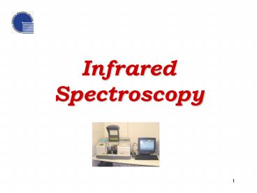
Infrared Spectroscopy PowerPoint PPT Presentation
Title: Infrared Spectroscopy
1
Infrared Spectroscopy
2
What is spectroscopy?
- The study of the interaction between radiation
and matter as a function of wavelength (?). - Historically, spectroscopy referred to the use of
visible light dispersed according to its
wavelength. - Later, the concept was expanded greatly to
comprise any measurement of a quantity as
function of either wavelength or frequency. - A plot of the response as a function of
wavelength or more commonly frequency is
referred to as a spectrum
3
Spectrometry Spectrometer
- Spectrometry - the spectroscopic technique used
to assess the concentration or amount of a given
species. - Spectrometer - instrument that performs such
measurements. (a.k.a spectrograph) - Spectrometry is often used in physical and
analytical chemistry for the identification of
substances through the spectrum emitted from or
absorbed by them.
4
Types of spectroscopy
- Electromagnetic spectroscopy involves
interactions of matter with electromagnetic
radiation, such as light. - Electron spectroscopy involves interactions with
electron beams. - Mass spectrometry involves the interaction of
charged species with magnetic and/or electric
fields - Acoustic spectroscopy involves the frequency of
sound.
5
Measurement process
- Most spectroscopic methods are differentiated as
either atomic or molecular, based on whether or
not they apply to atoms or molecules. - They also can be classified on the nature of
their interaction - Absorption spectroscopy
- Emission spectroscopy
- Scattering spectroscopy
6
Measurement process
- Absorption spectroscopy uses the range of the
electromagnetic spectra in which a substance
absorbs. - (E.g. Atomic absorption spectroscopy,
Infrared spectroscopy, Nuclear magnetic resonance
(NMR) spectroscopy) - Emission spectroscopy uses the range of
electromagnetic spectra in which a substance
radiates (emits). - Scattering spectroscopy measures the amount of
light that a substance scatters at certain
wavelengths, incident angles, and polarization
angles. One of the most useful applications of
light scattering spectroscopy is Raman
spectroscopy.
7
Infrared Spectroscopy
- Infrared spectroscopy - a technique used to
identify chemical compounds based on how infrared
radiation is absorbed by the compounds' chemical
bonds. - The most common technique used is absorption
spectroscopy. - Infrared spectroscopy exploits the fact that
molecules have specific frequencies at which they
rotate or vibrate corresponding to discrete
energy levels.
8
Infrared Absorption
For a molecule to show infrared absorpsions it
must possess a specific feature an electric
dipole moment of the molecule must change during
the vibration. A dipole moment, µ is
defined as the charge value multiplied by the
separation distance between positive and negative
charges. µ qd (C.m)
9
Infrared Absorption
The infrared spectrum of a sample is collected by
passing a beam of infrared light through the
sample. Examination of the transmitted light
reveals how much energy was absorbed at each
wavelength. From this, a transmittance or
absorbance spectrum can be produced, showing at
which IR wavelengths the sample absorbs.
Analysis of these absorption characteristics
reveals details about the molecular structure of
the sample.
10
Infrared Absorption
Simple spectra are obtained from samples with few
IR active bonds and high levels of purity. More
complex molecular structures lead to more
absorption bands and more complex spectra. The
technique has been used for the characterization
of very complex mixtures.
11
Modes of Vibration
The interactions of infrared radiation with
matter may be understood in terms of changes in
molecular dipoles associated with vibrations.
Vibrations can involve either change in bond
length (stretching) or bond angle (bending).
Some bonds can stretch in-phase (symmetric
stretching) or out-of-phase (asymmetric
stretching) Bending vibrations can be in-plane
(scissoring, rocking) or out-of-plane (wagging,
twisting) bending vibrations.
12
Vibrations
- Atoms in the molecule are subjected to number of
vibrations.
13
Modes of Vibration
- The degree of vibrational freedom for polyatomic
molecules containing (N) atoms is given by 3N 5
(linear molecules) and 3N 6 (non-linear
molecules). - Two other concepts are also used to explain the
frequency of vibrational modes - the stiffness of the bond and
- the masses of the atoms at each end of the bond
14
Modes of Vibration
The stiffness of the bond can be characterized by
proportionality constant termed the force
constant, k. The reduced mass, µ provides a
useful way of simplifying our calculations by
combining two-bodies problem as one-body. (1/µ)
m1m2/(m1m2) The relation between force
constant, the reduced mass and the frequency of
absorption is ? (1/2p)?(k/µ) or ?
(1/2pc)?(k/µ)
15
Theoretical principles
- In infrared spectroscopy wavelength is measured
in wave numbers which have the units cm-1 - IR radiation does not have enough energy to
induce electronic transitions as seen with UV.
Absorption of IR is restricted to compounds with
small energy differences in the possible
vibrational and rotational states. - For a molecule to absorb IR, the radiation must
interact with the electric field caused by
changing dipole moment
16
Calibration
www.internationalcrystal.net
www.chemistry.oregonstate.edu/courses/ch361-464/ch
362/irinstrs.htm - 9
- This device is precisely calibrated by using
polystyrene calibration film. - Size of peaks ? amount of material
17
Background Spectrum
- This background spectrum can be used to compare
with the sample measurement to determine
transmittance - Peaks in this region are characteristic of
specific kinds of bonds, thus can be used to
identify whether a specific functional group is
present.
18
Example of C-H(functional group) spectra
- Peaks in the region of (3000- 3100) cm-1
indicates that sp2 hybridized C-H bond are
present in the sample - And peaks in range of (2800-3000)cm-1 indicates
that sp3 hybridized C-H bond are present in a
sample
19
Acids 1650-1700cm-1 Esters 1740-1750cm-1
Aldehydes 1720-1750cm-1 Ketones
1720-1750 cm-1 Amides1650-1715 cm-1
20
FTIR Spectrum of Sample (98 N,N-Dimethylamphetami
ne Hydrochloride)
21
What information can FT-IR provide
- It can determine the amount of components in a
mixture - It can determine the quality or how consistent a
sample is - It can identify unknown materials
22
How FTIR works?
- Source Infrared energy is emitted from a glowing
black-body source. Ends at the Detector - Interferometer beam enters the interferometer
where the spectral encoding takes place - Interferogram signal then exits the
interferometer - Beamsplitter takes the incoming beam and divides
it into two optical beams - Sample beam enters the sample compartment where
it is transmitted through or reflected off of the
surface of the sample - Detector The beam finally passes to the detector
for final measurement - Computer measured signal is digitized and sent
to the computer where the Fourier transformation
takes place - Moving mirror in the interferometer is the only
moving part of the instrument - Fixed mirror
23
Is FT-IR Qualitative, Quantitative, or
Comparative?
- Qualitative
- -Because each different material is a unique
combination of atoms, no two compounds produce
the exact same infrared spectrum - -the size of the peaks in the spectrum is a
direct indication of the amount of material
present
- Quantitative and Comparative
- -Since it is sensitive, accurate, and has
software algorithms, the quantitative methods can
be developed and calibrated easily to perform
various analysis - -FT-IR is comparative due to the background
spectrum that is compared to the sample
24
Advantages/disadvantages
- Speed
- Sensitivity
- Mechanical simplicity
- Internally calibrated
- Destructive
- Too sensitive that it would detect the smallest
contaminant
25
Forensic Lab use
- A Forensic Scientist would use FT-IR to identify
chemicals in different types of samples - Paints
- Polymers
- Coatings
- Rugs
- Contaminants
- Explosive residues
26
Sample Preparation - Gaseous
Gaseous samples require little preparation beyond
purification, but a sample cell with a long
pathlength (typically 5-10 cm) is normally
needed. The walls are of glass or brass. Longer
pathlengths are necessary to analyse complex
mixtures and trace impurities. The cell
pathlength can be measured by the method of
counting interference fringes. L n/2(?1 ?2)
27
Sample Preparation - Liquids
Liquid samples use solution cells. Two types
of solution cells permanent and demountable.
Permanent cell The pathlength need to be
calibrated regularly if quatitative work is to be
undertaken. Diffucult to clean and can be damaged
by water. Demountable cell Easy to maintain as
it can be readily dismantled and cleaned. The
windows can be repolished, a new spacer supplied
and the cell reassembled.
28
Sample Preparation - Solids
- Solid samples can be prepared in four major ways.
- Crush the sample with a mulling agent in a
marble or agate mortar, with a pestle. A thin
film of the mull is applied onto salt plates and
measured. - Grind a quantity of the sample with a specially
purified salt (usually potassium bromide) finely
(to remove scattering effects from large
crystals). This powder mixture is then crushed in
a mechanical die press to form a translucent
pellet through which the beam of the spectrometer
can pass.
29
Sample Preparation - Solids
- The third technique is the Cast Film technique,
which is used mainly for polymeric materials. The
sample is first dissolved in a suitable, non
hygroscopic solvent. A drop of this solution is
deposited on surface of KBr or NaCl cell. The
solution is then evaporated to dryness and the
film formed on the cell is analysed directly. - The final method is to use microtomy to cut a
thin (20-100 micron) film from a solid sample.
This is one of the most important ways of
analysing failed plastic products for example
because the integrity of the solid is preserved.
30
Typical Method
- A beam of infrared light is produced and split
into two separate beams. - One is passed through the sample, the other
passed through a reference which is often the
substance the sample is dissolved in. - The beams are both reflected back towards a
detector, however first they pass through a
splitter which quickly alternates which of the
two beams enters the detector. - The two signals are then compared and a printout
is obtained. - A reference is used for two reasons
- To prevent fluctuations in the output of the
source affecting the data. - To allow the effects of the solvent to be
cancelled out (the reference is usually a pure
form of the solvent the sample is in).
31
Typical Method
32
Uses and Applications
Infrared spectroscopy is widely used in both
research and industry . It is applied for
detection and identification of different
elements/compounds in solving problems in the
fields of forensics, medicine, oil industry,
atmospheric chemistry, polymer degradation,
pharmacology, etc. Among the more common
spectroscopic methods used for analysis is FTIR
spectroscopy, where chemical bonds can be
detected through their characteristic infra-red
absorption frequencies or wavelengths.
33
Uses and Applications
The instruments are now small, and can be
transported, even for use in field trials. With
increasing technology in computer filtering and
manipulation of the results, samples in solution
can now be measured accurately (water produces a
broad absorbance across the range of interest,
and thus renders the spectra unreadable without
this computer treatment). Some machines will
also automatically tell you what substance is
being measured from a store of thousands of
reference spectra held in storage.
34
Analysis of Polymers
Many polymer degradation mechanisms such as UV
degradation and oxidation, amongst many other
failure modes, can be investigated using
infra-red spectroscopy.
35
UV Radiation
Many polymers are attacked by UV radiation at
vulnerable points in their chain structures.
Polypropylene suffers severe cracking in
sunlight unless anti-oxidants are added. The
point of attack occurs at the tertiary carbon
atom present in every repeat unit, causing
oxidation and finally chain breakage.
Polyethylene is also susceptible to UV
degradation, especially those variants which are
branched polymers such as LDPE. The branch
points are tertiary carbon atoms, so polymer
degradation starts there and results in
embrittlement.
36
UV Radiation
IR spectrum showing carbonyl absorption due to UV
degradation of polyethylene
37
Oxidation
Polymers are susceptible to attack by atmospheric
oxygen, especially at elevated temperatures
encountered during processing to shape. Many
process methods such as extrusion and injection
moulding involve pumping molten polymer into
tools, and the high temperatures needed for
melting may result in oxidation unless
precautions are taken. Oxidation tends to start
at tertiary carbon atoms because free radicals
here at more stable, so last longer and are
attacked by oxygen. The carbonyl group can be
further oxidised to break the chain, so weakening
the material by lowering the molecular weight,
and cracks start to grow in the regions affected.
38
Oxidation
IR spectrum showing carbonyl absorption due to
oxidative degradation of polypropylene

