
Skin innervation of the face PowerPoint PPT Presentation
1 / 56
Title: Skin innervation of the face
1
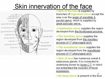
Skin innervation of the face
- Skin of the face is supplied by branches of
trigeminal nerve except the area over the angle
of mandible parotid gland, which is supplied by
great auricular nerve.. - Ophthalmic nerve supplies the region developed
from the frontonasal process. - The maxillary nerve supplies the region developed
from the maxillary process of 1st pharyngeal
arch. - The mandibular nerve supplies the region
developed from the mandibular process of 1st
pharyngeal arch. - Skin of face has numerous sweat sebaceous
glands. It is connected to underlying bones by
loose C.T, in which are embedded the muscles of
facial expression. - No deep fascia is present in the face.
2
Ophthalmic Nerve
- It supplies skin of forehead,
upper eyelid, conjunctiva, side of the nose, it
has 5 branches - 1-Lacrimal N. supplies skin conjunctiva of
lateral part of upper eyelid. - 2-Supraorbital N. winds at supraorbital notch,it
supplies skin conjunctiva on the central part
of upper eyelid skin of forehead. - 3-Supratrochlear N. it lies medial to
supraorbital N. it supplies skin conjunctiva on
medial part of upper eyelid skin of forehead. - 4-Infratrochlear N. leaves orbit to supply skin
conjunctiva on the medial part of upper eyelid
skin of adjoining part of the side of the nose. - 5-External nasal N. leaves nose to supply skin
on the side of the nose down as far as the tip.
3
Maxillary Nerve
- It supplies the skin of posterior part of the
side of nose, lower eyelid, cheek, upper lip,
lateral side of the orbit. It has 3
branches - 1-Infraorbital N. is a direct continuation of
maxillary N. it leveas orbit via infraorbital
foramen. It gives numerous small branches to
supply skin of lower eyelid cheek, side of
nose, the upper lip. - 2-Zygomaticofacial N. passes onto face via a
small foramen on lateral side of zygomatic bone
to supply skin over the cheek. - 3-Zygomaticotemporal N. passes through a small
foramen on the posterior part of zygomatic bone
to supply skin of temple.
4
Mandibular Nerve
- It supplies skin of lower lip, lower part of
face, temporal region part of the auricle
side of scalp. It has 3 branches - 1-Mental N. emerges from the mental foramen of
mandible to supply skin of lower lip chin. - 2-Buccal N. enters the face from under cover of
the masseter. It passes over the buccinator. It
supplies skin m.m of cheek. - 3-Auriculotemporal N. leaves upper border of
parotid gland , between superficial temporal
vessels auricle to supply skin of auricle,
external auditory meatus, outer surface of
tympanic membrane skin of scalp above auricle.
5
Arterial Supply of the Face
- Facial artery arises from external carotid
artery.
-it reaches face by piercing deep fascia at
the lower border of the mandible and then curving
up to the face close to anteroior border of
masseter, here its pulse can be easily felt.
-it then passes upwards in a tortuous course
over the mandible and buccinator towards the
angle of mouth.
-it then ascends along side of nose to
the medial angle of eye, where it anastomoses
with the terminal branches of the ophthalmic
artery. - Superficial temporal artery the smaller
terminal branch of external carotid artery within
the parotid gland.It ascends in front of auricle
to supply the scalp.
6
Arterial Supply of the Face
- Transverse facial artery a branch of
superficial temporal artery of external carotid
artery, within the parotid gland.It runs forward
across the cheek just above parotid duct. - Infraorbital artery it is the terminal part of
maxillary artery (one of terminal branches of
external carotisd artery), it enters face via
infraorbital foramen. - Mental artery branch of inferior alveolar from
maxillary from ext.c.artery, ,it enters face via
mental foramen of mandible. - Zygomaticofacial zygomaticotemporal arteries,
from superficial temporal artery. - Lacrimal artery from ophthalmic artery, of
internal carotid artery. - Supraorbital Supratrochlear arteries branches
of ophthalmic artery, of internal carotid artery
, supply skin of forehead.
7
Branches of Facial artery
1- Submental artery arises at the lower border
of the body of mandible to supply skin of chin
lowe lip. 2- Inferior labial artery
arises near angle of mouth to run medially in the
lower lip and anastomoses with its fellow of
opposite side. 3-
Superior labial artery runs medially in the
upper lip and gives branches to the septum ala
of nose. 4- Lateral nasal artery supplies skin
on the side dorsum of nose.
8
Venous Drainage of Face
- Facial vein
-is formed at the medial angle of eye by union
of supraorbital supratrochlear veins.
-it is connected to cavernous
sinus through superior ophthalmic vein. This
connection is of great clinical importance
because it provides a pathway for spread of
infection from face to cavernous sinus.
-It
descends behind the facial artery to the lower
border of body of mandible.
-It crosses with the facial artery
superficial to submandibular gland.
It is joined by anterior division of
retromandibular vein to form common facial vein
to end into the internal jugular vein.
9
Tributaries of Facial vein
- It recevies tributaries that correspond to the
branches of facial artery. - It is joined to pterygoid venous plexus ( a
venous network lying around pterygoid muscles) by
deep facial vein and to the cavernous sinus by
superior ophthalmic vein. - Transverse facial vein joins superficial temporal
vein within the parotid gland.
10
Lymph Drainage of the Face
- Lymph from forehead anterior part of face
drains into submandibular L.Ns., a few buccal
lymph nodes may be present along course of these
lymph vessels. - Lateral part of face lateral parts of eyelids
drin into parotid L.Ns. - Lower lip chin are drained into submental
L.Ns.
11
Facial Nerve
- It emerges from stylomastoid foramen to enter the
parotid gland , it supplies all muscles of facial
expression. it does not supply the skin ,It runs
within substance of parotid gland, it divides
into 5 terminal branches
1- Temporal branch emerges
from upper border of gland to supply anterior
superior auricular muscles, frontal belly of
occipitofrontalis, orbicularis oculi and
corrugator supercillii.
2- Zygomatic branch emerges from anterior
border of parotid gland to supply orbicularis
oculi.
12
Facial Nerve
3- Buccal branch emerges from anterior border
of parotid gland below parotid duct to supply
buccinator ms.of upper lip nostril.
4- Mandibular branch emerges from anterior
border of parotid gland to supply ms. of lower
lip. 5- Cervical branch
emerges from lower border of parotid gland , it
descends in the neck to supply platysma muscle
depressor anguli oris muscle.
13
(No Transcript)
14
Skin Fascia of the Face
- Skin of face has numerous sweat sebaceous
glands. - It is connected to the underlying bones by loose
connective tissue (superficial fascia), in which
are embedded muscles of facial expression. - No deep fascia in the face.
15
Muscles of Face (muscles of facial
expression)
- They are called ms. Of expression because they
pull skin of face to produce various expressions. - They are arranged in groups around the eye, nose
mouth. - They have bony origin.
- They are inserted into skin of face (no deep
fascia in face). - They are supplied by branches of facial N.,
Except levator P.S. by occulomotor N. (striated
ms.) sympathetic N. (smooth ms.).
16
Muscles of Face
A) Muscles of eyelids
1- levator palpebrae superioris (the dilator ms.
of eyelids, lying in the orbital
cavity). 2-Orbicularis oculi (the sphincter ms of
eyelids). 3-Corrugator supercilii (deep to
orbicularis oculi). 4-Occipitofrontalis (ms.
of scalp).
B) Muscles of Nose
1-Procerus. 2-Compressor dilator naris.
17
Muscles of Face
- C) Muscles of Lips
- Sphincter muscle of the lips
- Orbicularis Oris.
- Dilator muscles of the lips
- 1-Levator labii superioris alaeque nasi.
2-Levator labii
superioris. 3-Depressor labii
inferioris.
4-Zygomaticus minor.
5-Zygomaticus major.
6-Levator anguli Oris (deep to zygomatic ms.).
7-Depressor anguli
Oris. 8-Risorius.
9-Mentalis.
- D) Muscles of Cheek
- Buccinator
18
Muscles of Face (muscles of facial
expression)
- 3 large muscles
1- Buccinator m. (ms. of cheek).
2- Orbicularis oculi
m. 3- Orbicularis
oris m. - Many small muscles
1- Dilator ms. of lips (separate lips)
-Levator labii superioris alaeque nasi, levator
labii superioris.
-Zygomaticus minor major.
-Levator anguli oris, risorius depressor anguli
oris.
-Depressor labii inferioris mentalis.
origin bones
fascia around oral aperature.
Insertion into
substance of lips.
19
2- Corrugator supercilli -It
lies deep to orbicularis oculi. origin
superciliary arch (bone).
Insertion skin of eyebrow.
Action vertical wrinkles of forehead, as
in frowning.
3- Compressor naris dilator naris
origin
maxilla. Insertion the fibres are continuous
with those of opposite side in front of the
bridge of nose to form aponeurosis of bridge of
nose.
Action compesses widens nasal cartilages and
aperature.
4- Procerus
- It is continuous with the
medial part of occipito-frontalis ms.
Origin nasal bone.
Insertion medial part of
skin of eyebrow. Action wrinkles skin of nose.
20
- Orbicularis oculi
1- Orbital part
Origin medial palpebral ligament
adjoining bone.
Insertion The fibres have no lateral
attachment, it loops return to origin.
Action closes
eyelids by throwing skin around orbit into folds
to protect eyeball.
2- Palpebral part
Origin medial
palpebral ligament.
Insertion lateral palpebral raphe
skin of eyelids.
Action closes palpebral
fissure of eyelids gently (sleep) and dilates
lacrimal sac.
21
- Orbicularis oris
Origin maxilla, mandible deep skin.
Insertion encircles oral orifice to be inserted
to the m.m lining the inner surface of lips.
Action compresses the lips together to
close the mouth (sphincter muscle of lips).
22
Muscle of Cheek Buccinator Muscle
- Origin from outer surface of maxilla mandible
opposite the molar teeth from pterygomandibular
ligament. - Insertion
1-upper fibres into upper lip. 2-lower
fibres into lower lip. 3-middle fibres
decussate at the angle of mouth. - N.supply buccal branch of facial N.
- Action 1-
it compresses the cheeks lips against the teeth
to prevent accumulation of food in vestibule of
mouth.
2- it is used in wistling, when cheeks
are distended with air.
23
Muscle of Cheek Buccinator Muscle
- It is covered on outside by buccopharyngeal
fascia buccal pad of fat. - Its deep surface is lined by buccal mucosa.
- It is pierced by
1-parotid duct , opposite upper 2nd molar tooth.
2-Buccal branch of
mandibular nerve (sensory) to supply m.m of cheek
on the inner surface of buccinator muscle.
24
Facial muscle Paralysis
- The facial ms. Are innervated by facial N.
- Cause Damage to facial N. (by a tumor in
internal acoustic meatus or parotid galnd) /or
operation or infection in middle ear / or
perineuritis, Bells palsy in facial nerve canal. - Results Lower motor neuron lesion which
involves distortion of face drooping of lower
eyelid angle of mouth will sag on the affected
side. /But Upper motor neuron lesion is due to
lesion of pyramidal tract and here the upper face
is normal because the neurons supplying this part
receive corticobulbar fibres from both cerebral
cortices.
25
(No Transcript)
26
The Cranial Cavity
- Contents of cranial cavity 1-
the brain.
2-meninges of brain (dura, arachnoid pia
mater) from outside inwards.
3-blood vessels of brain
meninges.
4-parts of cranial nerves.
5-Blood venous sinuses.
6- Hypophysis cerebri (pituitary gland).
27
Dura Mater of Brain
- It is a thick dense membrane which consists of 2
layers (outer inner). - The 2 layers are attached together except at
blood venous sinuses. - Outer endosteal layer
-it covers inner surface of bones of skull.
-it is firmily attached to sutures of skull and
to foramen magnum, it does not extend through
foramen magnum . - Inner meningeal layer
-it covers brain and continuous with dura mater
of spinal cord through foramen magnum.
-it sends tubular sheaths around cranial nerves
as they pass through foramina in skull.
-it sends 4 septa into cranial cavity to
divide cavity into spaces, these septa stabilize
the brain within the cavity during movement of
head.
28
Dural Septa 1- Falx Cerebri
- It is a sickle-shaped fold of dura that descends
in the midline between 2 cerebral hemispheres. - Attachment
-its narrow anterior end is attached to
internal frontal crest crista galli.
-its wide posterior end is attached to upper
surface of tentorium cerebelli. -venous
sinuses in falx cerebri 1-
superior sagittal sinus lies in its upper
convex fixed border. 2-
inferior sagittal sinus lies in its lower
concave free margin. 3-
straight sinus lies at line of attachment of
posterior end of falax with tentorium cerebelli.
29
2- Tentorium cerebelli
- It is a crescentic fold of dura that roofs
posterior cranial fossa. - It separates the occipital lobe of cerebrum above
from cerebellum below. - Its free border
-is concave and forms a gap called,
tentorial notch, for passage of midbrain
it crosses above the attached border of
tentorium to be fixed at the 2 anterior clinoid
processes.
-at the point of crossing of the
free attached borders the trochlear
oculomotor Ns. Pierce the tentorium to enter
lateral wall of cavernus sinus.
-at the apex of petrous
temporal bone the inferior layer of tentorium
is invaginated anteriorly beneath the sup.
Petrosal sinus to form a recess called trigeminal
cave which contains the trigeminal ganglion.
30
2- Tentorium cerebelli
- Attached border
-it is convex and directed peripherally.
-its posterior part is attached to the
lips of transverse sulcus.
-its anterolateral part is
attached to the lips of groove for superior
petrosal sinus (at the upper border of petrous
temporal bone).
-Its anterior
end crosses below the free border and is attached
to the 2 posterior clinoid processes. - In the median plane
-the superior layer of tentorium cerebelli
is attached to falx cerebri.
-the inferior layer of tentorium
cerebelli is attached to falx cerebelli.
31
2- Tentorium cerebelli
- Venous sinuses in the tentorium cerebelli
1-straight sinus at the line of attachment of
posterior end of falx cerebri with tentorium
cerebelli.
2-transverse sinus in the posterior part of
the attached border.
3-superior petrosal sinus in the
anterolateral part of attached border.
32
3-Falx Cerebelli
- It is a small sickle-shaped fold of dura placed
in median plane below tentorium cerebelli. - Its free anterior border projects forwards
between the 2 cerebellar hemispheres. - Its posterior border is attached to the internal
occipital crest. - It contains the occipital sinus in its posterior
fixed border.
33
4-diaphragma sellae
- It is a small circular fold of dura.
- It forms the roof of sella turcica.
- It has a centeral opening for passage of the
stalk of hypophysis cerebri
(pituitary stalk).
34
Dural Nerve Supply
- Trigeminal N.
- Vagus N.
- First 3 cervical nerves.
- Sympathetic fibres around the meningeal arteries.
35
Dural Arterial Supply
- Internal carotid artery.
- Maxillary artery. Middle meningeal artery, it is
the most important branch. - Ascending pharyngeal artery.
- Occipital artery.
- Vertebral artery.
Meningeal Veins
- Middle meningeal vein follows the branches of
middle meningeal artery and drains into the
pterygoid venous plexus or sphenoparietal sinus.
36
Middle meningeal artery
- It is a branch of 1st part of maxillary artery.
- It reaches middle cranial fossa through foramen
spinosum to lie between the meningeal endosteal
layers of dura. - It passes forwards and laterally grooving the
squamous part of temporal bone. Then it divides
into 1-large anterior (frontal) branch.
2-small posterior (parietal) branch. - It divides into anterior posterior branches
opposite a point 20mm above center of zygomatic
arch.
37
Extradural hemorrhage
- This is intracranial hemorrhage outside the dura
mater. - It results mostly from injury of middle
meningeal artery, usually occuring in the region
of the pterion (at anteroinferior part of the
parietal bone) as a result of a blow over the
side of the head. - A collection of blood,
(extradural hematoma) occurs between the dura and
the skull bones, stripping off the periosteum of
inner table of the bone. - The intracranial pressure rises producing local
pressure on the motor area of brain. - Blood may pass out through the fracture to form a
soft swelling under the temporalis ms
38
Arachnoid mater
- It is a delicate, impermeable membrane covering
the brain, lying between pia mater dura mater. - It is separated from the dura by subdural space,
and from the pia by subarachnoid space, which is
filled with cerebro-spinal fluid. - The arachnoid projects into the venous sinuses to
form arachnoid villi, they are most numerous at
superior sagittal sinus, aggregations of
arachnoid villi are called arachnoid
granulations, where C.S.F diffuses into
bloodstream. - Cerebral arteries veins cranial nerves lie in
subarachnoid space.
39
Cerebrospinal fluid
- It is produced by the choroid plexuses within
lateral, 3rd 4th ventricles of brain. - It passes via 3 foramina in roof of 4th ventricle
to circulate in subarachnoid space, upward over
surfaces of cerebral hemispheres and downward
around spinal cord. - The spinal subarachnoid space extends down as far
as 2nd sacral vertebra. - The fluid enters bloodstream by passing into
arachnoid villi and diffuses into venous sinuses.
40
Pia mater
- It is a thin vascular membrane that closely
covering the brain. - It extends over the cranial nerves and fuses with
their epineurium. - The cerebral arteries enter the brain carrying a
sheath of pia mater.
41
(No Transcript)
42
Venous Sinuses
- They are blood channels between the endosteal
meningeal layers of dura mater. - The walls of sinuses are lined by endothelium.
- They receive tributaries from the brain, the
diploe of skull, the orbit, the internal ear. - They differ from the veins in having no valves,
or muscles in their walls. So they do not
contract when they are ruptured and bleeding is
controlled only by pressure. - Single sinuses are
1-superior sagittal, 2-inferior sagittal,
3-straight sinus 4-intercavernus sinus.
5-Occipital sinus.
- Paired sinuses are
1-transverse sinuses.
2-sigmoid sinuses.
3-cavernus sinuses.
4-superior inferior
petrosal sinuses.
5-sphenoparietal sinuses.
43
Superior Sagittal Sinus
- It lies in the upper fixed border of falx
cerebri. - It begins in front at frontal crest foramen
cecum, where it receives vein from nasal
cavity, then runs backward grooving vault of
skull. - At the internal occipital protuberance, it is
dilated to form the confluence of the sinuses,
here it deviates to one side (usually the right)
to become the right transverse sinus. - It is connected to the opposite transverse sinus
and it receives the occipital sinus.
44
Tributaries and communications of the Superior
Sagittal Sinus
- The sinus communicates with 2-3 venous lacunae on
each side. - Numerous arachnoid villi granulations project
into the lacunae, which also receive the diploic
meningeal veins. - It receives also the cerebral veins.
- It communicates with veins of scalp by emissary
veins passing through the parietal foramina.
45
Inferior Sagittal Sinus
- It lies in the free lower border of falx cerebri.
- It runs backward to join great cerebral vein at
free border of tentorium cerebelli to form
straight sinus.
- Straight Sinus
- It lies at the junction of falx cerebri with
tentorium cerebelli. - It is formed by union of inferior sagittal sinus
great cerebral vein. - It ends by turning to left to form the left
transverse sinus.
46
Transverse Sinuses
- Are paired sinuses, begin at the internal
occipital protuberance. - Right sinus is usually continuous with the
superior sagittal sinus, and left sinus is
continuous with the straight sinus. - Each sinus occupies the attached margin of
tentorium cerebelli, grooving the occipital bone. - They receive the superior petrosal sinuses,
cerebral cerebellar veins, diploic veins. - They end by turning downward as sigmoid sinuses.
47
Sigmoid Sinuses
- Are direct continuation of transverse sinuses.
- Each sinus turns downward and medially and
grooves the mastoid part of temporal bone, here
it lies behind mastoid antrum. - Finally, it passes through jugular foramen to
join the internal jugular vein.
48
Occipital sinus
- It is a small sinus occupying the attached border
of falax cerebelli. - It begins near foramen magnum, where it
communicates with vertebral veins and drains into
the confluence of the sinuses. - It connects the beginning of transverse sinus
with the end of sigmoid sinus. - May be single or paired.
49
Cavernus sinuses
- Are lie in middle cranial fossa on each side of
body of sphenoid bone ( hypophyseal
fossa). - Each sinus extends from superior orbital fissure
anteriorly, to apex of petrous temporal bone
posteriorly. - Inside the sinus
1- internal carotid artery surrounded by
sympathetic plexus.
2- abducent nerve. - In the lateral wall of cavernus sinus
1-oculomotor nerve.
2-trochlear nerve.
3-ophthalmic nerve of trigeminal N.
4-maxillary nerve of
trigeminal N.
50
Cavernus sinuses
- Tributaries
1-superior inferior ophthalmic veins.
2-cerebral veins.
3- sphenoparietal sinus, along
posterior margin of lesser wing of sphenoid.
4-central vein of retina. - Each sinus drains posteriorly into superior
inferior petrosal sinuses and inferiorly
into pterygoid venous plexus. - The 2 sinuses communicate with each other by
anterior posterior intercavernous sinuses,
which run in diaphragma sellae. - Each sinus has important communication with
facial vein through superior ophthalmic vein.
51
Superior inferior Petrosal sinuses
- Are small sinuses lying on the superior
inferior borders of petrous part of temporal
bone. - The superior sinus drains the cavernus sinus into
transverse sinus. - The inferior sinus drains the cavernus sinus into
internal jugular vein.
52
Hypophysis Cerebri
(Pituitary gland)
- It is the master endocrine gland, which attached
to brain by the infundibulum. - Site sella turcica of sphenoid bone
(hypophyseal fossa). - It has 2 lobes, anterior lobe or adenohhypophysis
and posterior lobe or neuro-hypophysis. - Superiorly diaphragma sellae, which has a
central aperature to allow passage of
infundibulum. - Inferiorly body of sphenoid sphenoid air
sinuses. - Laterally cavernus sinus.
- Posteriorly dorsum sellae, basilar artery,
pons. - Blood supply superior inferior hypophyseal
branches of internal carotid artery / veins
drains into intercavernous sinuses.
53
Diploic Veins
- They are thin- walled, valveless veins lying in
the diploe of skull (between the inner outer
tables of the bones of skull). - They communicate with the meningeal veins
dural venous sinuses. - They include frontal, temporal occipital
diploic veins.
54
Emissary Veins
- They are small valveless veins, pass via foramina
in the skull and connect the dural venous sinuses
with veins outside the skull. - Function they help to equalize the pressure in
veins outside the skull and in the dural venous
sinuses. - Their danger infection outside the skull may
spread along the emissary veins to reach the
dural venous sinuses and produce septic thrombi.
55
Intracranial part of Internal Carotid Artery
- It enters the cranial cavity through the carotid
canal in petrous part of temporal bone. - It passes into foramen lacerum to enter the
cavernus sinus. - It lies with the abducent nerve on the floor of
cavernus sinus. - It pierces the roof of cavernus sinus to lie on
the medial side of anterior clinoid process and
divide into anterior middle cerebral arteries. - Branches
1-superior inferior hypophyseal arteries to
pituitary gland. 2-
meningeal branches.
56
(No Transcript)
Recommended
