
Chapter 19 The Cardiovascular System: The Blood PowerPoint PPT Presentation
1 / 70
Title: Chapter 19 The Cardiovascular System: The Blood
1
Chapter 19The Cardiovascular System The Blood
2
Fluids of the Body
- Cells of the body are serviced by 2 fluids
- blood
- composed of plasma and a variety of cells
- transports nutrients and wastes
- interstitial fluid
- bathes the cells of the body
- Nutrients and oxygen diffuse from the blood into
the interstitial fluid then into the cells - Wastes move in the reverse direction
- Hematology is study of blood and blood disorders
3
Functions of Blood
- Transportation
- O2, CO2, metabolic wastes, nutrients, heat
hormones - Regulation
- helps regulate pH through buffers
- helps regulate body temperature
- coolant properties of water
- vasodilatation of surface vessels dump heat
- helps regulate water content of cells by
interactions with dissolved ions and proteins - Protection from disease loss of blood
4
Physical Characteristics of Blood
- Thicker (more viscous) than water and flows more
slowly than water - Temperature of 100.4 degrees F
- pH 7.4 (7.35-7.45)
- 8 of total body weight
- Blood volume
- 5 to 6 liters in average male
- 4 to 5 liters in average female
- hormonal negative feedback systems maintain
constant blood volume and osmotic pressure
5
Techniques of Blood Sampling
- Venipuncture
- sample taken from vein with hypodermic needle
syringe - median cubital vein (see page 717)
- why not stick an artery?
- less pressure
- closer to the surface
- Finger or heel stick
- common technique for diabetics to monitor daily
blood sugar - method used for infants
6
Components of Blood
- Hematocrit
- 55 plasma
- 45 cells
- 99 RBCs
- lt 1 WBCs and platelets
7
Blood Plasma
- 0ver 90 water
- 7 plasma proteins
- created in liver
- confined to bloodstream
- albumin
- maintain blood osmotic pressure
- globulins (immunoglobulins)
- antibodies bind to foreignsubstances called
antigens - form antigen-antibody complexes
- fibrinogen
- for clotting
- 2 other substances
- electrolytes, nutrients, hormones, gases, waste
products
8
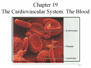
Formed Elements of Blood
- Red blood cells ( erythrocytes )
- White blood cells ( leukocytes )
- granular leukocytes
- neutrophils
- eosinophils
- basophils
- agranular leukocytes
- lymphocytes T cells, B cells, and natural
killer cells - monocytes
- Platelets (special cell fragments)
9
Hematocrit
- Percentage of blood occupied by cells
- female normal range
- 38 - 46 (average of 42)
- male normal range
- 40 - 54 (average of 46)
- testosterone
- Anemia
- not enough RBCs or not enough hemoglobin
- Polycythemia
- too many RBCs (over 65)
- dehydration, tissue hypoxia, blood doping in
athletes
10
Blood Doping
- Injecting previously stored RBCs before an
athletic event - more cells available to deliver oxygen to tissues
- Dangerous
- increases blood viscosity
- forces heart to work harder
- Banned by Olympic committee
11
Formation of Blood Cells
- Most blood cells types need to be continually
replaced - die within hours, days or weeks
- process of blood cells formation is hematopoiesis
or hemopoiesis - In the embryo
- occurs in yolk sac, liver, spleen, thymus, lymph
nodes red bone marrow - In adult
- occurs only in red marrow of flat bones like
sternum, ribs, skull pelvis and ends of long
bones
12
Hematopoiesis
13
Stages of Blood Cell Formation
- Pluripotent stem cells
- .1 of red marrow cells
- replenish themselves as they differentiate into
either myeloid or lymphoid stem cells - Myeloid stem cell line of development continues
- progenitor cells(colony-forming units) no longer
can divide and are specialized to form specific
cell types - example CFU-E develops eventually into only red
blood cells - next generation is blast cells
- have recognizable histological characteristics
- develop within several divisions into mature cell
types - Lymphoid stem cell line of development
- pre-B cells prothymocytes finish their develop
into B T lymphocytes in the lymphatic tissue
after leaving the red marrow
14
Hemopoietic Growth Factors
- Regulate differentiation proliferation
- Erythropoietin (EPO)
- produced by the kidneys increase RBC precursors
- Thrombopoietin (TPO)
- hormone from liver stimulates platelet formation
- Cytokines are local hormones of bone marrow
- produced by some marrow cells to stimulate
proliferation in other marrow cells - colony-stimulating factor (CSF) interleukin
stimulate WBC production
15
Medical Uses of Growth Factors
- Available through recombinant DNA technology
- recombinant erythropoietin (EPO) very effective
in treating decreased RBC production of end-stage
kidney disease - other products given to stimulate WBC formation
in cancer patients receiving chemotherapy which
kills bone marrow - granulocyte-macrophage colony-stimulating factor
- granulocyte colony stimulating factor
- thrombopoietin helps prevent platelet depletion
during chemotherapy
16
Red Blood Cells or Erythrocytes
- Contain oxygen-carrying protein hemoglobin that
gives blood its red color - 1/3 of cells weight is hemoglobin
- Biconcave disk 8 microns in diameter
- increased surface area/volume ratio
- flexible shape for narrow passages
- no nucleus or other organelles
- no cell division or mitochondrial ATP formation
- Normal RBC count
- male 5.4 million/drop ---- female 4.8
million/drop - new RBCs enter circulation at 2 million/second
17
Hemoglobin
- Globin protein consisting of 4 polypeptide chains
- One heme pigment attached to each polypeptide
chain - each heme contains an iron ion (Fe2) that can
combine reversibly with one oxygen molecule
18
Transport of O2, CO2 and Nitric Oxide
- Each hemoglobin molecule can carry 4 oxygen
molecules from lungs to tissue cells - Hemoglobin transports 23 of total CO2 waste
from tissue cells to lungs for release - combines with amino acids in globin portion of Hb
- Hemoglobin transports nitric oxide super nitric
oxide helping to regulate BP - iron ions pick up nitric oxide (NO) super
nitric oxide (SNO) transport it to from the
lungs - NO causing vasoconstriction is released in the
lungs - SNO causing vasodilation is picked up in the lungs
19
RBC Life Cycle
- RBCs live only 120 days
- wear out from bending to fit through capillaries
- no repair possible due to lack of organelles
- Worn out cells removed by fixed macrophages in
spleen liver - Breakdown products are recycled
20
Recycling of Hemoglobin Components
- In macrophages of liver or spleen
- globin portion broken down into amino acids
recycled - heme portion split into iron (Fe3) and
biliverdin (green pigment)
21
Fate of Components of Heme
- Iron(Fe3)
- transported in blood attached to transferrin
protein - stored in liver, muscle or spleen
- attached to ferritin or hemosiderin protein
- in bone marrow being used for hemoglobin
synthesis - Biliverdin (green) converted to bilirubin
(yellow) - bilirubin secreted by liver into bile
- converted to urobilinogen then stercobilin
(brown pigment in feces) by bacteria of large
intestine - if reabsorbed from intestines into blood is
converted to a yellow pigment, urobilin and
excreted in urine
22
Erythropoiesis Production of RBCs
- Proerythroblast starts to produce hemoglobin
- Many steps later, nucleus is ejected a
reticulocyte is formed - orange in color with traces of visible rough ER
- Reticulocytes escape from bone marrow into the
blood - In 1-2 days, they eject the remaining organelles
to become a mature RBC
23
Feedback Control of RBC Production
- Tissue hypoxia (cells not getting enough O2)
- high altitude since air has less O2
- anemia
- RBC production falls below RBC destruction
- circulatory problems
- Kidney response to hypoxia
- release erythropoietin
- speeds up development of proerythroblasts into
reticulocytes
24
Normal Reticulocyte Count
- Should be .5 to 1.5 of the circulating RBCs
- Low count in an anemic person might indicate bone
marrow problem - leukemia, nutritional deficiency or failure of
red bone marrow to respond to erythropoietin
stimulation - High count might indicate recent blood loss or
successful iron therapy
25
WBC Anatomy and Types
- All WBCs (leukocytes) have a nucleus and no
hemoglobin - Granular or agranular classification based on
presence of cytoplasmic granules made visible by
staining - granulocytes are neutrophils, eosinophils or
basophils - agranulocytes are monocyes or lymphocytes
26
Neutrophils (Granulocyte)
- Polymorphonuclear Leukocytes or Polys
- Nuclei 2 to 5 lobes connected by thin strands
- older cells have more lobes
- young cells called band cells because of
horseshoe shaped nucleus (band) - Fine, pale lilac practically invisible granules
- Diameter is 10-12 microns
- 60 to 70 of circulating WBCs
27
Eosinophils (Granulocyte)
- Nucleus with 2 or 3 lobes connected by a thin
strand - Large, uniform-sized granules stain orange-red
with acidic dyes - do not obscure the nucleus
- Diameter is 10 to 12 microns
- 2 to 4 of circulating WBCs
28
Basophils (Granulocyte)
- Large, dark purple, variable-sized granules stain
with basic dyes - obscure the nucleus
- Irregular, s-shaped, bilobed nuclei
- Diameter is 8 to 10 microns
- Less than 1 of circulating WBCs
29
Lymphocyte (Agranulocyte)
- Dark, oval to round nucleus
- Cytoplasm sky blue in color
- amount varies from rim of blue to normal amount
- Small cells 6 - 9 microns in diameter
- Large cells 10 - 14 microns in diameter
- increase in number during viral infections
- 20 to 25 of circulating WBCs
30
Monocyte (Agranulocyte)
- Nucleus is kidney or horse-shoe shaped
- Largest WBC in circulating blood
- does not remain in blood long before migrating to
the tissues - differentiate into macrophages
- fixed group found in specific tissues
- alveolar macrophages in lungs
- kupffer cells in liver
- wandering group gathers at sites of infection
- Diameter is 12 - 20 microns
- Cytoplasm is a foamy blue-gray
- 3 to 8 o circulating WBCs
31
WBC Physiology
- Less numerous than RBCs
- 5000 to 10,000 cells per drop of blood
- 1 WBC for every 700 RBC
- Leukocytosis is a high white blood cell count
- microbes, strenuous exercise, anesthesia or
surgery - Leukopenia is low white blood cell count
- radiation, shock or chemotherapy
- Only 2 of total WBC population is in circulating
blood at any given time - rest is in lymphatic fluid, skin, lungs, lymph
nodes spleen
32
Emigration Phagocytosis in WBCs
- WBCs roll along endothelium, stick to it
squeeze between cells. - adhesion molecules (selectins) help WBCs stick to
endothelium - displayed near site of injury
- molecules (integrins) found on neutrophils assist
in movement through wall - Neutrophils macrophages phagocytize bacteria
debris - chemotaxis of both
- kinins from injury site toxins
33
Neutrophil Function
- Fastest response of all WBC to bacteria
- Direct actions against bacteria
- release lysozymes which destroy/digest bacteria
- release defensin proteins that act like
antibiotics poke holes in bacterial cell walls
destroying them - release strong oxidants (bleach-like, strong
chemicals ) that destroy bacteria
34
Monocyte Function
- Take longer to get to site of infection, but
arrive in larger numbers - Become wandering macrophages, once they leave the
capillaries - Destroy microbes and clean up dead tissue
following an infection
35
Basophil Function
- Involved in inflammatory and allergy reactions
- Leave capillaries enter connective tissue as
mast cells - Release heparin, histamine serotonin
- heighten the inflammatory response and account
for hypersensitivity (allergic) reaction
36
Eosinophil Function
- Leave capillaries to enter tissue fluid
- Release histaminase
- slows down inflammation caused by basophils
- Attack parasitic worms
- Phagocytize antibody-antigen complexes
37
Lymphocyte Functions
- B cells
- destroy bacteria and their toxins
- turn into plasma cells that produces antibodies
- T cells
- attack viruses, fungi, transplanted organs,
cancer cells some bacteria - Natural killer cells
- attack many different microbes some tumor cells
- destroy foreign invaders by direct attack
38
Differential WBC Count
- Detection of changes in numbers of circulating
WBCs (percentages of each type) - indicates infection, poisoning, leukemia,
chemotherapy, parasites or allergy reaction - Normal WBC counts
- neutrophils 60-70 (up if bacterial infection)
- lymphocyte 20-25 (up if viral infection)
- monocytes 3 -- 8 (up if fungal/viral
infection) - eosinophil 2 -- 4 (up if parasite or allergy
reaction) - basophil lt1 (up if allergy reaction or
hypothyroid)
39
Bone Marrow Transplant
- Intravenous transfer of healthy bone marrow
- Procedure
- destroy sick bone marrow with radiation
chemotherapy - donor matches surface antigens on WBC
- put sample of donor marrow into patient's vein
for reseeding of bone marrow - success depends on histocompatibility of donor
recipient - Treatment for leukemia, sickle-cell, breast,
ovarian or testicular cancer, lymphoma or
aplastic anemia
40
Platelet (Thrombocyte) Anatomy
- Disc-shaped, 2 - 4 micron cell fragment with no
nucleus - Normal platelet count is 150,000-400,000/drop of
blood - Other blood cell counts
- 5 million red 5-10,000 white blood cells
41
Platelets--Life History
- Platelets form in bone marrow by following steps
- myeloid stem cells to megakaryocyte-colony
forming cells to megakaryoblast to megakaryocytes
whose cell fragments form platelets - Short life span (5 to 9 days in bloodstream)
- formed in bone marrow
- few days in circulating blood
- aged ones removed by fixed macrophages in liver
and spleen
42
Complete Blood Count
- Screens for anemia and infection
- Total RBC, WBC platelet counts differential
WBC hematocrit and hemoglobin measurements - Normal hemoglobin range
- infants have 14 to 20 g/100mL of blood
- adult females have 12 to 16 g/100mL of blood
- adult males have 13.5 to 18g/100mL of blood
43
Hemostasis
- Stoppage of bleeding in a quick localized
fashion when blood vessels are damaged - Prevents hemorrhage (loss of a large amount of
blood) - Methods utilized
- vascular spasm
- platelet plug formation
- blood clotting (coagulation formation of fibrin
threads)
44
Vascular Spasm
- Damage to blood vessel produces stimulates pain
receptors - Reflex contraction of smooth muscle of small
blood vessels - Can reduce blood loss for several hours until
other mechanisms can take over - Only for small blood vessel or arteriole
45
Platelet Plug Formation
- Platelets store a lot of chemicals in granules
needed for platelet plug formation - alpha granules
- clotting factors
- platelet-derived growth factor
- cause proliferation of vascular endothelial
cells, smooth muscle fibroblasts to repair
damaged vessels - dense granules
- ADP, ATP, Ca2, serotonin, fibrin-stabilizing
factor, enzymes that produce thromboxane A2 - Steps in the process
- (1) platelet adhesion (2) platelet release
reaction (3) platelet aggregation
46
Platelet Adhesion
- Platelets stick to exposed collagen underlying
damaged endothelial cells in vessel wall
47
Platelet Release Reaction
- Platelets activated by adhesion
- Extend projections to make contact with each
other - Release thromboxane A2 ADP activating other
platelets - Serotonin thromboxane A2 are vasoconstrictors
decreasing blood flow through the injured vessel
48
Platelet Aggregation
- Activated platelets stick together and activate
new platelets to form a mass called a platelet
plug - Plug reinforced by fibrin threads formed during
clotting process
49
Blood Clotting
- Blood drawn from the body thickens into a gel
- gel separates into liquid (serum) and a clot of
insoluble fibers (fibrin) in which the cells are
trapped - If clotting occurs in an unbroken vessel is
called a thrombosis - Substances required for clotting are Ca2,
enzymes synthesized by liver cells and substances
released by platelets or damaged tissues - Clotting is a cascade of reactions in which each
clotting factor activates the next in a fixed
sequence resulting in the formation of fibrin
threads - prothrombinase Ca2 convert prothrombin into
thrombin - thrombin converts fibrinogen into fibrin threads
50
Overview of the Clotting Cascade
- Prothrombinase is formed by either the intrinsic
or extrinsic pathway - Final common pathway produces fibrin threads
51
Extrinsic Pathway
- Damaged tissues leak tissue factor
(thromboplastin) into bloodstream - Prothrombinase forms in seconds
- In the presence of Ca2, clotting factor X
combines with V to form prothrombinase
52
Intrinsic Pathway
- Activation occurs
- endothelium is damaged platelets come in
contact with collagen of blood vessel wall - platelets damaged release phospholipids
- Requires several minutes for reaction to occur
- Substances involved Ca2 and clotting factors
XII, X and V
53
Final Common Pathway
- Prothrombinase and Ca2
- catalyze the conversion of prothrombin to
thrombin - Thrombin
- in the presence of Ca2 converts soluble
fibrinogen to insoluble fibrin threads - activates fibrin stabilizing factor XIII
- positive feedback effects of thrombin
- accelerates formation of prothrombinase
- activates platelets to release phospholipids
54
Clot Retraction Blood Vessel Repair
- Clot plugs ruptured area of blood vessel
- Platelets pull on fibrin threads causing clot
retraction - trapped platelets release factor XIII stabilizing
the fibrin threads - Edges of damaged vessel are pulled together
- Fibroblasts endothelial cells repair the blood
vessel
55
Role of Vitamin K in Clotting
- Normal clotting requires adequate vitamin K
- fat soluble vitamin absorbed if lipids are
present - absorption slowed if bile release is insufficient
- Required for synthesis of 4 clotting factors by
hepatocytes - factors II (prothrombin), VII, IX and X
- Produced by bacteria in large intestine
56
Hemostatic Control Mechanisms
- Fibrinolytic system dissolves small,
inappropriate clots clots at a site of a
completed repair - fibrinolysis is dissolution of a clot
- Inactive plasminogen is incorporated into the
clot - activation occurs because of factor XII and
thrombin - plasminogen becomes plasmin (fibrinolysin) which
digests fibrin threads - Clot formation remains localized
- fibrin absorbs thrombin
- blood disperses clotting factors
- endothelial cells WBC produce prostacyclin that
opposes thromboxane A2 (platelet adhesion
release) - Anticoagulants present in blood produced by
mast cells
57
Intravascular Clotting
- Thrombosis
- clot (thrombus) forming in an unbroken blood
vessel - forms on rough inner lining of BV
- if blood flows too slowly (stasis) allowing
clotting factors to build up locally cause
coagulation - may dissolve spontaneously or dislodge travel
- Embolus
- clot, air bubble or fat from broken bone in the
blood - pulmonary embolus is found in lungs
- Low dose aspirin blocks synthesis of thromboxane
A2 reduces inappropriate clot formation - strokes, TIAs and myocardial infarctions
58
Anticoagulants and Thrombolytic Agents
- Anticoagulants suppress or prevent blood clotting
- heparin
- administered during hemodialysis and surgery
- warfarin (Coumadin)
- antagonist to vitamin K so blocks synthesis of
clotting factors - slower than heparin
- stored blood in blood banks treated with citrate
phosphate dextrose (CPD) that removes Ca2 - Thrombolytic agents are injected to dissolve
clots - directly or indirectly activate plasminogen
- streptokinase or tissue plasminogen activator
(t-PA)
59
Blood Groups and Blood Types
- RBC surfaces are marked by genetically determined
glycoproteins glycolipids - agglutinogens or isoantigens
- distinguishes at least 24 different blood groups
- ABO, Rh, Lewis, Kell, Kidd and Duffy systems
60
ABO Blood Groups
- Based on 2 glycolipid isoantigens called A and B
found on the surface of RBCs - display only antigen A -- blood type A
- display only antigen B -- blood type B
- display both antigens A B -- blood type AB
- display neither antigen -- blood type O
- Plasma contains isoantibodies or agglutinins to
the A or B antigens not found in your blood - anti-A antibody reacts with antigen A
- anti-B antibody reacts with antigen B
61
RH blood groups
- Antigen was discovered in blood of Rhesus monkey
- People with Rh agglutinogens on RBC surface are
Rh. Normal plasma contains no anti-Rh
antibodies - Antibodies develop only in Rh- blood type only
with exposure to the antigen - transfusion of positive blood
- during a pregnancy with a positive blood type
fetus - Transfusion reaction upon 2nd exposure to the
antigen results in hemolysis of the RBCs in the
donated blood
62
Hemolytic Disease of Newborn
- Rh negative mom and Rh fetus will have mixing of
blood at birth - Mom's body creates Rh antibodies unless she
receives a RhoGam shot soon after first delivery,
miscarriage or abortion - RhoGam binds to loose fetal blood and removes it
from body before she reacts - In 2nd child, hemolytic disease of the newborn
may develop causing hemolysis of the fetal RBCs
63
Transfusion and Transfusion Reactions
- Transfer of whole blood, cells or plasma into the
bloodstream of recipient - used to treat anemia or severe blood loss
- Incompatible blood transfusions
- antigen-antibody complexes form between plasma
antibodies foreign proteins on donated RBC's
(agglutination) - donated RBCs become leaky (complement proteins)
burst - loose hemoglobin causes kidney damage
- Problems caused by incompatibility between
donors cells and recipients plasma - Donor plasma is too diluted to cause problems
64
Universal Donors and Recipients
- People with type AB blood called universal
recipients since have no antibodies in plasma - only true if cross match the blood for other
antigens - People with type O blood cell called universal
donors since have no antigens on their cells - theoretically can be given to anyone
65
Typing and Cross-Matching Blood
- Mixing of incompatible blood causes agglutination
(visible clumping) - formation of antigen-antibody complex that sticks
cells together - not the same as blood clotting
- Typing involves testing blood with known antisera
that contain antibodies A, B or Rh - Cross-matching is to test by mixing donor cells
with recipients serum - Screening is to test recipients serum against
known RBCs having known antigens
66
Anemia Not Enough RBCs
- Symptoms
- oxygen-carrying capacity of blood is reduced
- fatigue, cold intolerance paleness
- lack of O2 for ATP heat production
- Types of anemia
- iron-deficiency lack of absorption or loss of
iron - pernicious lack of intrinsic factor for B12
absorption - hemorrhagic loss of RBCs due to bleeding
(ulcer) - hemolytic defects in cell membranes cause
rupture - thalassemia hereditary deficiency of hemoglobin
- aplastic destruction of bone marrow
(radiation/toxins)
67
Sickle-cell Anemia (SCA)
- Genetic defect in hemoglobin molecule (Hb-S) that
changes 2 amino acids - at low very O2 levels, RBC is deformed by changes
in hemoglobin molecule within the RBC - sickle-shaped cells rupture easily causing
anemia clots - Found among populations in malaria belt
- Mediterranean Europe, sub-Saharan Africa Asia
- Person with only one sickle cell gene
- increased resistance to malaria because RBC
membranes leak K lowered levels of K kill the
parasite infecting the red blood cells
68
Hemophilia
- Inherited deficiency of clotting factors
- bleeding spontaneously or after minor trauma
- subcutaneous intramuscular hemorrhaging
- nosebleeds, blood in urine, articular bleeding
pain - Hemophilia A lacks factor VIII (males only)
- most common
- Hemophilia B lacks factor IX (males only)
- Hemophilia C (males females)
- less severe because alternate clotting activator
exists - Treatment is transfusions of fresh plasma or
concentrates of the missing clotting factor
69
Disseminated Intravascular Clotting
- Life threatening paradoxical presence of blood
clotting and bleeding at the same time throughout
the whole body - so many clotting factors are removed by
widespread clotting that too few remain to permit
normal clotting - Associated with infections, hypoxia, low blood
flow rates, trauma, hypotension hemolysis - Clots cause ischemia and necrosis leading to
multisystem organ failure
70
Leukemia
- Acute leukemia
- uncontrolled production of immature leukocytes
- crowding out of normal red bone marrow cells by
production of immature WBC - prevents production of RBC platelets
- Chronic leukemia
- accumulation of mature WBC in bloodstream because
they do not die - classified by type of WBC that is
predominant---monocytic, lymphocytic.
Recommended
