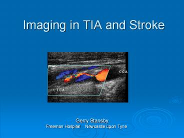
Imaging in TIA and Stroke Gerry Stansby Freeman Hospital PowerPoint PPT Presentation
1 / 37
Title: Imaging in TIA and Stroke Gerry Stansby Freeman Hospital
1
Imaging in TIA and Stroke
- Gerry Stansby
- Freeman Hospital Newcastle upon Tyne
2
1. Imaging for TIA 2. Imaging for stroke 3.
Imaging techniques Brain imaging Carotid
imaging
3
5.1 TIA MRI/MRA brain for those patients
requiring it available seven days a week with
Contrast Enhanced MRA (CEMRA) for first-line
carotid imaging. This requires MR software for
diffusion weighted and gradient echo imaging and
CEMRA, and a pump injector for CEMRA and
Carotid imaging seven days a week, which will
ideally include CEMRA and duplex ultrasound, and
CT angiography. 5.2 Stroke 24 hour access to
CT Rapidly accessible MRI, with the features
described above, for those patients who require
it and Ability to undertake more complex
imaging examinations for stroke subtypes as
required
4
(No Transcript)
5
ABCD2 SCORE
The ABCD2 score identifies groups who are at high
risk of stroke after transient ischaemic attack
(TIA).
The score is based on
Age 60 years (1 point)
Blood pressure systolic gt140mm Hg and/or
diastolic 90mm Hg (1 point)
Clinical features unilateral weakness (2
points), speech impairment without weakness (1
point)
Duration 60 min (2 points) or 10-59 min (1
point)
Diabetes (1 point).
6
(No Transcript)
7
(No Transcript)
8
(No Transcript)
9
(No Transcript)
10
ABCD2 lt4
ABCD2 4
TIA
Specialist assessment/ investigation within 1 week
Specialist assessment/ investigation within 24
hours
Best medical therapy
Best medical therapy
Yes
Yes
No
No
Vascular territory/ pathology unknown
Vascular territory/ pathology unknown
Brain imaging (DWI) within 1 week symptom onset
Urgent brain imaging (DWI) within 24
hours symptom onset
Carotid imaging if the patient is a candidate
for carotid intervention within 1 week of symptom
onset
Carotid imaging if the patient is a candidate
for carotid intervention within 1 week of symptom
onset
NICE 2008
11
Carotid Imaging Why?
To assess severity of stenosis (carotid
bifurcation) To identify candidates for Carotid
Endarterectomy
To exclude an embolic source elsewhere or other
pathology (tandem lesions, arch atheroma)
Department of Health Implementing the National
Stroke Strategy an Imaging Guide 2008
12
(No Transcript)
13
Carotid imaging should ideally be performed at
initial assessment and should not be delayed for
more than 24 hours after first clinical
assessment of TIA for those at higher risk of
stroke (i.e. ABCD2 score gt_ 4) or in those with
non-cardioembolic carotid-territory minor stroke.
Those people who are found to have carotid
stenosis will require carotid intervention within
48 hours of presentation. For those people at
lower risk of subsequent stroke (i.e. ABCD2 score
lt4), carotid imaging should be performed within
seven days. If they are found to have carotid
stenosis, they should have carotid intervention
within two weeks of presentation.
14
Carotid Imaging
Severity of stenosis
Indications to intervene are based on randomized
trials in which the severity of stenosis was
assessed on selective catheter angiography
15
Carotid Imaging
Modalities
- Duplex ultrasound
- TOF MRA
- CEMRA
- CTA
- Arch aortography
16
Carotid Imaging
Any imaging test for carotid stenosis should be
able at least to categorize patients into
- lt50
- 50-69
Categories reflect cut-off points at
which treatment decisions are made
- 70-99
- 100 (occlusion)
17
Duplex Ultrasound
Primary Screening Modality
Advantages
Low cost
Accessible / generally widely available
Non-invasive
18
Carotid Imaging
Department of Health Implementing the National
Stroke Strategy an Imaging Guide 2008
19
Duplex Ultrasound
Disadvantages general
Duplex is often performed inconsistently within
a given laboratory, and there is non-uniformity
in practice from one laboratory to the next. In
many settings, interpretive criteria for carotid
stenosis are either indiscriminately applied or
the interpreters are uncertain about exactly how
to make the diagnosis of carotid stenosis
Grant EG et al. Carotid ArteryStenosis
Gray-Scale and Doppler US Diagnosis Society
of Radiologists in Ultrasound Consensus
Conference. Radiology 2003 229340-346.
20
(No Transcript)
21
Duplex Ultrasound
Audit of intra and inter scanner differences
The authors concluded that there were differences
in Peak Systolic Velocity (PSV) measurement
between scanners and within scanners that could
affect categorisation of a carotid stenosis.
Twenty years on from early work on the subject,
little has changed and physical limitations of
ultrasound still combine with physiological
differences to limit the accuracy of velocity
measurements and their use in defining the degree
of carotid stenosis
Deane C et al. Society for Vascular Technology of
Great Britain Ireland, Manchester, November 2007
22
Longitudinal View
JV
23
Grading of disease
24
Criteria for stenosis
- absolute velocities - PSV and EDV in ICA
- St Marys ratio
25
St Marys Ratio
ICA
CCA
- ICA (peak systolic velocity)
- CCA (end diastolic velocity)
Dhanjil et al (1997) J Vascular Technology
21237-240
26
Accuracy of St Marys Ratio
Using St Marys Ratio of 14 70 stenosis
sensitivity 0.92 specificity 0.92 PPV
0.93 NPV 0.91 accuracy 0.92
27
Problems with measurements
- CCA disease
- proximal disease - innominate / cardiac
- contralateral disease
- calcification
28
ICA Occlusion
- absent EDV in distal CCA
- Settings
- low velocity scale - CDU and Doppler
- reduce B-mode gain
29
Report Form
30
Carotid Imaging
Results of the meta-analysis for all stenosis
groups and imaging modalities
Wardlaw JM et al. Lancet 2006367 1503
31
Carotid Imaging
Confirmation of severity of stenosis
Systematic Review
Catheter angiography vs. Duplex ultrasound Time
of flight MRA CEMRA CTA
Wardlaw JM et al. Lancet 2006367 1503
32
DSA MRA CTA
33
Carotid Imaging
Executive Summary (Department of Health)
Department of Health Implementing the National
Stroke Strategy an Imaging Guide 2008
34
Trickle Flow
35
CA Stenting Overview anatomic imaging from
arch origins to the circle of Willis
36
(No Transcript)
37
(No Transcript)

