
Image Analysis: - PowerPoint PPT Presentation
1 / 50
Title:
Image Analysis:
Description:
By grey level (pixel value) By size (# of contiguous pixels within a certain value range) ... First step is create a binary image based on some cut off value for pixel intensity. ... – PowerPoint PPT presentation
Number of Views:76
Avg rating:3.0/5.0
Title: Image Analysis:
1
Image Analysis
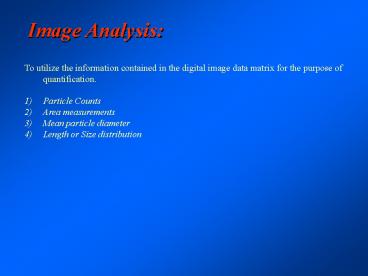
- To utilize the information contained in the
digital image data matrix for the purpose of
quantification. - Particle Counts
- Area measurements
- Mean particle diameter
- Length or Size distribution
2
Image Analysis
Underlying the principles of image analysis the
operator must remember one essential fact.
3
Image Analysis
Underlying the principles of image analysis the
operator must remember one essential fact.
Computers are STUPID!
4
Image Analysis
Dog or Cat?
Any four year old can tell the difference but
even the most sophisticated computers would have
difficulty making the distinction.
5
First step in image analysis is to define those
features that you wish to analyze so that the
computer can know what data in the image is
significant.
Thresholding
- By grey level (pixel value)
- By size ( of contiguous pixels within a certain
value range) - By shape (round vs. elongate)
6
First step is create a binary image based on some
cut off value for pixel intensity.
7
First step is create a binary image based on some
cut off value for pixel intensity.
8
A binary image can also be adjusted to cover a
subset of pixel values.
9
In cases where the objects of interest are quite
distinct it can be relatively straightforward to
distinguish them based on pixel value alone.
10
Sometimes simple thresholding is insufficient in
defining those features one wishes to count,
especially if the objects are touching each other
making it difficult to distinguish one object
from another.
11
Adjusting the contrast of the image may help the
operator identify the objects of interest but the
computer would still have difficulty identifying
the objects.
12
The operator can use a marking tool to identify
the objects of interest.
13
This image may be easily thresholded and made
into a binary image in which the number of
objects may be easily counted.
14
The creation of a binary image is only part of
what needs to be done.
15
If Objects appear to be connected, even by a
narrow bridge, then those two objects will be
considered as a single object by the computer.
16
The connections can be dissolved by performing an
erosion operation which will remove the
peripheral pixels in a pixel by pixel manner.
17
This will reduce the area occupied by the objects
but pixels can be added back by a process known
as dilation so that objects are restored to
nearly their original size without reconnection.
18
Free image analysis programs are available for
downloading from the National Institutes of
Health. They include many basic and some
sophisticated capabilities.
rsb.info.nih.gov/nih-image NIH Image
(Mac) rsb.info.nih.gov/ij ImageJ (PC)
19
Both NIH-Image and ImageJ have similar
capabilities although the layouts are quite
different.
20
First step is to crop the image so that only the
area of interest is used.
21
Sometimes the image contrast must be inverted if
the objects of interest are dark.
22
Next the image must be thresholded to create a
binary image.
23
Using the Analyze Particles under the Analyze
window all particles greater than 1 pixel and
smaller than 999999 will be counted
24
Over 1300 particles are counted, most of which
are only 1 pixel in size
25
If we raise the minimum size to 5 adjacent pixels
and rerun the analysis
26
If we raise the minimum size to 5 adjacent pixels
and rerun the analysis we get many fewer
particles, only the 166 ones of interest
27
The output of the particle analysis can be
exported as a file that can be uploaded into a
spreadsheet program such as Excel and analyzed.
28
When placed in a spreadsheet the data can be
analyzed in many different ways including size
distribution, average size, percent area, etc.
29
Using this information one can refine the
analysis looking for a way to distinguish single
pores (72-92) vs. double pores (100-152)
30
An average area measurement can now be calculated
for each single pore (85.5) and a ratio of single
vs. double pores (52) can be determined.
31
Depending on how the image is to be used the
operator can choose to collect the image in very
high contrast. This will make the subsequent
thresholding of the image much easier.
32
Side SE detector
The choice of detector can also affect the image
analysis
In-lens SE detector
33
Side SE detector
Especially after the image is thresholded
In-lens SE detector
34
If one can calculate the pixel size, then
accurate size and area measurements are possible.
35
Sophisticated image analysis can recognize
patterns and detect and identify such complex
patterns as those contained in diffraction
patterns.
36
Measurements in Three Dimensions
What if we wanted to measure the precise height
difference between two objects in an SEM image?
37
Measurements in Three Dimensions
First we must create a stereo pair image. Even
though a conventional SEM image has a great depth
of field it is still a two dimensional image.
38
Measurements in Three Dimensions
Steps in creating a stereo pair image (Bozzola
Russell p. 221-224) True 3-D imaging requires
that the object be viewed from two different
angles at the same time. A person with sight in
only one eye can have excellent depth perception
but cannot see something in 3-D.
39
Measurements in Three Dimensions
The first step is to create a stereo pair of
images in which the specimen is eucentricaly
tilted 8-12 degrees between pictures.
40
Measurements in Three Dimensions
To aid the observer in visualizing the stereo
pair the left hand view can be colorized blue
and the right hand view made red and
superimposed on one another
41
Measurements in Three Dimensions
These Red/Blue images are known as anaglyph
projections and can be quite dramatic
42
Quartz Crystals
43
Measurements in Three Dimensions
44
Measurements in Three Dimensions
MeX is a software product to compute and analyze
digital elevation models (DEMs) from stereoscopic
scanning electron microscope (SEM) images. MeX
opens up the third dimension to the SEM users. In
order to determine the topography of
microstructures MeX is the ultimate tool when
other means have come to an end. Using MeX you
can measure profiles, roughness values, area
parameters and even volumes of your specimen from
SEM images.
www.alicona.com/
45
Measurements in Three Dimensions
First one defines an area for a digital elevation
map (DEM).
www.alicona.com/
46
Measurements in Three Dimensions
The creation of a DEM is computationally
intensive and starts by building a wire-frame
model which can then be surfaced rendered
www.alicona.com/
47
Measurements in Three Dimensions
Once created the DEM can be viewed from many
angles and color coded to emphasize features such
as height.
www.alicona.com/
48
Measurements in Three Dimensions
Compare the DEM with an anaglyph projection
www.alicona.com/
49
Measurements in Three Dimensions
Profiles can produce accurate measurements for
objects that vary in height
www.alicona.com/
50
Measurements in Three Dimensions
One can even measure volumes using this
software. Unlike ImageJ MeX is not free!
www.alicona.com/
Recommended
CrystalGraphics Presentations


