
Tissue Types in the Human PowerPoint PPT Presentation
1 / 72
Title: Tissue Types in the Human
1
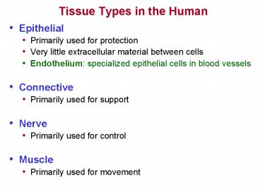
Tissue Types in the Human
- Epithelial
- Primarily used for protection
- Very little extracellular material between cells
- Endothelium specialized epithelial cells in
blood vessels - Connective
- Primarily used for support
- Nerve
- Primarily used for control
- Muscle
- Primarily used for movement
2
Epithelial Tissue
- Cells are polyhedral (many sided) with little
interstitial space - Covers the outermost layer of tissue (skin)
- Skin
- Covers innermost and layer of most organs and
cavities - Lungs, GI tract, Urinary tracts, Reproductive
tracts, - One side always exposed to
- Body exterior READING FOR EPITHLEIUM
- Organ tract or cavity
- Makes up the exocrine and endocrine glands
- Exocrine (excreting) sweat glands, digestive
glands, mammary glands - Endocrine (hormones) thyroid, pancreas,
adrenal cortex - Cells have high regeneration potential but are
avascular - Rely on perfusion for O2 supply
- Many epithelial cells rest on a Basement
Membrane - Basement Membrane Basal Laminae Reticular
Laminae - Basal Laminae flat sheet of nonliving
adhesive-like collagen and glycoprotein - secreted by the epithelial cells themselves
- Reticular Laminae foundation for the Basil
Laminae
3
Adjectives Describing Epithelial Tissue
- Squamous (meaning scale) - flat cells
- Cuboidal - cells as tall as they are wide
- Columnar - tall and column shaped
- Simple - having a single layer of cells
- Stratified - having stacked layers
- Transitional dome surface cells
- - capable of stretching (bladder)
- Ciliated - cilia on the exposed surface
- Examples you should remember
- SIMPLE SQUAMOUS EPITHELIUM
- Permeable cell structure - used for filtration
and gas exchange - Examples capillaries, alveoli, kidney glomeruli
- STRATIFIED SQUAMOUS EPITHELIUM
- Used for protection
- basil cells (cells next to basement membrane) may
be cuboidal - Examples skin, inside of mouth, vagina
- CILIATED COLUMNAR EPITHELIUM
- Used to move substances along a particular
direction using the cilia - Examples upper respiratory tract, fallopian tubes
Transitional
Stratified Squamous
Stratified Cuboidal
Pseudostratified Columnar
4
Examples of Epithelial Tissue
Simple Squamous Epithelium Artery Endothelium
Stratified Squamous Epithelium Human Skin
- Orange and brown/green covering Adventicia
- Blue Actin in smooth muscle
- Green Elastic basal membrane (Basil Laminae)
- Innermost Orange Arterial Endothelium
Ciliated Columnar Epithelium Tracheal Lung Tissue
Cilia
Ciliated Columnar Epithelial Cells
5
Diseases of Epithelial Tissue
Simple Squamous Epithelium Arterial Endothelium
Dysfunction The Beginnings of Atherosclerosis
Tear in endothelial wall (injury - dysfunction)
Monocyte (Macrophage)
Cholesterol crystal deposits
Red blood cell
Foam cell (Lipid
filled macrophages)
Ciliated Columnar Epithelium Trachea Tissue from
a SMOKER
Fat deposits
Note Lack of Cilia
Note Disorganization of Columnar Epithelial Cells
6
Connective Tissue
- Matrix - non-living component of connective
tissue - Ground Substance
- Proteoglycan aggregates (PGA) - pine tree shaped
molecules - Glycosaminoglycans - neg charged binds Na
K attract H20 - Hyaluronic Acid - negative charged slippery
polysaccharride - Condroitin sulfate
- Fluid - H2O, gasses, nutrients for cells (H2O
facilitates tissue turgor) - Minerals - Calcium salts
- Adhesive glycoproteins hold PGAs together to
membranes - Chondronectin (cartilage), osteonectin (bone),
fibronectin (fibrous tissue) - Laminin (holds epithelial cells to basement
membrane) - Fibers
- Collagen, Elastin, and Reticular Fibers READING
ON CONNECTIVE TISSUE - Cells - living component of connective tissue
- Blast Cells, Cyte Cells, Clast Cells
- Macrophages and white blood cells
- Mast cells containing Heparin Histamine
- Adipose tissue
7
Proteoglycans
Electron Micrograph of actual Proteoglycan
Aggregate
Other Glycosaminoglycans Dermatin sulfate
Keratin Sulfate
8
Types of Fibers
Collagen
- Fibrous protein in connective tissue structure
- Derived from Greek word meaning to glue
together - Constitutes about 50 of the proteins in man
- Present to some degree in all human organs
- Collagen has a finite life span after which it is
degraded to the constituent amino acids and
replaced by new fibers. - Has high tensile strength
- 4.5 pound load needed to break collagen fiber 1
mm thick - Maximal strength of scar collagen is about 75
of the original tissue
READING FOR COLLAGEN STRUCTURE FUNCTION
9
Collagen Fibers
10
Collagen Fiber (Fibril)
Each collagen molecule (also called a
tropocollagen) is connected to others via
PYRIDINIUM CROSS-LINK BONDS. These bonds are
degraded in Ehlers-Danlos Syndrome, Osteogenesis
Imperfecta, and metastatic bone cancer and can
serve as biomarkers which will be elevated in the
urine.
Microfibril
A Collagen Molecule (Tropocollagen)
Collagen Structure
Each chain connected to the other two with
Hydrogen Bonds
Alpha Helix Chains Within Each Collagen Molecule
Collagen Molecule (Tropocollagen)
Fibril
Although Hydrogen bonds are weak, the stacked
intertwined formation of the triple helix give
collagen remarkable strength.
Microfibril
Fiber
Tropocollagen Helix
Individual Amino Acid Bonds Are Reinforced With
Hydrogen Bonds
11
Diseases that Affect Collagen
- Overproduction of Collagen Fibers
- Lung Fibrosis (Cystic Fibrosis) excess
glandular secretions (mucous) - Caused by a mutation in CTFR gene r product of
this gene is ion channel - This channel is important in creating sweat,
digestive juices, and mucous - High salt content in sweat is usually present in
CF kids - Life-span used to be limited to 20-30 years.now
possibly 40-50 years - Obstructions and fluid in lungs r breathing
disorders numerous infections - Obstructions in pancreas r d digestive enzymes r
d nutrient absorption - Malnutrition r d growth READING FOR CYSTIC
FIBROSIS - Liver Cirrhosis irreversible scarring (fiber
deposition) in the liver - Common causes Hepatitis-C Hepatitis-B,
alcoholism - Alcohol blocks normal metabolism of protein,
fats, and carbs r injury - Cirrhosis r edema ascites (fluid in peritoneal
space) - Liver cannot make Albumin r blood looses osmotic
(sucking) pressure - Cirrhosis r u infection risk, jaundice, bruising
bleeding, portal hypertension - Cirrhosis will elevate Aminotransferase enzymes
READING FOR LIVER CIRRHOSIS - ALT, AST, GGT(aka SGOT- large elevations
associated with alcoholism) - Atherosclerotic heart disease
12
Diseases that Affect Collagen
- Autoimmune Disorders that Damage Collagen
- Lupus Erythematosus - production of auto
antibodies that target body tissue - 90 of Lupus patients will experience joint and
muscle pain - Pain caused by collagen damage and destruction
- Collagen damage and inflammation can occur
anywhere in the body - Most common areas affected skin, articular
tissue, lungs, blood vessels, liver ,
kidneys, and nervous system - Course of the disease is unpredictable with
attacks and remissions - Most common in women ages 18 40 READING
FOR LUPUS
- Insufficient Collagen
- Ehlers-Danlos syndrome - rubber man -
contortionist disease - Weakening of collagen cross-link bonds
- Osteogenesis Imperfecta - brittle bones
- Scurvey -Vitmain C deficiency
- Too few hydrogen bonds in the formation of the
collagen molecule - Inferior tissue formation in bones, blood
vessels, skin, and teeth
13
Therapy for Common Collagen Diseases
- Cystic Fibrosis
- Clearance techniques for excess lung secretions
- Pancreatic enzyme replacement for pancreatic duct
obstruction - Healthy diet and exercise
- Drugs
- Ibuprofin slows rate of decline of pulmonary
function - Corticosteroids d inflammation in lungs,
joints, and vasculature - Antibiotics to treat and reduce the incidence
of lung infections - Rotation of drugs to prevent development of
resistance - Immunoglobulin experimental research (currently
in human trials phase) - Lupus
- Drugs
- Disease Modifying Anti Rheumatic Drugs (DMARDS)
d flare-ups - NSAIDs, corticosteroids and other
immunosuppresants - Hydroxychloroquine (commonly used as an
antimalarial drug) - Experimental drugs
- Stem cell infusion very promising
- Ultraviolet Radiation Therapy (not yet approved
in U.S.)
14
Other Types of Fibers
- Reticular Fibers
- Actually very fine collagen fibers
- Usually form a network
- Fill space between other tissues organs
- Contained in the reticular laminae
- Elastic Fibers
- Contain protein called ELASTIN
- Elastin molecules look like coiled springs
- Return to original shape after distortion
- Found in arteries, skin, alveoli of lungs
(prominent in skin) - Synthesized by only fetal and juvenile
fibroblasts - Loss of elastin with age contributes to
development of wrinkles - Degraded elastin found in Aortic aneurysm COPD
- Malformed elastin is found in aortic stenosis
(genetic predisposition)
15
- Elastin Fibers
- Stretched or taught
- Relaxed
16
Types of Connective Tissue
- Fibroconnective Tissue
- Cartilage
- Bone
READING ON CONNECTIVE TISSUE 2
17
Types of Connective Tissue
- Fibro connective Tissue - matrix composed mostly
fibers - Areolar -Loose connective tissue serves as
packing material - Component of some basal membranes (attaches
underlying structures) - Separates muscles - allows for muscles to slide
over each other - Fibers in extracellular matrix are collagen
elastin - Extracellular matrix is not well organized
- Has more cells than Dense Regular or Dense
Irregular - Reticular - fibers form a soft internal skeleton
for tissues and organs - Fibers are collagen elastin
- Adipose - highly vascular insulator, shock
absorber energy store - Cells account for 90 of tissue mass (little
matrix present) - Dense regular - closely packed parallel collagen
fibers few cells - Found where tension is exerted in a particular
direction - Examples tendons, ligaments
- Dense irregular - closely packed non-directional
collagen few cells - Forms sheets where tension is exerted in many
directions - Examples dermis of skin, muscle fascia, organ
nerve coverings - Elastic - composed of mostly elastin fibers
- Examples vocal cords, ligamenta flava
(vertebral connective tissue)
18
Areolar Tissue
19
Adipose Tissue X 200 ( bv blood vessel ) (
arrow adipocyte nucleus )
bv
20
Reticular Tissue
21
Dense Regular Tissue
(Tendons Ligaments)
Horse Tendon x100 arrow orientation of
collagen fibers
22
Dense Irregular Tissue
(Dermis of Skin Muscle Fascia)
23
Types of Connective Tissue
- Cartilage - matrix mostly fibers ground
substance - Avascular (slow to heal) and not innervated
- Composition of cartilage matrix
- Ground substance chondroitin sulfate
hyaluronic acid - Collagen (the main fiber) elastin
- Perichondrium surrounding tissue from which
nutrients diffuse - Limits cartilage thickness - nutrients must
diffuse entire tissue thickness - Gives rise to chondrocytes
- Types of cartilage
- Hyaline - tough flexible - much matrix / few
cells - shock absorber - Covers ends of long bones (articular cartilage
eroded in OA) - Forms skeleton of trachea and bronchi
- Fibrocartilage - less firm than hyaline - more
cells and fibers - Similar in structure to dense regular tissue
(tendon) - Transitional tissue between tendon and articular
hyaline cartilage - Component of joint capsules transitions to
spongy knee menisci - Elastic - contains more elastin fibers than other
types of cartilage - forms ear pinna epiglottis
READING FOR CARTILAGE
24
Hyaline Cartilage X 250 arrows Perichondrial
borders
Hyaline Cartilage Perichondrium on the
left Chondrocytes form in the
perichondrium and move out into the tissue
25
Fibrocartilage
26
Elastic Cartilage (note numerous chondrocytes and
elastic fibers)
27
Types of Connective Tissue
- Bone - matrix mostly calcium and phosphate
- 65 of bone weight is calcium hydroxyapatite
- Calcium phosphate, calcium hydroxide, calcium
carbonate - Tropocollagen subunits giving bone elasticity and
fracture resistance - Bone collagen d with age r u fracture risk
- Highly vascular and well innervated
- Contains lymph channels
- Functions in mineral storage and blood cell
production - Bone remodeling - deposition and resorption -
negative feedback - d blood Ca r u Parathormone (PTH) r u
osteoclast activity - u blood Ca r u Calcitonin r u
osteoblast activity - Red marrow contains hematopoietic tissue -
produces blood cells - Influences on Bone Growth
- Levels of Ca, Phosphorous, Vitamin D, HGH,
estrogen, testosterone - READING FOR BONE
28
Review of Bone Histology
Epiphyseal Plates
Haversian System (osteon)
Compact (Cortical) Bone
Haversian Canal (contain blood vessels)
Lamellae (concentric rings of hard bone)
Marrow
Osteocytes in Lacunae
Endosteum
Periosteum
Volkmans Canal
Canaliculi (connecting tunnels)
Trabecular (Cancellous) Bone
29
Growth Plates (epiphyseal plates) in Long Bone
EPIPHYSEL INJURIES IN CHILDREN
EPIPHYSEL INJURIES IN CHILDREN 2
30
Growth Plate (epiphyseal plate) in Long Bone
Hematopoietic Tissue
zone of resting hyaline cartilage
zone of proliferation
zone of hypertrophy
zone of calcification
ossified bone
Epiphysis (bone end)
Diaphysis (bone shaft)
Length Increase (Growth) Occurs Toward Diaphysis
Chondrocytes
Chondrocytes die upon calcification - blood
vessels from diaphysis grow into the area
Chondrocytes divide and stack on top of one
another
Red Bone Marrow
31
Bone Diseases Treatments
- Pagets Disease - enlarged deformed bones
- u bone resorption and deposition r weak bones,
bone pain, u fractures, arthritis - Affects about 1 of adults have - rarely
diagnosed in people under 40 years of age - Symptoms pain, pinched nerves r tingling
numbness, leg bowing, hip knee pain - Bones most often affected spine, femur, pelvis,
skull, clavicle, humerus. - Treated with Bisphosphonate drugs (see
osteoporosis treatment)
- Osteoporosis - d bone density r fracture
predisposition - Osteopenia bone loss not as severe as
Osteoperosis - u bone resorption in the presence of normal bone
metabolism - d both cortical (thick) and trabecular (porous)
bone density, but affects cortical more - Women start losing bone density about age 40, men
at age 60 - Over 28 million people in the U.S. have
osteoporosis - 80 of this 28 million are women
- Many women experience up to a 20 d in bone mass
by 5 to 7 years after menopause - 1 in 2 women and 1 in 4 men over age 50 will have
an osteoporosis - related fracture in their lifetime.
- Common Fracture Sites thoracic vertebra,
- distal radius (close to wrist), femur
neck (Hip Fracture)
READING FOR OSTEOPOROSIS
32
Bone Diseases Treatments
- Osteoporosis continued)
- Causes
- Prolonged treatment with corticosteroids
- Anorexia nervosa
- Inadequate diet, especially during pregnancy and
breast feeding - Amenorrhea r u estrogen metabolism disturbances r
bone loss (Osteopenia) - Treatment
- Estrogen replacement (for postmenopausal women)??
- Calcium supplementation
- Vitamin D supplementation
- Bisphosphonate drugs (also called diphosphonates)
- FOSAMAX, ACTONEL, BONIVA, AREDIA, RECLAST d
osteoclast activity - Also used to treat bone cancer other bone
weakening diseases - Calcitonin, FORTEO (Teriparatide) a PTH analog
r u osteoblast activity - Intermittent exposure to PTH will u osteoblast
activity more that osteoclast activity - Weight bearing exercise
- Osteomyelitis - bone inflammation destruction
- Caused by bacteria and fungi spreading from other
infection sites - Symptoms fever, localized warmth swelling,
localized pain - Treated with antibiotics
33
Bone Diseases Treatments
- Osteoarthritis - Degenerative changes in
cartilage bone - Loss of articular cartilage (proteoglycan loss r
water loss r d compliance) - Roughening, pitting, destruction in hyaline
cartilage r u joint stiffness - Most common in hands, hips, and knees
- May result in the formation of osetophytes (bone
spurs) or nodes (Bouchards nodes) - 80 90 of people over age 65 have some
evidence of osteoarthritis - Cause
- Genetics (60), infection, endocrine disorders,
joint injury, overuse - Fracture or Ligament Injury r bad joint alignment
instability r u wear and tear - Treatment READING FOR OSTEOARTHRITIS
- Exercise - helps maintain ROM, healthy cartilage,
strength and reduces pain - Rogind et.al. 1998 Gur et.al. 2002
- Immobilization can worsen the course of the
disease - Weight loss for OA in weight bearing joints
- NSAIDS and COX2 inhibitors for pain (COX 2
inhibitors ???) - Injections of HYALURONIN (hyaluronic acid) or new
artificial injectible materials - Corticosteroid injections may be useful when
inflammation is present - Joint replacement (when conservative therapy
fails) - Tissue engineering to regenerate cartilage has
had some success
34
Osteoarthritis of the Knee
Knee Arthroplasty
Bone on bone in this resected tibial plateau
Bouchards nodes
Osteophytes
35
Bone Diseases Treatments
- Rheumatoid Arthritis (RA) Autoimmune
inflammatory disease - May be related to genetic factors
- Usually occurs between ages 25 55 and affects
mostly young and - middle age females - may fluctuate
substantially in severity - Rheumatoid factor (autoantibody) globulins r
immune complexes - Immune complexes activate the compliment system r
inflammation - Involves synovial membranes of joints (most
common manifestation) - Inflammation leads to swelling thickening of
synovial membrane (u ESR) - Joints most often affected wrists, fingers,
knees, feet, and ankles - May possibly affect READING FOR
RHEUMATOID ARTHRITIS - Heart endocarditis, pericarditis, CHF, valvular
fibrosis, MI - - RA and other autoimmune disease patients
have an u risk for CHD - Lungs fibrosis and pleural effusion
- Kidneys amyloidosis (deposition of insoluble
proteins in organ tissue)
36
Bone Diseases Treatments
Rheumatoid Arthritis
Comparison of Normal Rheumatoid Joint
Normal Joint
Rheumatoid Joint
Infiltration of numerous cell types
Synovial Membrane
Pannus (granulation tissue)
Leukocyte
Macrophage
Interdigitating Cell (traps antigens)
Cartilage
Plasma Cell (B-lymphocyte)
Neutrophil
Capillary Bud (angiogenesis)
Immune Complex
Inflamed Synovial Membrane
37
Bone Diseases Treatments
Rheumatoid Arthritis
38
Bone Diseases Treatments
- Treatments for Rheumatoid Arthritis
- NSAIDs
- COX2 inhibitors
- Corticosteroids
- Disease-Modifying Anti-Rheumatic Drugs (DMARDs)
- Methotrexate
- d TNF, neutrophils, histamine, lymphocyte number
function - d growth of certain cells in blood, skin, GI
tract, immune system - Cytotoxic inhibits metabolism rd immune
function - Developed in 1940s as chemotherapy for Leukemia
- Sulfasalazine - d immune function
- Hydroxychloroquine an antimalarial drug
- Gold salt injections
- Exercise to maintain joint mobility
- Physiotherapy, physical therapy, water exercise
- Surgery synovectomy or joint replacement
- Gene therapy injection of genes that produces
desired protein via a vector (vector various
types of viruses)
39
New Anti-Arthritic Drugs Biological Agents
(or Biologics)
- Tumor Necrosis Factor (TNF) blockers
- Must be given by subcutaneous injection or IV
HUMIRA adalimumab REMICADE infliximab
ENBREL etanercept
- Mechanism (Effects)
- Binds to TNF
- Prevents attachment to its receptor
- Inhibits inflammatory mediators
- d inflammation in joint r d pain
15,000 - 45,000 / year (2008)
- Indications TNFa
- Rheumatoid Arthritis
- Effective in 70 of patients who
- have not responded to Methotrexate
- Ankylosing Spondylitis
- Psoriasis
- Psoriatic Arthritis- 12 of people with
- psoriasis have psoriatic arthritis
- Chrons Disease - autoimmune
- inflammatory bowel disease
- Adverse effects
- Immunosuppression !!
- u risk of infection !!
- Tuberculosis common
- Allergic reactions
- KINERET Anakinra
- Mechanism Injectable man-made protein that
blocks interleukin-1 (IL-1) - IL-1 r cartilage degradation, u bone resorption
- Adverse Reactions Injection site reactions,
systemic infections (d immunity) - malignancies, neutropenia
40
Blood Body Fluids
- 62.5 of total body fluid is intracellular
(contained within cells) and is abbreviated ICF - 37.5 of total body fluid is extracellular
(outside of cells blood, interstitial fluid,
etc) abbreviated ECF - Average human blood volume is about 5 Liters
- 3 Liters plasma 2 Liters RBCs, WBCs etc
- Hematocrit (crit) packed RBC volume about
45 of total volume for men, 40 for women - Anemia crit Hb
- Anemia can lead to fatigue weakness and may
be caused by - Colon Cancer (bleeding), IBS (bleeding),
Bleeding Ulcer, Chrons Disease (bleeding) - Other Cancers
- u cytokines (TNFa, IL-1 r d EPO synthesis r d
RBC count - Chemotherapy Radiation for cancer
- RBC destruction d RBC production
- Kidney disease
- d Erythropoietin (EPO) Uremia r d RBCs and d
platelet function - Blood Components
- Formed elements RBCs, WBCs, Platelets
- Plasma Plasma Serum - clotting proteins such
as fibrinogen (Serum Plasma Proteins) - Elevated fibrinogen levels r u risk of stroke
and atherosclerotic disease (tests available)
41
Blood Body Fluids
- Red blood cells (Erythrocytes) - formed in bone
marrow - Production and homeostasis regulated by tissue
oxygenation - d tissue O2 r u erythropoietin from kidneys
liver r u RBCs - White blood cells (Leukocytes)
- Primary effector against infection tissue
damage - WBCs engulf foreign substances lysozomal
enzymes digest them - Inadequate circulatory or lymphatic function r
abscess - GRANULOCYTES (Polymorphonuclear cells) -
granulated WBCs - Neutrophils - 62 of WBCs - 1st to travel
arrive at injury kamikazi phagocytotic - Eosinophils - 2 of WBCs - destroy parasites -
involved in allergies - Basophils - heparin
- AGRANULOCYTES phagocytotic non-granulated WBCS
- Monocytes - 5.3 of WBCs - become lysosome
filled macrophages - Play important role in remove dust and necrotic
tissue in lungs - Macrophages - monocytes that have left the
circulation long lived - Macrophages release cytokines and compliment
proteins (inflam. mediators) - Lymphocytes - Tcells Bcells - 30 of WBCs
- Function in acquired immunity (antigen r B cells
r antibody production)
42
Macrophage (in the box) Arrow Pedicle for
locomotion
Monocytes
43
large arrow Basophil small arrow Neutrophil
Arrow Eisonophil
44
Blood Body Fluids
- Platelets - Thrombocytes
- Sticky cells that function in all aspects of
hemostasis - Plasma
- Water (90 of plasma volume)
- Metabolic by products lactic acid, urea,
creatinine, etc. - Nutrients glucose, FFAs, lipids, cholesterol,
vitamins - Electrolytes sodium, potassium, magnesium,
bicarbonate, etc. - Gasses oxygen, nitrogen, carbon dioxide, etc.
- Fibrinogen other clotting proteins
45
Blood Body Fluids
- Plasma (continued)
- Proteins Total Blood Protein Albumin
Globulin - Albumin - (60) manufactured by the liver
- Maintains oncotic pressure
- Transports FFAs, thyroid other hormones,
bilirubin (heme catabolite) - Functions as a free radical scavenger
(antioxidant) - Globulins (36) (globulin injections r u
immunity r new CF therapy) - Alpha transport biliruben steroids
- Beta transport copper and iron, form
lipoproteins (mostly LDL) - Gamma (or immunoglobulin) Ig contains
antibodies - - Produced by immune system in response to
infection, allergic reaction - - Provide short term disease protection (GG
injections are possible) - NOTE d hepatic function r d Albumin / Globulin
ratio - Fibrinogen - (4) functions in hemostasis (the
clotting process) - Enzymes catalyzes physiological reactions (PFK,
citrate synthase.) - Antibacterial Proteins CAP18, LL37
- Protein Hormones Insulin, HGH, LH, FSH,
ADH(vasopresin),
46
The Basic Unit of Nerve Tissue The Neuron
READING FOR NERVE TISSUE
47
Skeletal Muscle or Motor Unit Action Potential
Excitatory post-synaptic potentials
48
Nerve Tissue X 200 Large arrow Soma (cell
body) Small arrow axon body Box Axon Hillock
49
Propagation of a Neural Impulse
Receptor Operated Na / K channels
Membrane yet to be depolarized
Repolarization
Depolarization
K
Voltage Gated Ca Channels
Na
EPSPs
Na
K
Soma
Volatage Gated Na Channels
Dendrites
Axon Hillock
50
Inhibition of Neural Transmission via GABA the
inhibitory neurotransmitter
GABA - gamma
aminobutyric acid
Inhibitory GABA receptors exist on the post
synaptic structure of the dendrite. Activation
of these receptors permits Chloride ions to enter
the dendrite and nullifies EPSP's. This takes
the neuron further from depolarization
axon of
inhibitory
neuron
axon
collateral
GABA
released
- Tranquilizing drugs of the benzodiazapine family
(VALIUM, ATIVAN, XANAX, LIBRIUM, RESTORIL,
HALCION, CLONOPIN) bind to receptors in the brain
and enhance the affinity of GABA for its
receptor. This further promotes the inward
chloride current, which reduces anxiety and
promotes a calming effect.
Chlorine channels
Excitatory
open in response to
axon
GABA release and
chloride ions enter
the neural cell.
Chloride ion hyperpolarizes the cell ( less
depolarization) so AP's traveling down the axon
are
inhibited. This causes less Calcium to enter the
terminal bouton, resulting in less acetylcholine
release (neuronal transmission is retarded).
51
Higher Neural Processing Centers
Sensory - Motor Structure Signal transmission
Sensory Neuron
Motor Neuron
Axon Hillock
Efferent Signal
Afferent Signal
Effector Muscle
Free Nerve Endings (type of receptor)
52
Regeneration of Peripheral Nervous Tissue
schwann cell columns
axonal sprouts
53
Nerve Diseases Associated Therapy
- Multiple Sclerosis - demyelination of nerve
tissues several types of MS exist - Causes autoimmune factors (exact antigen not
identified), virus triggers (not proven),
possible genetic predisposition, various forms of
physical trauma. - Symptoms weakness, numbness (pins needles),
loss of balance, loss of coordination, bowel
bladder dysfunction, muscle spasticity, optic
nerve neuritis - symptoms are epidsodic
- Therapies
- Immunomodulator drugs d inflammatory effects of
lymphocytes - inhibit cytokines r d inflammation, also d number
of episodes - Interferon b (1a 1b) - AVONEX BETASERON
REBIF (1st line drugs) - Glatiramer Acetate - COPAXONE (2nd line drug)
- Immunosuppressants dd inflammatory effects of
lymphocytes, d inflammation (3rd line drug) - Mitoxantrone, Cyclophosphamide,
Methylprednisolone, ACTH - Danger of infection from compromised immune
system - Weakness therapies dopaminergic drugs similar
to drugs for Parkinsons patients, exercise
(water exercise swimming most beneficial) - Spacticity therapies reflex inhibitors, ex a2
agonists (inhibit spinal motor neurons), muscle
relaxer drugs - Incontinence therapies muscarinic achetylcholine
receptors antagonists (relaxes bladder dutrussor
muscle), antidiuretic hormone analogs - Tremmor therapies anticonvulsant drugs
- Visual problem therapies corticosteroids to
reduce ocular inflammation
24,000 / year (2008)
54
MRI of the brain showing a plaque associated with
Multiple Sclerosis
55
Nerve Diseases Associated Therapy
- Parkinsons - loss of production of the
neurotransmitter dopamine in the basal ganglia
(loss of 80 of dopamine producing cells) r
disruption of balance between dopamine and Ach r
d voluntary movement control - Causes free radical damage (theory), toxins
(theory), age related d in dopamine producing
neurons, genetic predisposition, repeated head
trauma (boxing), illegal drug use, hydrocephalus
(CSF accumulation in ventricle of the brain),
encephalitis -most often viral-(inflammation of
white and gray brain matter). - Symptoms resting tremor - pill rolling motion
(70), bradykinesia (inability to generate
movement), rigidity, postural instability,
difficulty rising from sitting position,
shuffling gait. - Therapy
- Levodopa u dopamine levels in brain (current
gold standard of treatment) - Stem cells r u Dopamine neurons r likely 1st or
2nd disease cured by SC - Catechol-O-methyltransferase inhibitors
(inhibits levodopas peripheral metabolism r
more available for transport across blood brain
barrier) - Dopamine agonists stimulate post-synaptic
dopamine receptors - Monoamine Oxidase B inhibitors slow dopamine
neuron degeneration - Embryonic tissue transplantation not very
successful so far - DBS Surgery brain pacemaker sends e- to parts
of brain - promising
READING FOR PARKINSONS
READING FOR STEM CELL RESEARCH
56
PET Scan showing reduced uptake of injected
flurodopa (radioactive dopamine) in the dopamine
producing neurons in the brain of a Parkinsons
Patient
57
Presynaptic Terminal Bouton
Neuromuscular Junction
Synaptic Vesicles
READING FOR NM JUNCTION
Acetylcholine Receptors
Synaptic cleft
Acetylcholine
Mitochondria (ATP Producer) (Ca Reservoir)
Acetylcholinesterase
T-tubule
Saroplasmic Reticulum
(Site of Ca storage)
Ca
Myosin
Actin
ANIMATION EXPLAINING NM JUCTION FUNCTION
H
I
A
Z disk
58
Motor End Plates (Skeletal Muscle)
59
Neuromuscular Junction ( a Motor Neuron )
60
Action of Selected Toxins Drugs Around the NM
Junction
Local anesthetics Tetrodotoxin (puffer fish)
Batrachotoxin (S.A. frog)
1 frog toxin to kill 50 men
Black Widow Venom (Latrodectism)
blocks AP transmission
B toxin blocks Ach release T toxin blocks inhib.
n.t. release
Botulinium toxin (B) Tetanus toxin (T)
u Ach release
blocks Ach receptors
Cobra / Mamba snake Curare
resp. muscles affected
Ach-ase inhibitors Nerve gas (Sarin, VX)
Neostigmine - treat MG
Dantroline muscle relaxer used in MS treatment
d CA release from SR
Blocks AP transmission within muscle
Quinine (antimalarial drug) muscle relaxer
61
Muscle Diseases Associated Therapy
- Fibromyalgia- pain in muscles and connective
tissues - Theoretical Causes Thyroid problems, over growth
of yeast, bacteria, trauma, stress,
neurotransmitter hormone malfunction,
infection, immune system dysfunction, autonomic
nervous system malfunction - Symptoms Aching, un-refreshed by sleep, GI
problems, fatigue, - anxiety depression, d energy, presence
of pain trigger points - Symptoms may be chronic better one day, worse
the next - Disease is often associated with other co-morbid
conditions - Chronic Fatigue Syndrome Irritable bowel
syndrome - Migraine Headache Restless leg syndrome
- TMJ syndrome Depression
- Therapy symptom control, stress reduction,
exercise, antidepressants, NSAIDs, growth
hormone therapy, psychiatric help
READING FOR FIBROMYALGIA
62
Muscle Diseases Associated Therapy
- Muscular Dystrophy - an inherited disorder
characterized by progressive proximal muscle
weakness with destruction of muscle fibers and
replacement with connective tissue - Diagnosed between 2 5, wheelchair by 10 or 12,
death in 20s - Blood creatine kinase is elevated (indicator of
muscle damage) - Some are mildly retarded
- Causes genetic related absence of Dystrophin, a
muscle membrane protein - Initial Symptoms waddling gait, falls,
difficulty standing, difficulty climbing or
descending stairs r muscle wasting, contractures,
cardiac involvement, respiratory muscle weakness
with complications (respiratory infections). - Therapy daily steroids produce long term symptom
improvement, exercise should be continued as long
as possible, surgery may be done to release
contractures, pneumonia vaccine (prophylactic),
physical therapy to delay development of
contractures. - Contractures any condition that affects
mobility or range of motion of a joint - Usually involves fiber deposition in skin,
fascia, muscle or joint capsule
READING FOR MUSCULAR DYSTROPHY
63
Somata Sensory Neuron Types and Function
Classification Diameter Velocity Receptor
Function / Sensation Aa (a efferents)
Largest Highest Reflexes Joint
sensation Ia II - - - - - - - - - -
- - - - Spindle Ib - - - - - - - -
- - - - - - - - GTO Ab (g efferents)
Spindle Fine touch Kinesthesia
Merkels disks Joint sensation
Meisners corpuscle Deep tissue sensation
Pacinian corpuscle Deep pressure
Vibration Ruffinis end organs Touch
Pressure Hair end organs Touch Ad
(g efferents) Cold receptors
Temp(cold) Crude touch Free nerve
endings Fast (sharp) pain C (unmylenated) Smalles
t Slowest Warm receptors Temp(warm)
Crude touch Free nerve endings
Slow(aching) pain Joint muscle aches,
Itch Deep tissue pain, pressure
READING FOR SENSORY NEURON TYPES
64
Muscle Spindles, GTOs, and the Myotatic Stretch
Reflex
READING FOR MUSCLE SPINDLES GTOS
- Muscle Spindles
- Detect change in muscle length and rate of change
in muscle length - Arranged in parallel with actual muscle fibers
- Muscle involved in fine motor activity 120
spindles per gram of muscle - Muscles involved in bulk movement 5
spindles per gram of muscle - Myotatic Stretch Reflex (MSR)
- Smooths out or dampens movements, body
stabilization during fine motor tasks - Overactive MSR r hyperactivity of motor cortex r
cortex lesion (stroke, tumor, injury) - Muscle spasticity (muscle always receives
contraction signals r u tone) - Positive Babinski sign, clasped knife sign, d
limb strength dexterity - Underactive MSR r lower motor neuron lesion(s)
(multiple sclerosis, neuropathy) - Loss of strength, d tone, wasting atrophy,
muscle twitches - Golgi Tendon Organs
- Detect both magnitude and rate of tension
development in a muscle - Arranged in series with muscle units
- Less active, less numerous, and slower to react
than spindles - Autogenic inhibition (inverse myotatic reflex)
- Excessive stretch / tension r activation r GTO
signal overrides a motor activity
READING FOR UPPER MOTOR NEURON LESIONS
READING FOR LOWER MOTOR NEURON LESIONS
65
- g motor efferent motor neuron
- (co-activated with a motor neruon)
Type II afferent
Type Ia afferent
a motor efferent motor neuron
Connective Tissue Capsule
Lymphatic Space
Extrafusal Muscle Fibers
The Muscle Spindle
Intrafusal Muscle Fibers
66
Cell Body of Sensory Neuron
Peripheral Nerve
Mechanisms of the Myotatic Stretch Reflex
Cell Body of Motor Neuron
Aa motor neuron
Quadricep Motor Units
Type Ia afferent neuron
Muscle Spindle
Stretch Receptors
PatellerTendon
67
Type Ia afferent
Quadriceps
The Myotatic Stretch Reflex
a Motor Neuron
Muscle Spindle
Tendon of Quadriceps
68
The Golgi Tendon Organ
Striated Skeletal Muscle
Tendon Organ Capsule
Type Ib afferent Sensory Nerve Fiber (Myelinated)
Distribution of Golgi Tendon Organ
Tendon
Bone Insertion
69
a
Muscle Spindle Golgi Tendon Organ Structure
Function
Note some authors suggest that the GTO does not
function in the reflex protection of the agonist
muscle
70
Etiology Treatment of Exercise Induced Muscle
Cramps
- Factors thought to contribute to Fatigue and
subsequent EIMCs - Exercising in hot, humid environments
- Dehydration
- Electrolyte deficiencies (possibly from
malnutrition also) - Hyponatremia, hypokalemia, hypocalcaemia,
hypomagnesaemia - Overexertion (Fatigue) r muscle hypoxia
- Disturbances in carbohydrate (hypoglycemia), fat
, or protein metabolism - Nutritional deficiencies
- Inadequate amounts of electrolytes, vitamins
B1(Thiamine), B5 (Pantothenic acid)
- Hypothesized cramp mechanisms supported by the
recent literature - Fatigue r u spindle firing rate d GTO firing
rate r u a motor neuron reflex activity - Overproduction of reflex a motor neuron
activity in a shortened muscle r CRAMP - Further exact mechanisms remain to be
elucidated
READING FOR CRAMPS
71
Factors Thought to Contribute to Idiopathic Leg
Cramps (No consensus on exact etiology)
- Metabolic disturbance
- hyponatraemia, hypokalaemia, hyperkalaemia,
hypocalcaemia, hypomagnesaemia, hypoglycaemia - Chronic or severe acute diarrhea
- Pregnancy, especially in the late months
- Cirrhosis of the liver
- Renal dialysis, possibly owing to plasma volume
contraction - Thyroid disease
- Hyperthyroid myopathy (autoimmune disease) may
be associated with cramps - Hypothyroidism is associated with weakness and
painful muscle spasms - d protein turnover / metabolism d carbohydrate
metabolism - Heavy alcohol ingestion, may induce severe
muscle cramps. - Lead toxicity
- Disorders of the lower motor neurons, including
amyotrophic lateral sclerosis (ALS), MS,
polyneuropathies involving the motor neurons,
recovered poliomyelitis, peripheral nerve injury
and nerve root compression
72
Treatment and Prevention for Cramps
- Treatment for Cramps
- Stretching - activation of GTO helps relax the
muscle - Movement - walking allows muscle spindles to
"reset" - Massage
- Cold application - reduces a motor neuron
activity - Transcutaneous electrical nerve stimulation
- Quinine (anti-malaria, antipyretic, analgesic
drug) used for chronic cramping - d excitability of motor endplate u refractory
period of skeletal muscle - Preventing Cramps
- Stretching
- Correction of muscle weaknesses and strength
imbalances - Conditioning to prevent the onset of fatigue
- Strength training - reduced spindle sensitivity
r d reflex a motor activity - Proper nutrition before and during event
- replacement of fluid and energy substrate during
exercise r d fatigue - Some common drugs that might cause or increase
the risk for cramps - b-blockers, Ca channel blockers, b2 agonists
(asthma), Phenothiazines (psychosis) - Danozol (cortocosteroid for endometriosis),
Lithium, Co
Recommended
