
A Biologically Based Doseresponse Model for Ethanolinduced Developmental neurotoxicity - PowerPoint PPT Presentation
1 / 6
Title:
A Biologically Based Doseresponse Model for Ethanolinduced Developmental neurotoxicity
Description:
... CC11) whereas in the rhesus monkey it is 60 days long transversing at least ... Figure 2. Cell cycle length changes over time in the mouse, rat and monkey. ... – PowerPoint PPT presentation
Number of Views:81
Avg rating:3.0/5.0
Title: A Biologically Based Doseresponse Model for Ethanolinduced Developmental neurotoxicity
1
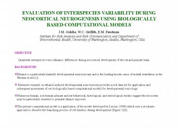
EVALUATION OF INTERSPECIES VARIABILITY DURING
NEOCORTICAL NEUROGENESIS USING BIOLOGICALLY BASED
COMPUTATIONAL MODELS
- J.M. Gohlke, W.C. Griffith, E.M. Faustman
- Institute for Risk Analysis and Risk
Communication and Department of Environmental
Health, University of Washington, Seattle,
Washington, USA
- OBJECTIVE
- Quantitate interspecies toxicodynamic
differences during neocortical development of the
rat and primate brain. - BACKGROUND
- Ethanol is a particularly harmful developmental
neurotoxicant and is the leading known cause of
mental retardation in the Western world (1). - Extensive research on ethanol-induced
developmental neurotoxicity provides a rich data
set for application and subsequent assessment of
our biologically based computational models for
developmental toxicology. - Numerous human, non-human primate and rat
behavioral, histological, and stereological
studies suggest the neocortex may be particularly
sensitive to prenatal ethanol exposure. - The present computational model is a application
of the model developed by Leroux (1996) which
uses a stochastic approach to describe the
branching process of cell kinetics during
development (Figure 1)(2).
2
Figure 1. The anatomy of neocortical
neuronogenesis and how it relates to model
building. a. A mammalian nervous system at the
5 vesicle stage in lateral view showing
progenitor cells (orange)are generated in the
pseudostratefied ventricular epithelium (PVE).
During G1 newly generated cells either stay in
the proliferative population (P fraction) or
become postmitotic (Q fraction-blue cells) and
begin migration through the intermediate zone
(IZ) to the cortical plate (CP). In the mouse,
the neuronogenesis period is six days long and
traverses eleven cell cycles (CC1-CC11) whereas
in the rhesus monkey it is 60 days long
transversing at least 28 cell cycles b. Basic
model framework from Leroux (1996) which was
modified as a model for neocortical
neuronogenesis where Type X cells represent
neuronal progenitor cells in the PVE (orange
circles) and Type Y cells represent postmitotic
neurons leaving the PVE (blue circles).
3
- I. KEY DIFFERENCES BETWEEN RODENT AND PRIMATE
NEOCORTICAL NEUROGENESIS
Figure 2. Cell cycle length changes over time in
the mouse, rat and monkey. During rodent
neurogenesis the cell cycle lengthens over time,
whereas in the rhesus monkey the cell cycle
length peaks at E60 and thereafter shortens.
Data was derived from references 3-5.
- Key Points
- The length of neocortical neurogenesis is
increased in the primate (6 days in the mosue
compared with 60 days in the rhesus monkey). - The cell cycle length is longer during primate
neurogenesis (between 2 to 4 times on average). - The time dependent change in the cell cycle is
different in rodents and the rhesus monkey (See
Fig. 2). - The founder cell population is larger in the
primate (approximately 4 times larger than the
mouse founder cell population).
4
- II. APPLICATION OF MODEL TO MOUSE, RAT AND
PRIMATE CONTROL DATASETS
Figure 3. Comparison of mouse and rat
neocortical neuronal output predictions. This
figure shows a plot of our model predictions for
the normal mouse (?), rat (?), and rhesus monkey
(?) neuronal output (total Y cells). For
comparison we plotted stereological estimates of
total neurons in the neocortex of the adult mouse
and rat adjusted for a 35-50 reduction in
postnatal cells due to normal processes of cell
death ( ? ). The length of the vertical line
represents the variation due to differences in
the mean estimates from different stereological
studies (9-11) (12-16) and variation due to
35-50 reduction (17) of population in the cell
death period.
- Key Point
- Our model predictions closely match
stereologically determined neuronal counts in the
mouse, rat, and monkey.
5
- APPLICATION OF RAT ETHANOL NEUROTOXICITY DATA TO
PRIMATE MODEL - Ethanol exposure (maternal BEC 150 mg/dl)
during neocortical neurogenesis lengthens the
cell cycle prematurely in the rat (4). - The time-dependent effect of dose on the
neocortical progenitor division rate is based on
an in vitro study of cell cycle inhibition (18)
and is modeled using the equation - rate untreated baseline e
(drpdose) - where drp is the time-dependent dose-response
parameter
- Key Points
- At human blood ethanol concentrations (BEC)
that occur after 3-5 drinks (150 mg/dl), our
model predicts a 30-35 neocortical cellular
deficit by the end of neurogenesis in the rat
model, which match independent stereological
studies on ethanol-induced cellular loss (19). - At BECs ranging from 15 to 50 mg/dl (occuring
after .5 to 1.5 drinks) the model predicts a gain
in neurons over normal. - Application of rat toxicity data to the primate
neurogenesis model indicates primates may be more
sensitive to ethanol induced neocortical neuronal
loss during neurogenesis at BECs above 75 mg/dl.
Figure 4. Prediced ethanol dose-response curve
for necortical cell loss in the rat and monkey
model. Rat toxicity data showing a lengthening
of the cell cycle what applied to our monkey
model. The differences in the dose response
curves highlight toxicodynamic differences across
species may cause differences in susceptibility.
6
- CONCLUSIONS
- We have developed stochastic models for mouse,
rat and primate neocortical neurogenesis allowing
for cross species comparisons of toxicodynamic
processes. - We have applied rodent toxicity data to the new
primate model to compare the neurodevelopmental
impacts of ethanol on neocortical development
across species. - Our model predictions indicate dynamic
differences in normal neocortical neurogenesis
may enhance the suseptibility of the primate
brain to neuron loss after exposure to ethanol
during neocortical neurogenesis.
Acknowledgements This study was supported by the
Center for Child Environmental health Risks
research through EPA grant R826886 and NIEHS
1PO1ES09601.
Recommended
CrystalGraphics Presentations


