
NH2 PowerPoint PPT Presentation
1 / 50
Title: NH2
1
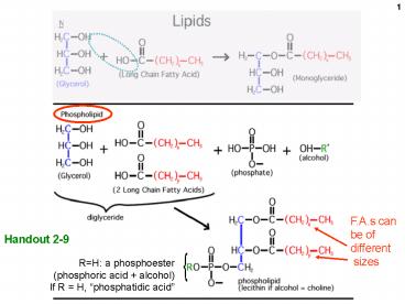
Handout 2-9
NH2
2
HO
HO
Handout 2-9
3
HO
HO CH2CH2NH3 (alcohol ethanolamine)
HO
Handout 2-9
4
Biological membranes are phospholipid bilayers
5
Incidentally, note the functional groups we have
met so far Hydroxyl Amine Amide Carboxyl Carbony
l Aldehyde Ketone Ester Carboxylic acid
ester Phosphoester Phosphodiester And Glycosi
dic bonds CC double bonds (cis and trans)
6
Handout 2-9
NH2
7
HO
HO
Handout 2-9
8
HO
HO CH2CH2NH3 (alcohol ethanolamine)
HO
Handout 2-9
9
Biological membranes are phospholipid bilayers
10
Incidentally, note the functional groups we have
met so far Hydroxyl Amine Amide Carboxyl Carbony
l Aldehyde Ketone Ester Carboxylic acid
ester Phosphoester And Glycosidic bonds CC
double bonds (cis and trans)
11
PROTEINS
Amino acids (the monomer of proteins)
12
At pH 7, ,most amino acids are zwitterions
(charged, but electrically neutral)
13
pH7
H
Equilibrium state of the carboxyl group lies far
towards the ionized molecule at pH7
14
OH- ( -H)
R OH
/ H3N - C CO H
R O- /
H2N - C CO H
R O-
/ H3N - C CO H
pH Net charge
1 1
11 -1
7 0
H
15
Numbering (lettering) amino acids
e-amino group
e
d
?
ß
alpha-carboxyl (attached to the a-carbon)
alpha-amino
alpha-carbon
16
Shown uncharged (as on exams)
17
(No Transcript)
18
(No Transcript)
19
H ? H 50 at pH7
20
Ball and stick physical model of an amino acid
21
Amino acids in 3 dimensions
- Asymmetric carbon (4 different groups attached)
- Stereoisomers
- Rotate polarized light
- Optical isomers
- Non-superimposable
- Mirror images
- L and D forms
From Sadava text
22
(No Transcript)
23
Condensation of amino acids to form a
polypeptide (must be catalyzed)
dehydration again
24
Parts of a polypeptide chain
25
Handout 3-3
The backbone is monotonous
It is the side chains that provide the variety
26
Polypeptides vs. proteins
- Polypeptide amino acids connected in a linear
chain (polymer) - Protein a polypeptide or several associated
polypeptides (discussed later) - Often used synonymously
- Peptide (as opposed to polypeptide) is smaller,
even 2 AAs (dipeptide)
27
The backbone is monotonous
(Without showing the R-groups)
It is the side chains that provide the variety
28
Proteins do most of the jobs in the cell E.g.,
egg albumin, hemoglobin, keratin, estrogen
receptor,immunoglobulins (antibodies), enzymes
(e.g., beta-galactosidase) Each is a polymer or
assemblage of polymers made up of amino
acidsEach particular protein polymer
(polypeptide) has a unique sequence of amino
acids . . . . and an English name.Each molecule
of a particular protein has the same sequence of
amino acids. E.g., met-ala-leu-leu-arg-glu-leu-v
al- . . . . How is this sequence determined?
29
Primary (1o) Structure the sequence of the
amino acids in the polypeptide chain
30
Determining the sequence
Carboxypeptidase hydrolyzes the peptide bond
One way use an enzyme (an old method, but
useful for teaching)
,
identify
e.g., . arg-leu-leu-val-gly-ala-gly-phe-trp-lys-g
lu-asp-ser . arg-leu-leu-val-gly-ala-gly
-phe-trp-lys-glu-asp
. arg-leu-leu-val-gly-ala-gly-phe
-trp-lys-glu
31
METHODS . . .
AA mixture (ala, glu, lys
(-)
()
Anode
Cathode
32
A paper electrophoresis apparatus
33
Handout 3-4
Side view
AAs applied at lower end
34
After stopping the paper chromatography
and staining for the amino acids
Rf 0.82 0.69 0.45 0.27 0.11
1.00
front
35
Paper chromatography apparatus
36
- Treatment of a polypeptide with trypsin
- Trypsin is a proteolytic enzyme.
- It catalyzes cleavage (hydrolysis) after lysine
and arginine residues
Polypeptide chain
37
But the order of the subpeptides is unknown. . .
. The sequence is reconstructed by noting the
overlap between differently produced subpeptides
Trypsin (lys, arg)
(1)
Chymotrypsin (trp, tyr, phe)
N
C
(2)
38
The order of the subpeptides is unknown. The
sequence is reconstructed by noting the overlap
between differently produced subpeptides
Trypsin (lys, arg)
(1)
Chymotrypsin (trp, tyr, phe)
N
C
(2)
39
The order of the subpeptides is unknown. The
sequence is reconstructed by noting the overlap
between differently produced subpeptides
Trypsin (lys, arg)
(1)
Chymotrypsin (trp, tyr, phe)
N
C
(2)
40
Fingerprinting a protein analysis of the
sub-peptides (without breaking them down to
their constituent amino acids)
Sub-peptides
No further digestion to amino acids left as
sub-peptides
41
Oligopeptides behave as a composite of their
constituent amino acids
-
-
Net charge -1 moves toward the anode in paper
electrophoreses Fairly hydrophobic (5/6)
expected to move moderately well in paper
chromatography
Nomenclature ala-tyr-glu-pro-val-trp or
AYEPVW or alanyl-tyrosyl-gluta
myl-prolyl-valyl-tryptophan
42
Hb
In fingerprinting, these spots contain peptides,
not amino acids
trypsin
Negatively charged
------valine------ (sickle)
Positively charged
More hydrophobic
------glutamate----- (normal)
More hydrophilic
Negatively charged
Positively charged
Negatively charged
Positively charged
43
Every different polypeptide has a different
primary structure (sequence). Every polypeptide
will have different arrangement of spots after
fingerprinting.
44
3-dimensional structure of proteins
One given purified polypeptide
- Molecule 1 N-met-leu-ala-asp-val-val-lys-....
- Molecule 2 N-met-leu-ala-asp-val-val-lys-...
- Molecule 3 N-met-leu-ala-asp-val-val-lys-...
- Molecule 4 N-met-leu-ala-asp-val-val-lys-...
etc.
clothesline . . .
45
Information for proper exact folding (How does a
polypeptide fold correctly?)
Predicting protein 3-dimensional structure
Determining protein 3-dimensional structure
Where is the information for choosing the correct
folded structure?
Is it being provided by another source or does
it reside in the primary structure itself?
46
Renaturation of a hard-boiled egg
Denature by heat
X
Cool, renature?
ovalbumin
Too long to sort out
Cool, entangled
Tangle, gel. Probably due to non-productive hydro
phobic interactions
47
urea
- chaotropic agent
- used at very high concentrations (e.g., 7 M)
- gentler, gradual denaturation, renaturation
48
Renaturation of ribonuclease after urea
urea, denature
- urea, renature
- ??
native ribonuclease active enzyme compact
denatured ribonuclease inactive enzyme random
coil
49
Slow denaturation of ribonuclease by urea
O Urea
H2N-CNH2
Ribonuclease in the bag is denatured
Macromolecules (protein here) cannot permeate
bag material
Small molecules (H20, urea) can.
Urea will move from areas of high
concentration to areas of low concentration
50
Christian Anfinsen
PRIMARY STRUCTURE DETERMINES TERTIARY STRUCTURE.
urea, denatures
- urea, renatures
The Anfinsen Experiment
Recommended
